
Abstract
Understanding the structure and function of the meniscus is critical to understanding its role in overall knee joint function. Injury to, or removal of, meniscal tissue may be associated with articular cartilage wear, knee instability, and, ultimately, the progression of osteoarthritis. While every effort is made for preserving and/or repairing damaged meniscal tissue, in some cases, the meniscus is not amenable to repair after injury. For appropriately indicated patients with symptomatic meniscal deficiency, meniscus allograft transplantation is an excellent surgical solution aimed at reducing pain and improving function. Indications for meniscus allograft transplantation are limited, and concomitant procedures such as osteotomy for malalignment, ligamentous, and/or articular cartilage restoration may be necessary in order to ensure an optimal result following meniscus allograft transplantation. Surgical techniques for meniscus allograft transplantation are variable and include soft-tissue fixation versus bone plug fixation versus bone bridge fixation. Outcomes following meniscus allograft transplantation are generally good to excellent, though reoperation rates are relatively high. The purpose of this article is to provide a concise review of recently published data on meniscus allograft transplantation, with a focus on recent outcomes studies.
Introduction
The medial and lateral menisci play a critical role in overall knee function. Injury to, or removal of, meniscal tissue may be associated with articular cartilage degeneration, knee instability, and, ultimately, the progression of osteoarthritis. Multiple authors have reported on the potentially devastating implications of meniscal deficiency, and every attempt should be made to preserve meniscal tissue in the setting of meniscus injury, particularly in the young and/or athletic patient population [1]. Unfortunately, damaged meniscus tissue is not always amenable to repair, and meniscectomy is unavoidable. For some patients, meniscus allograft transplantation (MAT) is a viable surgical solution that aims to reduce pain and improve function. First described in 1972 by Zuker and colleagues, the utilization of MAT has increased substantially, with continuous improvements in patient selection and surgical technique. Over the past two decades, there has been an exponential increase in data describing surgical anatomy, indications, techniques, and outcomes associated with MAT. The purpose of this article is to provide a concise review of recently published data on MAT, with a focus on recent outcomes studies.
Anatomical considerations
Comprised primarily of water (~75 % weight) and fibrocartilage consisting of type I collagen (~65 % dry weight), the menisci are wedge-shaped, semilunar structures that function to provide joint lubrication and nutrition, assist in shock absorption and load transmission during impact, and contribute to global knee stability. The orientation of the collagen fibers within the menisci is important to understand, as their anatomy directly impacts their function. Circumferential fibers function to resist hoop stresses, while radial fibers function to resist shear stress and maintain the integrity of the circumferential fibers. During weight-bearing, the menisci increase joint contact area and thus dissipate the compressive forces at the articular surface. In addition to distributing load during weight-bearing, the menisci deepen the tibial sockets and thereby function as secondary stabilizers to knee translation, especially the posterior horn of the medial meniscus.
During normal gait, the articular surface of the knee bears up to 6× body weight, with 70 % or more through the medial tibial plateau. The lateral meniscus is C-shaped structure that carries approximately 70 % of the lateral tibial-femoral compartment load during weight-bearing. The medial meniscus is a U-shaped structure with a wider separation between its anterior and posterior horns compared to the lateral meniscus and carries approximately 40 % of the medial tibial-femoral compartment load during weight-bearing. This difference in tibial plateau coverage, with the lateral meniscus covering proportionally more of the plateau compared to the medial meniscus, may account for the rapid deterioration of the lateral compartment articular surfaces following lateral meniscectomy [3, 21, 28, 41]. Joint contact forces, particularly in flexion, increase as meniscal deficiency occurs [41]. Specifically, loss of 20 % of meniscal tissue has been shown to lead to a 350 % increase in joint contact forces. For appropriately indicated patients (see indications section below), MAT is helpful in restoring native joint anatomy and biomechanics and is associated with reduced joint contact pressures compared to the menisectomized state [4, 18, 33].
Meniscal vascular anatomy is critical to understand, as the arterial supply to the menisci is directly related to the ability of a repaired meniscus, or MAT, to heal. The inferior medial and inferior lateral geniculate arteries provide a rich vascular network to the outer 10 to 30 % of the medial and lateral menisci, respectively, allowing for healing of transplanted allograft to the native meniscal rim and capsule following MAT.
Meniscal root anatomy is also important to understand, not only in the setting of an isolated MAT, but more importantly, in the setting of MAT with concomitant anterior cruciate ligament (ACL) reconstruction. The lateral meniscus anterior and posterior roots are in close proximity to each other; thus, the authors recommend utilizing the bone bridge technique for lateral MAT. In contrast, the medial meniscal roots, as described above, are further apart from each other; thus, medial MAT can be safely performed with either a bone bridge or with bone plugs (see “Surgical technique” section below). Overall, a thorough appreciation for meniscal anatomy and biomechanics is helpful when considering patients for MAT.
Indications and contraindications
A variety of factors must be considered when evaluating a potential candidate for MAT as a joint preservation strategy [23, 38–40]. MAT is a technically demanding procedure, and surgeon familiarly with the technique is critical. Further, not all patients with meniscal deficiency are candidates for MAT, and understanding appropriate indications is critical for ensuring a successful outcome. In the experience of the senior author, after performing more than 600 MATs over 18 years of clinical practice, general indications for MAT include:
-
Patients younger than 50 years old with a chief complaint of pain limiting their desired activities
-
BMI < 35 kg/m2
-
Previous meniscectomy (or non-viable meniscus state) with pain localized to the affected compartment
-
Normal or correctable coronal and sagittal alignment
-
Normal or correctable ligamentous stability
-
Normal or correctable articular cartilage
-
Willingness to comply with rehabilitation protocol
-
Realistic post-surgical activity expectations
Contraindications for MAT are controversial, as the available literature discussing contraindications is very limited. In the experience of the senior author, diffuse femoral and/or tibial articular cartilage wear and radiographic evidence of arthritis typically indicate that the patient is not a candidate for joint preservation with MAT, as the native joint anatomy will not provide a suitable environment to support the transplanted tissue. Further, caution should be exerted when considering MAT in patients with inflammatory conditions and obesity, though the literature is limited in these patient populations. Finally, MAT performed as a prophylactic measure in the absence of appropriate symptoms is highly controversial and not advocated by the senior author [12].
Patients with focal chondral defects can be consider for MAT with concomitant cartilage restoration, in either a staged or simultaneous fashion. In addition, patients with ligamentous deficiency and/or malalignment can also be considered for MAT, as long as those concomitant pathologies are addressed. The patient with clear evidence of meniscal insufficiency in the setting of a focal chondral defect, typically on the femoral side, is perhaps among the most challenging to treat. In these cases, it is difficult to determine if the patient’s symptoms are attributable to the meniscal deficiency, to the chondral defect, or to both pathologies. Advanced imaging with magnetic resonance imaging (MRI), which may show evidence of subchondral bone marrow edema underlying the cartilage defect, may indicate that pain, at least in part, is due to the chondral defect, and the surgeon should take this into consideration when performing MAT. Overall, each patient should be evaluated on a case-by-case basis, taking into account surgeon experience, patient symptoms, patient expectations, and concomitant pathologies.
Patient evaluation
History
A thorough history on any patient being considered for MAT should be performed. Careful attention should be paid to original injury mechanism, prior surgical procedures and timing of recent procedures, prior non-operative treatment attempts including injections and/or therapy, and current symptoms. Currently, there is no evidence to support “prophylactic” MAT; thus, patients with a history of meniscectomy without pain, swelling, mechanical symptoms, or functional deficits should not be considered for MAT. A typical history may include a patient with a sports-related injury who underwent one or more prior meniscectomies, with initial improvement, followed by unicompartmental joint pain associated with swelling and possibly mechanical symptoms. Most often, these symptoms are exacerbated by activity and the joint is not typically painful at rest. It can be helpful to assess prior arthroscopic images, if available, to better determine the status of the tibial and femoral articular surfaces.
Physical examination
Following the history, a comprehensive physical examination of both knees should be performed. Careful analysis of alignment and gait mechanics is critical, as abnormalities may impact clinical decision-making. For example, a patient with a clear varus deformity on visual inspection may not benefit from a medial MAT performed in isolation, even if the medial compartment articular cartilage is pristine, and concomitant and sometimes isolated high tibial osteotomy (HTO) may be required to protect the meniscus allograft from overload following transplantation. Visual inspection, palpation, range-of-motion, and ligamentous examinations should be performed in a systematic fashion. Patients may have a minor effusion and will typically have unicompartmental joint line tenderness to palpation. Range of motion and ligamentous stability are almost always normal, except in patients with concurrent ligamentous insufficiency.
Imaging
Imaging of affected knee is helpful in the evaluation of patients undergoing or being considered for MAT. A standard radiographic series, including anterior-posterior (AP), lateral, 45° posterior-anterior flexion weight-bearing and axial views, should be obtained. In addition, a standing long-leg mechanical axis view of both legs should be obtained. Advanced imaging with MRI can be helpful to evaluate for focal chondral defects and associated subchondral bone marrow edema. While ligamentous integrity should be appreciable on the physical examination, MRI can be helpful for evaluation of the ACL and other ligamentous structures.
Preoperative planning: allograft considerations
Once a patient has been deemed appropriate for MAT, the surgeon must complete the necessary steps to ensure an adequate graft is available.
Allograft sizing and matching
Meniscal allograft tissue is both compartment- and size-specific, and errors made during the matching process can lead to inferior outcomes. Specifically, allografts that are oversized have been associated with increased contact forces on the articular surface, whereas allografts that are undersized have been associated with increased contact forces along the allograft itself [9, 42]. Several sizing techniques have been described, and the senior author employs the technique as described by Pollard and colleagues [29], in which preoperative AP and lateral radiographs are utilized. Specifically, the AP view is used to measure meniscal width from the peak of the tibial eminence on the involve compartment to the medial tibial metaphyseal margin, and the lateral view is used to measure meniscal length from the anterior aspect of the tibia above the tibial tuberosity to the posterior plateau margin. After accounting for radiographic magnification, meniscal length measurements should be multiplied by 0.8 (medial) or 0.7 (lateral), to determine the true desired graft length.
Several authors [32, 34] have suggested incorporating donor gender, height, and weight as variables to improve allograft matching. In addition, other authors have suggested that allograft tissue from donors under age 45 is acceptable for transplantation [5].
Allograft harvesting and processing
Meniscal allografts should be harvested and frozen within 24 h of donor death. The most common preservation technique is the fresh-frozen method, which requires rapid cooling of the tissue to −80 °C. This method, while popular, has been shown to be associated with graft shrinkage, though the biomechanical consequences of this are unclear [13, 27]. Donors are assessed for communicable diseases, including hepatitis B and C, HIV, human T-lymphocytic virus, and syphilis, as well as for active infection with aerobic and anaerobic bacteria. While possible, disease transmission risks are minimal, especially with recent improvements in graft processing techniques. Current techniques utilize on aseptic, antibiotic soaks, as other strategies such as gamma irradiation and ethyl oxide compromise graft integrity [35]. In addition to processing techniques, the storage of the graft itself after harvest, and prior to transplantation, must be scrutinized, as an increased number of freeze-thaw cycles may be detrimental to graft function [22].
Surgical technique
There are a variety of reported techniques for MAT, most of which are arthroscopic-assisted or all-arthroscopic. Certain factors, including laterality and the need for concomitant procedures, dictate the specific technique employed by the surgeon for MAT. In general, graft fixation techniques include either all-suture fixation [17] or bone fixation. Bone fixation techniques are variable and include the use of bone plugs or the use of a bone bridge. Finally, bone bridge approaches are variable and include keyhole, trough, dove-tail, and bridge-in-slot techniques. The senior author utilizes a bridge-in-slot technique for the vast majority of his MAT procedures, and certainly for all lateral MAT procedures, due to the close proximity of the lateral meniscal anterior and posterior horns.
The senior author’s preferred technique for MAT has been previously published [7, 8, 25], with a summary of all critical steps as follows:
-
Anesthesia: general or spinal
-
Positioning: supine with foot of table down; operative leg in thigh holder with contralateral leg in a well-leg holder
-
Tourniquet: per surgeon preference
-
Landmarks: patella, patellar tendon, tibial tubercle, fibular head/neck
-
Diagnostic arthroscopy
-
Assess for concomitant pathologies
-
Debride remaining meniscal tissue in affected compartment to a stable rim of 1–2 mm of peripheral meniscal tissue
-
For medial MAT preparation ➔ take care to protect the medial ACL insertional fibers while attempting to visual medial tibial spine
-
-
Incision: anterior longitudinal incision to facilitate the mini-arthrotomy
-
Mini-arthrotomy: made through the patellar tendon, in line with its fibers
-
Accessory incision: posterolateral (for lateral MAT) or posteromedial (for medial MAT) incision is made (1/3 above, 2/3 below joint line) to facilitate the inside-out repair following insertion of the allograft
-
Take care to protect lateral collateral ligament and peroneal nerve during posterolateral incision (lateral MAT)
-
Take care to protect medial collateral ligament and saphenous nerve during posteromedial incision (medial MAT)
-
-
Tibial slot preparation
-
Use electrocautery to mark a line between the anterior and posterior meniscal horn insertions
-
Use a 4.5-mm burr to establish an initial slot along this line
-
The slot follows native tibial slope
-
The slot is the width and height of the burr itself
-
-
Use depth gauge to measure anterior-posterior length of tibial plateau
-
Insert guide pin just distal to, and parallel to, the initial slot
-
Use 8-mm cannulated reamer over the guide wire
-
Use box cutter to create tibial slot—typically 8 mm wide by 10 mm deep
-
Use rasp to smooth out all edges from the burr and box cutter
-
-
Allograft preparation: performed on the back table at any time during the case
-
Specimen arrives as a hemi-tibial plateau with attached meniscus
-
Thaw entire specimen in normal saline
-
Identify anterior and posterior horns
-
Create a bone bridge 10 mm deep and 7 mm wide (undersized by 1 mm compared to tibial slot, which will help facilitate graft passage)
-
Remove bone posterior to posterior meniscal horn attachment based on the distance between the posterior tibia and the posterior meniscal horn insertion
-
Keep bone anterior to anterior meniscal horn to maintain graft integrity during graft passage
-
Place a single 0-PDS suture (Ethicon, Blue Ash, OH) in a vertical mattress fashion at the junction of the posterior and middle thirds—this will be used as a traction stitch
-
-
-
Allograft passage
-
Arthroscope in same compartment portal (i.e., anterolateral portal for lateral MAT)
-
Zone-specific meniscal repair cannula in opposite portal (i.e., anteromedial portal for lateral MAT) with cannula aiming toward junction of posterior and middle thirds of remnant meniscal tissue
-
Pass flexible, nitinol passing wire through accessory incision (i.e., posterolateral incision for lateral MAT) and out the anterior arthrotomy
-
Insert PDS suture ends from traction stitch within the graft through the nitinol wire, and pull wire back through the accessory incision, bringing traction sutures (and allograft) into the joint through the anterior arthrotomy incision
-
Advance bone bridge into tibial slot under direct visualization
-
Confirm placement of tibial slot and reduction of meniscus in compartment
-
Secure bone bridge with a single 7 × 23 mm bioabsorbable interference screw with the knee in flexion (tap first)
-
Repair meniscus to native joint capsule using inside-out vertical mattress sutures (Fig. 1)
Fig. 1 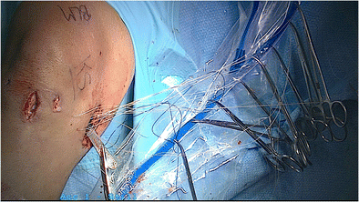
Intraoperative photograph demonstrating suture management prior to tying all sutures via the inside-out technique
-
All-inside fixation devices can be used posteriorly
-
8–10 sutures typically required
-
Vertical mattress sutures inferior and superior to graft reduce graft eversion
-
Tie sutures against capsule deep to superficial layers with the knee in extension to avoid creating a post-operative flexion contracture
-
-

Concomitant procedures
For patients with malalignment, focal chondral defects, and/or ligamentous insufficiency, concomitant osteotomy, articular cartilage restoration, and/or ligament reconstruction, respectively, must be performed in addition to MAT. These procedures can be performed prior to MAT as a staged surgery, or concurrently with MAT in a single procedure. If performing in a single procedure, MAT should be performed prior to osteotomy as well as prior to articular cartilage restoration. This is because the surgeon may need to place significant valgus or varus stress on the knee to accomplish the MAT which may compromise the osteotomy if performed first. In the setting of concomitant ACL reconstruction, a modified bridge-in-slot technique (with middle third of bone block removed) can be used, and the ACL graft can be a soft-tissue graft (instead of bone patellar tendon bone), to allow for a smaller tibial tunnel. For these cases, we recommend this sequence of steps:
-
Create the meniscal slot on the tibia
-
Drill the femoral and tibial ACL tunnels, and pass ACL graft and fix on femoral side
-
Use ACL tibial guide to drill two transtibial tunnels that exit into the meniscal slot
-
Place two stay sutures into meniscal allograft (one in posterior horn, one in anterior horn)
-
Use suture passer to pass these sutures through the two tibial tunnels
-
Reduce meniscal allograft in the slot using these two stay sutures
-
Tie sutures over a tibial bone bridge
-
Fix the ACL graft on the tibial side
-
Repair meniscus to capsule as described above
Complications
Complications following MAT are rare and are similar to those seen with standard meniscus repair. Potential complications include infection, neurovascular damage, stiffness, failure of healing, hardware irritation, reoperation, and re-tear. If the transplanted allograft is retorn, treatment is similar to that of a native meniscus and includes meniscectomy or repair, when indicated. In rare cases, revision MAT can be performed. Reoperation rates have been reported to be as high as 32 %, and reoperations within 2 years of the index MAT associated with ultimate failure [26]. Of note, reoperation is not indicative of failure, as the vast majority of reoperations are for debridement, and patients experience excellent outcomes following post-MAT arthroscopic debridement.
Rehabilitation
The senior author’s preferred approach to rehabilitation following MAT has been previously published [31]. In brief, patients are restricted to partial weight-bearing for 2 weeks with the knee locked in extension in a knee brace, with gentle range of motion allowed (0 to 90°). Weight-bearing is increased from weeks 3–8, and full weight-bearing and restoration of range of motion are expected by week 8. Running is allowed at 16 weeks, and return to full activity is allowed between 6 and 9 months following surgery.
Clinical outcomes
Since its first report in the 1970s, several dozen papers have been published that describe clinical outcomes following MAT [2, 7, 8, 11, 14, 19, 20, 25, 30, 31, 36, 37, 43]. Despite the volume of literature available, it is difficult to draw conclusions regarding the overall success of the procedure. This is for a variety of reasons, primarily due to the extreme heterogeneity in surgical technique, surgeon experience, and patient population. Variables including the presence or absence of concomitant procedures, including articular cartilage repair/restoration, realignment procedures, and/or ligament reconstruction, make it difficult to understand the relative contribution of the MAT compared to the additional procedure(s). Further, factors such as surgical technique (soft-tissue fixation versus bone plug versus bone bridge), compartment (medial versus lateral), and patient activity level (professional athlete versus not) are not often described in the abstracts of published papers, and careful scrutiny of the actual manuscript is necessary. Further, most studies describing MAT are Level IV, making the interpretation of MAT studies limited by the low level of evidence. Overall, we recommend caution when attempting to apply published results to any specific patient.
Overall, the short-, medium-, and long-term outcomes following MAT are encouraging. It should be remembered that patients undergoing MAT are typically in salvage situations, often having undergone one or more prior operations on the ipsilateral knee, with no other surgical or non-surgical solutions available. Despite the significant degree of pathology in their knees, these patients are typically young, high-demand, and with expectations to return to high levels of activities. In the largest study of MAT outcomes by a single surgeon to date, the overall survival rate of MAT is reported as 95 % at 5 years. Nearly one third of the patient population underwent a secondary surgery within the study period, with the vast majority of reoperations being arthroscopic debridement. While most patients undergoing secondary surgery still experienced excellent outcomes, undergoing surgery within 2 years of the index procedure was associated with an 8.4 odds ratio for future arthroplasty or revision MAT.
Two systematic reviews have recently been published discussing clinical outcomes of MAT, providing a more global overview of all available studies. In an analysis of 14 papers of MAT with bony fixation only, Hergan et al. [16] found overall good patient outcomes in patients undergoing MAT, with no differences noted in patients undergoing medial versus lateral MAT and no statistical differences between patients undergoing isolated MAT and those undergoing MAT with concomitant procedures. In a larger review, Elattar et al. [10] analyzed 44 papers consisting of a total of 1136 MATs in 1068 patients. Importantly, this review included a significantly heterogenous mix of patients over a period of three decades. Similar to the study by Hergan, the authors found clinical improvements in patients undergoing MAT, with “acceptable” complication and failure rates. An additional systematic review conducted by Harris et al. [15] analyzed the clinical outcomes of patients undergoing MAT with osteochondral autograft transfer, autologous chondrocyte implantation, osteochondral allograft transfer, or microfracture. Overall, the authors noted improved outcomes in all studies with an overall failure rate of 12 % at 36 months, but could not draw conclusions as to the impact of concomitant procedures on overall outcomes; as in several studies, the outcomes of combined procedures were equivalent to those of either procedure performed in isolation, whereas in other studies, patients undergoing combined procedures performed worse.
The outcomes of MAT in high-level athletes undergoing MAT are relatively limited, simply due to the low incidence of this procedure in this specific patient population.
Marcacci et al. [24] described the clinical outcomes following MAT in male professional soccer players, with 92 % of patients returning to soccer at an average 11 ± 3 months following surgery. While the authors described significant improvements outcome scores at 1 year, there were no significant improvements at final follow-up 3 years following surgery. In a separate study of “high-level” athletes participating in a variety of sports, Chalmers et al. [6] reported a 77 % return to play rate at an average 17 months following surgery. The authors reported significant improvements in nearly all outcomes scales at a follow-up of 3.3 years.
In addition to the heterogeneity of patient populations and surgical techniques within the available studies, part of the difficulty in interpreting MAT studies is understanding the criteria the authors use to define failure. Certainly, failure rates will change depending on the criteria applied, such as reoperation, revision MAT, conversion to arthroplasty, MRI-evidence of graft extrusion, and/or poor outcomes scores on validated knee outcomes assessment tools. Overall, clinical outcomes following MAT, whether performed in isolation or performed with concomitant procedures, and regardless of surgical technique employed or compartment affected, are acceptable, with most studies reporting improved clinical outcomes, regardless of the scoring system employed.
Summary
The clinical outcomes following subtotal meniscectomy are concerning, and MAT offers an acceptable surgical solution for appropriately indicated patients who are not amenable to meniscus repair. Future research efforts should be aimed at understanding the long-term outcomes of MAT, the ability of MAT to prevent or prolong knee arthroplasty, and the role, if any, of prophylactic MAT in the asymptomatic patient who has undergone meniscectomy.
References
Abrams GD, Frank RM, Gupta AK, Harris JD, McCormick FM, Cole BJ. Trends in meniscus repair and meniscectomy in the United States, 2005-2011. Am J Sports Med. 2013;41(10):2333–9.
Abrams GD, Hussey KE, Harris JD, Cole BJ. Clinical results of combined meniscus and femoral osteochondral allograft transplantation: minimum 2-year follow-up. Arthroscopy. 2014;30(8):964–70. e961.
Ahmed AM, Burke DL. In-vitro measurement of static pressure distribution in synovial joints—Part I: Tibial surface of the knee. J Biomech Eng. 1983;105(3):216–25.
Arnoczky SP, Warren RF, McDevitt CA. Meniscal replacement using a cryopreserved allograft. An experimental study in the dog. Clin Orthop Relat Res. 1990;252:121–8.
Bursac P, York A, Kuznia P, Brown LM, Arnoczky SP. Influence of donor age on the biomechanical and biochemical properties of human meniscal allografts. Am J Sports Med. 2009;37(5):884–9.
Chalmers PN, Karas V, Sherman SL, Cole BJ. Return to high-level sport after meniscal allograft transplantation. Arthroscopy. 2013;29(3):539–44.
Cole BJ, Carter TR, Rodeo SA. Allograft meniscal transplantation: background, techniques, and results. Instr Course Lect. 2003;52:383–96.
Cole BJ, Dennis MG, Lee SJ, et al. Prospective evaluation of allograft meniscus transplantation: a minimum 2-year follow-up. Am J Sports Med. 2006;34(6):919–27.
Dienst M, Greis PE, Ellis BJ, Bachus KN, Burks RT. Effect of lateral meniscal allograft sizing on contact mechanics of the lateral tibial plateau: an experimental study in human cadaveric knee joints. Am J Sports Med. 2007;35(1):34–42.
Elattar M, Dhollander A, Verdonk R, Almqvist KF, Verdonk P. Twenty-six years of meniscal allograft transplantation: is it still experimental? A meta-analysis of 44 trials. Knee Surg Sports Traumatol Arthrosc. 2011;19(2):147–57.
Farr J, Meneghini RM, Cole BJ. Allograft interference screw fixation in meniscus transplantation. Arthroscopy. 2004;20(3):322–7.
Frank RM, Yanke A, Verma NN, Cole BJ. Immediate versus delayed meniscus allograft transplantation: letter to the editor. Am J Sports Med. 2015;43(5):NP8–9.
Gelber PE, Gonzalez G, Lloreta JL, Reina F, Caceres E, Monllau JC. Freezing causes changes in the meniscus collagen net: a new ultrastructural meniscus disarray scale. Knee Surg Sports Traumatol Arthrosc. 2008;16(4):353–9.
Gomoll AH, Kang RW, Chen AL, Cole BJ. Triad of cartilage restoration for unicompartmental arthritis treatment in young patients: meniscus allograft transplantation, cartilage repair and osteotomy. J Knee Surg. 2009;22(2):137–41.
Harris JD, Cavo M, Brophy R, Siston R, Flanigan D. Biological knee reconstruction: a systematic review of combined meniscal allograft transplantation and cartilage repair or restoration. Arthroscopy. 2011;27(3):409–18.
Hergan D, Thut D, Sherman O, Day MS. Meniscal allograft transplantation. Arthroscopy. 2011;27(1):101–12.
Hunt S, Kaplan K, Ishak C, Kummer FJ, Meislin R. Bone plug versus suture fixation of the posterior horn in medial meniscalallograft transplantation: a biomechanical study. Bull NYU Hosp Jt Dis. 2008;66(1):22–6.
Jackson DW, Whelan J, Simon TM. Cell survival after transplantation of fresh meniscal allografts. DNA probe analysis in a goat model. Am J Sports Med. 1993;21(4):540–50.
Kang RW, Lattermann C, Cole BJ. Allograft meniscus transplantation: background, indications, techniques, and outcomes. J Knee Surg. 2006;19(3):220–30.
Lee AS, Kang RW, Kroin E, Verma NN, Cole BJ. Allograft meniscus transplantation. Sports Med Arthrosc. 2012;20(2):106–14.
Levy IM, Torzilli PA, Gould JD, Warren RF. The effect of lateral meniscectomy on motion of the knee. J Bone Joint Surg Am. 1989;71(3):401–6.
Lewis PB, Williams JM, Hallab N, Virdi A, Yanke A, Cole BJ. Multiple freeze-thaw cycled meniscal allograft tissue: a biomechanical, biochemical, and histologic analysis. J Orthop Res. 2008;26(1):49–55.
Lubowitz JH, Verdonk PC, Reid 3rd JB, Verdonk R. Meniscus allograft transplantation: a current concepts review. Knee Surg Sports Traumatol Arthrosc. 2007;15(5):476–92.
Marcacci M, Marcheggiani Muccioli GM, Grassi A, et al. Arthroscopic meniscus allograft transplantation in male professional soccer players: a 36-month follow-up study. Am J Sports Med. 2014;42(2):382–8.
Mascarenhas R, Yanke AB, Frank RM, Butty DC, Cole BJ. Meniscal allograft transplantation: preoperative assessment, surgical considerations, and clinical outcomes. J Knee Surg. 2014;27(6):443–58.
McCormick F, Harris JD, Abrams GD, et al. Survival and reoperation rates after meniscal allograft transplantation: analysis of failures for 172 consecutive transplants at a minimum 2-year follow-up. Am J Sports Med. 2014;42(4):892–7.
McDermott ID. What tissue bankers should know about the use of allograft meniscus in orthopaedics. Cell Tissue Bank. 2010;11(1):75–85.
Paletta Jr GA, Manning T, Snell E, Parker R, Bergfeld J. The effect of allograft meniscal replacement on intraarticular contact area and pressures in the human knee. A biomechanical study. Am J Sports Med. 1997;25(5):692–8.
Pollard ME, Kang Q, Berg EE. Radiographic sizing for meniscal transplantation. Arthroscopy. 1995;11(6):684–7.
Rue JP, Yanke AB, Busam ML, McNickle AG, Cole BJ. Prospective evaluation of concurrent meniscus transplantation and articular cartilage repair: minimum 2-year follow-up. Am J Sports Med. 2008;36(9):1770–8.
Saltzman BM, Bajaj S, Salata M, et al. Prospective long-term evaluation of meniscal allograft transplantation procedure: a minimum of 7-year follow-up. J Knee Surg. 2012;25(2):165–75.
Stone KR, Walgenbach AW, Turek TJ, Freyer A, Hill MD. Meniscus allograft survival in patients with moderate to severe unicompartmental arthritis: a 2- to 7-year follow-up. Arthroscopy. 2006;22(5):469–78.
Szomor ZL, Martin TE, Bonar F, Murrell GA. The protective effects of meniscal transplantation on cartilage. An experimental study in sheep. J Bone Joint Surg Am. 2000;82(1):80–8.
Van Thiel GS, Verma N, Yanke A, Basu S, Farr J, Cole B. Meniscal allograft size can be predicted by height, weight, and gender. Arthroscopy. 2009;25(7):722–7.
Vangsness Jr CT, Garcia IA, Mills CR, Kainer MA, Roberts MR, Moore TM. Allograft transplantation in the knee: tissue regulation, procurement, processing, and sterilization. Am J Sports Med. 2003;31(3):474–81.
Verbruggen D, Verschueren T, Tampere T, et al. Revision of meniscal transplants: long-term clinical follow-up. Knee Surg Sports Traumatol Arthrosc. 2014;22(2):351–6.
Verdonk PC, Verstraete KL, Almqvist KF, et al. Meniscal allograft transplantation: long-term clinical results with radiological and magnetic resonance imaging correlations. Knee Surg Sports Traumatol Arthrosc. 2006;14(8):694–706.
Verdonk R. Meniscal transplantation. Acta Orthop Belg. 2002;68(2):118–27.
Verdonk R, Almqvist KF, Huysse W, Verdonk PC. Meniscal allografts: indications and outcomes. Sports Med Arthrosc. 2007;15(3):121–5.
Verdonk R, Volpi P, Verdonk P, et al. Indications and limits of meniscal allografts. Injury. 2013;44 Suppl 1:S21–7.
Walker PS, Erkman MJ. The role of the menisci in force transmission across the knee. Clin Orthop Relat Res. 1975;109:184–92.
Wilmes P, Pape D, Kohn D, Seil R. The reproducibility of radiographic measurement of lateral meniscus horn position. Arthroscopy. 2007;23(10):1079–86.
Yanke AB, Chalmers PN, Frank RM, Friel NA, Karas V, Cole BJ. Clinical outcome of revision meniscal allograft transplantation: minimum 2-year follow-up. Arthroscopy. 2014;30(12):1602–8.
Compliance with ethical standards
Conflict of interest
Dr. Brian Cole has received research support from Aesculap/B.Braun, Cytori, Medipost, the National Institutes of Health (NIAMS & NICHD), and Zimmer. He has also received royalties from Arthrex, Inc., DJ Orthopaedics, Elsevier Publishing, Saunders/Mosby-Elsevier, and Slack Incorporated. In addition, he has received other financial or material support from Athletico, Ossur, Smith & Nephew, and Tornier and has been given stock or stock options from Carticept and Regentis.
Dr. Rachel Frank has nothing to disclose.
Human and animal rights and informed consent
This article does not contain any studies with human or animal subjects performed by any of the authors.
Author information
Authors and Affiliations
Corresponding author
Additional information
This article is part of the Topical Collection on Cartilage Repair Techniques in the Knee
Rights and permissions
About this article

Cite this article
Frank, R.M., Cole, B.J. Meniscus transplantation. Curr Rev Musculoskelet Med 8, 443–450 (2015). https://doi.org/10.1007/s12178-015-9309-4
Published:
Issue Date:
DOI: https://doi.org/10.1007/s12178-015-9309-4