
Abstract
There are four ways to achieve oxygenation: bag-mask-ventilation, ventilation using an extraglottic device, a tracheal tube and a surgical airway. The creation of a surgical airway is often viewed as the “last resort” in a dire “cannot intubate, cannot oxygenate” airway emergency. It is also incorporated into the difficult airway algorithms of various specialist societies. Despite its importance, there are many issues regarding timing of and the expertise of practitioners in performing a surgical airway. The aim of this chapter is to enhance the reader’s understanding of the anatomy of the cricothyroid membrane, the technique of performing a cricothyrotomy, and the importance of skill maintenance. The recent developments in ultrasound technology may help practitioners in identifying airway anatomy in patients with a predicted difficult airway. It should be emphasized that having to perform an open cricothyrotomy cannot be viewed as a personal failure to secure the airway by conventional methods. It is, after all, one of the four methods of ventilation and oxygenation.
Access provided by Autonomous University of Puebla. Download chapter PDF
Keywords
Introduction
Surgical airway is an invasive airway intervention incorporated into the difficult airway algorithms of various specialist societies [1–4]. It involves the creation of an infraglottic conduit to allow oxygenation and ventilation when the trachea could not be intubated through the glottis. Anesthesia practitioners favor cricothyrotomy for surgical airway access due to multiple reasons. Firstly, the airway is superficial at the level of the cricothyroid membrane, separated from the skin by subcutaneous tissue and the anterior cervical fascia. It is also easily identifiable with palpable landmarks such as the thyroid and cricoid cartilages. The trachea on the other hand, is situated deep in the neck and in close proximity with the thyroid gland and major vascular structures. Tracheotomy is associated with higher complication rates. As such, in an emergency setting, cricothyrotomy is preferred over tracheotomy due to its simplicity, speed and fewer complications.
This chapter will focus on the different techniques of cricothyrotomy, the associated complications and important issues surrounding the procedure.
History of Cricothyrotomy
In Virginia in December 1799…. the first President of the United States of America … lay struggling to breathe… The man kept shifting his position as he gasped for air. It was obvious the patient’s airway was severely compromised … One of the physicians present … was aware of tracheostomy but was disinclined to perform it, especially on such an important personage, because he believed the procedure to be futile… George Washington died from fully preventable suffocation due to an upper airway obstruction caused by bacterial epiglottitis [5].
Some of the earliest descriptions of a surgical airway access were found on Egyptian and Bronze Age artifacts as early as 3600 BC, but modern evolution of this medical procedure began only in the 1800s. The pandemic of morbus strangulatorius, or diphtheria, swept through Europe in the eighteenth and nineteenth centuries. Pierre Brentonneau, a French surgeon, first attempted to relieve the airway obstruction caused by this infectious laryngotracheobronchitis by tracheotomy in 1818. He finally succeeded in 1825 and in his paper published in 1826, he gave the disease the name diphterite (from the Greek word ‘diphtheria’, meaning ‘leather’), describing the appearance of pseudomembrane in the throat. Over the next two decades, Armand Trousseau routinely performed tracheotomy when required in patients suffering from diphtheria, and published his experience in 1851, where 127 out of 222 cases survived [6].
In 1909, Chevalier Jackson, an American laryngologist, described the first systematic approach to tracheotomy. He devised non-irritating and appropriately shaped tubes which helped to reduce mortality of the procedure to approximately 3 % [7]. In 1921, he published findings of a prohibitively high incidence of subglottic stenosis (92.9 %) in patients who had undergone high tracheotomy, or cricothyrotomy. He concluded that high tracheotomy should be abandoned and the only acceptable point of access was below the first tracheal ring (“low” tracheotomy) [8]. It was this landmark paper that subsequently condemned cricothyrotomy into oblivion.
Five decades later, the report by Brantigan and Grow in 1976 sparked a renewed interest in cricothyrotomy, challenging Jackson’s dogma against the procedure. The investigators reported their experience with 655 cardiovascular patients with cricothyrotomy tubes in place from hours to months and found no cases of chronic subglottic stenosis. Subsequent studies continued to refute Jackson’s findings [9, 10]. Sise et al. performed a prospective analysis of cricothyrotomy in 76 critically ill patients, the largest prospective observational study reported on this procedure. Morbidity and mortality of cricothyrotomy in adults were found to be similar to that reported for tracheotomy with only two documented cases of subglottic stenosis. They occurred in adolescent trauma patients, who underwent cricothyrotomy only after they had experienced complications associated with antecedent endotracheal intubation. Based on the findings, the authors recommended avoidance of cricothyrotomy in children and adolescences in view of the risk of subglottic stenosis [10]. In 2010, Talving et al. conducted a review of the 20 published series (17 retrospective reports and 3 prospective observational series) on cricothyrotomy between 1978 and 2008. Collectively, a total of 1134 patients were reviewed, including 368 trauma patients who underwent emergency cricothyrotomy, with a follow up ranged from 2 to 60 months. The rates of chronic subglottic stenosis among survivors were 2.2 % overall and 1.1 % in trauma patients. The authors concluded that cricothyrotomy after trauma is safe for initial emergency airway access, but they also acknowledged that routine conversion of cricothyrotomy to tracheotomy remains controversial and a well-designed prospective investigation is warranted [11].
The discrepancy between Jackson’s findings with the contemporary literature might be attributed to the following reasons. Firstly, many of Jackson’s patients were children, in whom the cricoid cartilage is known to be the narrowest part of the airway. Secondly, most of the cricothyrotomies were performed for inflammatory and infectious diseases, such as diphtheria, tuberculosis and epiglottitis. Thirdly, the Jackson’s technique for “high tracheotomy” involved the division of the cricoid and/or thyroid cartilages. Anatomical factors, disease process and traumatic surgical technique might have put these patients at a higher risk for subglottic stenosis.
The simplicity and relative safety of cricothyrotomy nowadays makes it an advantageous technique in an airway emergency.
Anatomy of the Glottis and Trachea
The cricothyroid membrane is a superficial structure, and familiarity of its anatomy and its surrounding structures is crucial to a fast, safe and successful cricothyrotomy.
The cricothyroid membrane (CTM) is a dense, fibroelastic, trapezoidal membrane or ligament. It arises from the cricoid cartilage and attaches to the thyroid and arytenoid cartilages. The CTM is bounded superiorly by the thyroid cartilage, inferiorly by the cricoid cartilage and laterally by the cricothyroid muscles (Fig. 13.1). The size of the CTM varies with adults, but is generally 9–10 mm high and 22–33 mm wide (at its widest border) [12]. The vocal cords are located approximately 1 cm above the CTM, so the risk of direct trauma during cricothyrotomy is relatively low. The CTM can be palpated as a depression below the laryngeal prominence of the thyroid cartilage (“Adam’s Apple”).

Anterior view of the cricothyroid membrane (ligament) (Reproduced, with permission, from Hung and Murphy [41])
The hyoid bone suspends the airway and is located above the thyroid cartilage. It is important to identify the hyoid bone so as to avoid misplacing the cannula or tube through the thyrohyoid membrane. In situations where the surface landmark is poorly palpable, the location of the hyoid bone is estimated to be at the midpoint of the horizontal distance between the mentum and the angle of mandible [13].
Important Vascular Structures
The superior thyroid artery, a branch of the external carotid artery, gives rise to the cricothyroid artery. The left and right cricothyroid arteries traverse the upper third of the CTM, where they anastomose in the midline (Fig. 13.2) [14]. To minimize the risk of arterial injury, it is recommended to puncture or make the incision at the lower third of the CTM and the incision should also not extend more than 1 cm laterally [12].

Vasculature of the cricothyroid membrane (Adapted, with permission from Walls and Murphy [42])
Major vascular structures, such as the carotid arteries and internal jugular veins, lie posterolateral to the cricoid cartilage, deep to the pretracheal fascia. Injury to these structures is unlikely if the cricothyrotomy is performed in the midline. However, the anterior jugular veins travel vertically down along the SCM muscle in the lateral aspect of the neck. A vertical skin incision and keeping it in the midline may reduce the risk of injuring these lateral structures [15, 16].
The pyramidal lobe of the thyroid gland is present in up to 65 % of the population [17]. In some individuals, it may extend as high as the hyoid bone. As the thyroid gland is rich in venous plexus, injury to the pyramidal lobe can lead to severe bleeding.
Anatomical Variations
Individual anatomic variations may pose technical difficulties in performing cricothyrotomy. Palpation of the CTM is more challenging in females due to a less prominent thyroid cartilage, and human dissection studies also show that the CTM is smaller in females [14]. Obesity may make landmark identification more difficult due to the presence of overhanging submental fat and increased neck circumference.
The airway anatomy of the pediatric population differs from the adult airway (Fig. 13.3). In young children and infants, the cricoid cartilage is located more cephalad in the neck than adults. The hyoid bone and cricoid cartilage are more prominent than the thyroid cartilage. In addition, the adiposity of their short necks makes landmark palpation more difficult and confusing. The CTM of young children and infants are also much narrower, with a mean height of only 2.6 mm in neonates [18]. The fragile mucosa is more susceptible to laceration and edema, which increase the risk of subglottic stenosis [12]. As such, open cricothyrotomy is not recommended in children under 10 years old, and needle cricothyrotomy or surgical tracheotomy are the preferred choices in this patient population.

Comparison of the pediatric and adult upper airway anatomy (Reproduced, with permission from Walls and Murphy [42])
Indications for Cricothyrotomy
The primary indication for an emergency cricothyrotomy (Table 13.1) is the inability to secure the airway by intubation or other non-invasive means via the transglottic route, i.e. a failed airway. Cricothyrotomy is considered the “last resort” in an emergency “can’t intubate, can’t oxygenate” (CICO) situation in various difficult airway guidelines, such as those published by the American Society of Anesthesiologists (ASA) and the Difficult Airway Society (DAS) in the UK [1–4]. However, the timing for this potentially life-saving manoeuvre often gets delayed due to the inability to recognize a failed airway and repeated, ineffective attempts at intubation or insertion of supraglottic devices [19]. In addition, the recent Fourth National Audit Project from the United Kingdom (NAP4) also identified poor planning, and lack of equipment as major contributory factors to poor outcomes [20]. Hence, it is crucial to have backup strategies when faced with a failed airway and promptly switch to alternative methods to minimize delay in establishing a surgical airway.
Although cricothyrotomy has been often taught as an emergency rescue technique in airway management, there are situations in which it should be considered as the primary airway of choice. An example would be a patient with severe maxillofacial injury, where it would be nearly impossible to intubate the trachea using the transglottic route. Another scenario would be a patient with severe upper airway edema (e.g. epiglottitis) or obstruction (e.g. tumor), where conventional techniques such as bronchoscopic and laryngoscopic intubation are unsuitable due to the distorted airway anatomy.
Contraindications to a Surgical Airway
When faced with an emergency CICO situation, and in the presence of severe hypoxemia, there are no true absolute contraindications to a surgical airway. However, there are various factors to consider when an emergency surgical airway is required (Table 13.2) [12].
Tracheal transection, cricoid cartilage and laryngeal fracture have been proposed as absolute contraindications to cricothyrotomy. Tracheotomy is the preferred method in these situations.
Obesity, burns to the neck, a large neck hematoma or infection, subcutaneous emphysema are relative contraindications as these pathologies may distort the airway anatomy and make surface landmark palpation much more difficult. Patients with systemic coagulopathy are more prone to bleeding and surgical airway should be performed with caution with careful hemostasis.
A surgical airway is generally not recommended for children less than 10 years of age [18]. The cricothyroid membrane is much narrower in young children, and the surface landmark is less prominent. The mucosa is also more friable and more susceptible to edema and laceration, leading to subglottic stenosis [21]. Hence, needle cricothyrotomy or a formal tracheotomy are the methods of choice in young children and infants.
Preparing for Cricothyrotomy
Adequate preparation is essential to ensure a successful cricothyrotomy. The following steps should apply to all techniques of cricothyrotomy:
-
(1)
Equipment
Airway practitioners should be familiar with the commercially available cricothyrotomy kits in their institutions. Kits for emergency surgical airway should be readily accessible and preferably located in a difficult airway cart.
-
(2)
Preparation
The procedure should be performed under aseptic conditions. If time permits, every effort should be made to ensure asepsis and proper infiltration of the surgical site with local anesthetic.
-
(3)
Patient positioning
While the patient with a potentially cervical spine injury should be placed in the supine position and the head and neck in a neutral position, it is necessary to extend the neck for proper exposure of the surgical landmarks. In patients with an anticipated difficult airway, practitioners may consider marking the site of the CTM before induction of anesthesia.
-
(4)
Identifying the cricothyroid membrane
Using the non-dominant hand, the laryngeal unit is stabilised by grasping the body of the thyroid cartilage between the thumb and middle finger (Fig. 13.4). This allows the index finger to move freely to palpate for the CTM. After identifying the laryngeal notch, the index finger moves inferiorly along the thyroid cartilage till it encounters a depression below the cartilage, which marks the location of the CTM. The hyoid bone, which lies above the thyroid cartilage, should also be identified, as wrongful insertion of a cricothyrotomy cannula through the thyrohyoid membrane will damage the vocal cords.
Fig. 13.4 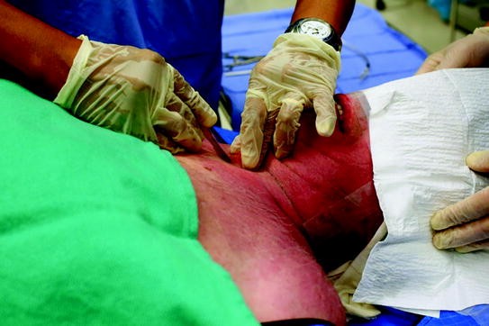
Stabilizing the cricothyroid membrane between the thumb and the middle finger
Should the surface landmarks be difficult to appreciate, the practitioner could estimate the level of the CTM by placing four fingers on the midline of the neck, with the last finger touching the suprasternal notch. The position of the index finger will coincide with the approximate location of the CTM.

Techniques
There are many methods of performing a cricothyrotomy. For simplicity, only the following methods of a surgical airway access will be outlined in this chapter:
-
(1)
Open cricothyrotomy
-
(2)
Seldinger cricothyrotomy
Open Cricothyrotomy
Equipment required to perform an open cricothyrotomy include a #11 scalpel, tracheal hook, Trousseau dilator and a small cuffed tracheal tube or a tracheostomy tube. The steps in performing this procedure are as follows:
-
(1)
A 5 cm vertical incision through the skin overlying the CTM is made in the midline and through the subcutaneous layer (Fig. 13.4).
-
(2)
Palpate for the cricothyroid membrane through the incision
-
(3)
Make a horizontal incision of the membrane along its lower border to avoid vascular injury.
-
(4)
Retract superiorly using a tracheal hook to stabilise the laryngeal unit. Traction in an upward and anterior direction brings the airway closer to the skin. The tracheal hook is then passed to an assistant, and the incision site through the cricothyroid membrane should be dilated using the index finger (Fig. 13.5).
Fig. 13.5 
Technique for open cricothyrotomy. Dilating the cricothyroid space with the index finger
-
(5)
Insert the Trousseau dilator to open up the cricothyroid space, and insert the small cuffed tracheal or tracheostomy tube in a caudad direction (Fig. 13.6)
Fig. 13.6 
Technique for open cricothyrotomy. Insertion of the tracheal tube with the aid of Trousseau dilator
-
(6)
Inflate the cuff and confirm proper tube placement by presence of end-tidal CO2 and auscultation
-
(7)
Remove the tracheal dilator
-
(8)
Secure the tube around the neck.


Seldinger Cricothyrotomy
Numerous commercial Seldinger cricothyrotomy sets are available. These kits come pre-assembled and most of them use a modified Seldinger technique, which is more familiar to anesthesia practitioners, hence more appealing. In general, these kits contain a scalpel blade, a syringe, an 18 G catheter over needle or introducer needle, a guidewire, a dilator and a cuffed airway catheter.
-
(1)
Make a vertical stab incision through the skin overlying the CTM
-
(2)
A puncture of the CTM in a caudad direction using the needle attached to a syringe (Fig. 13.7)
Fig. 13.7 
Technique for Seldinger cricothyrotomy. Aspiration of air confirms entry into the cricothyroid space
-
(3)
Positive air aspiration confirms entry into the intra-tracheal space
-
(4)
Remove the needle and thread the guidewire through the catheter caudally into the trachea (Fig. 13.8). Remove the cannula, leaving the guidewire in place
Fig. 13.8 
Technique for Seldinger cricothyrotomy. Passage of the guidewire through the cannula
-
(5)
Make a small cut in the skin along the guidewire. With the dilator loaded through the cuffed airway catheter, advance the dilator/catheter unit over the guidewire into the trachea (Fig. 13.9)
Fig. 13.9 
Technique for Seldinger cricothyrotomy. Railroading the cricothyrotomy cannula and dilator en bloc over the guidewire
-
(6)
Remove the guidewire and dilator and inflate the cuff. Confirm proper tube placement with the presence of end-tidal CO2 and auscultation
-
(7)
Connect the patient to a ventilator and commence ventilation


While it remains controversial, current recommendations view cricothyrotomy as a temporary life-saving measure. Conversion to a formal tracheotomy should be done once the patient is stabilized, usually within 72 h after cricothyrotomy.
Complications of Cricothyrotomy
The reported complication rates of cricothyrotomy vary depending on the technique used, the level of experience of the practitioner, the patient population, and the clinical situation. In general, the complication rates are higher for emergency cricothyrotomy (10–40 %) compared to their elective counterparts (6–8 %) [10, 13, 22].
Complications of cricothyrotomy can be divided into early and late complications, as illustrated in Table 13.3. McGill et al. reported an overall rate of 40 % in a study of 38 emergency cricothyrotomies. The most frequent problem identified was incorrect tube placement through the thyrohyoid membrane [13]. Bleeding is frequently associated with open cricothyrotomy and is usually due to superficial venous plexus injury, which can be easily controlled after securing the airway. Plexus injury can also be prevented by keeping the incision close to the midline and limiting its lateral extension. Severe bleeding is rarely encountered, although catastrophic and fatal hemorrhage due to laceration of the cricothyroid artery has been reported [13, 23, 24]. As the cricothyroid artery courses close to the thyroid cartilage, making the incision through the inferior half of the cricothyroid membrane for an open cricothyrotomy reduces the risk of hemorrhage. Caution should also be taken to avoid injury to the thyroid isthmus or its pyramidal lobe. Other early complications include prolonged procedure time, posterior tracheal wall perforation, aspiration of gastric content, and esophageal/mediastinal perforation.
Some complications are technique-related. Kinking of the guidewire is a common problem when performing the Seldinger technique and this increases the risk of tube misplacement. Laryngeal damage has been reported with open cricothyrotomy. McGill et al. reported a case of longitudinal fracture through the thyroid cartilage as a result of the insertion of an oversized tube, It is therefore recommended that the outer diameter of the tube should not exceed 8 mm [13, 25]. Reported long-term complications include subglottic stenosis, voice changes, scarring, infection, delayed bleeding, swallowing dysfunction, trachea-esophageal fistula and tracheomalacia [12]. Voice change is the most common complication, occurring in up to 50 % of cases [26]. Voice problems, such as dysphonia, hoarseness and weak voice, may be due to vocal cord paralysis from recurrent laryngeal nerve injury, vocal cord laceration, thyroid cartilage fracture or excessive traction on the thyroid cartilage [12]. Chronic subglottic stenosis occurs in approximately 2 % of cases [11] and is reported most frequently in long-term cricothyrotomy.
The mortality and morbidity of cricothyrotomy in adults are similar to that reported for tracheotomy [10]. Gillespie et al. also reported similar complication rates for emergency cricothyrotomy and tracheotomy, and no long-term complications were seen in the patients who received cricothyrotomy but were not subsequently converted to tracheotomy [27]. While conversion to a tracheotomy within 72 h of establishing cricothyrotomy has been advocated in view of the increased risk of subglottic stenosis, this remains a highly controversial subject which requires future investigations.
Other Considerations
Timing
Cricothyrotomy is indicated when maximal efforts at non-invasive methods have failed to relieve hypoxemia. Unfortunately, this decision is often delayed. The American Society of Anesthesiologists Closed Claims Project revealed that in two-thirds of the claims where airway emergency occurred, a surgical airway was obtained too late to avoid a poor outcome [19]. These findings were echoed in the Fourth National Audit Project (NAP4) in the UK, which showed that anesthetists failed to alter behavior even when faced with an airway crisis, favoring repeated attempts at tracheal intubation via direct laryngoscopy, despite knowledge that this strategy is rarely successful [20]. Although cricothyrotomy is placed as the final step in difficult airway algorithms, it must be instituted early to be an effective rescue option. Most reports of the timing of performing cricothyrotomy do not include “time taken to act” and “time taken to prepare” in their outcome measurements. Factoring the extra time required for preparation, cricothyrotomy should begin, and not just being considered, by the time hemodynamic instability supervenes. Surgeons should be present in the operating room, ready to perform an emergency tracheotomy in known challenging patients to minimize the delay of an airway rescue [28].
Training
A survey published in 2005 showed that most anesthetists preferred needle cricothyrotomy, and favored wire-guided (Seldinger) technique over open cricothyrotomy [29]. However, the NAP4 study revealed alarmingly low success with surgical airways performed by anesthetists, with a failure rate of 64 %. Success rates of narrow bore cricothyrotomy, wide bore cannula and open cricothyrotomy were 37, 57 and 100 % respectively. On the other hand, all surgical airways done by surgeons were successful. These observations concur with the findings of studies that reflected the poor ability of physicians to correctly identify the cricothyroid membrane in patients. Aslani et al. studied the accuracy of locating the cricothyroid membrane of fifty-six female patients by anesthetists and obstetrical trainees. Of the 112 identification attempts, only 30 % were correct, while only one accurate identification was made among 30 attempts in obese patients [30]. Elliott et al. showed that only 30 % of the attempts made by anesthetists accurately marked the area over the cricothyroid membrane, of which a mere 10 % were over its center point [31]. The authors conducted a similar study where 61 participants were asked to palpate for the cricothyroid membrane of 6 male and 6 female subjects. Out of the 186 attempts recorded, the overall success rate was only 42.5 %, with better success seen in male subjects (males 55.4 % vs. females 29.8 %, p = 0.001) [32]. Because cricothyrotomy is a life-saving core skill for the management of a CICO situation, these are disturbing findings.
The reasons for failure include problems with technique, training and equipment used. The rarity of having to perform a cricothyrotomy makes it a challenge to maintain the skill and to perform it proficiently when required. Training and confidence of practitioners in performing an open surgical airway need to be addressed. Many practitioners have little experience with the open technique and hence are uncomfortable with it, but those who have received training on manikins are more confident in using it in patients [29]. Systematic education using a combination of didactic teaching and hands-on practice will help improve practitioner skills and confidence [33, 34]. One study using a manikin model suggested that five percutaneous dilatational cricothyrotomies were required to reach a quick (<40 s), consistent, and successful performance [35]. Practitioners also need regular refresher training and practice on manikins, simulators, cadavers and/or animal models to maintain basic proficiency. The optimum retraining interval is unclear. However, one study demonstrated sustained skill retention for up to 6–8 months, and recommended training be repeated at 6 monthly intervals or less in order to maintain adequate skill level [36].
Other opportunities for practice and trainee education include the transtracheal injection of local anesthetics through a cricothyroid membrane puncture, as part of the topicalization process for an awake intubation [37]. Elective cricothyrotomies and percutaneous dilational tracheotomies in the operating room or ICU also offer similar opportunities for familiarization with airway anatomy and surgical skills.
Ultrasound of the Airway
With ultrasound machines now readily available in the operating rooms, emergency rooms and the ICU, there is an increased interest in the use of ultrasound to identify airway anatomy and to locate the cricothyroid membrane. Various techniques have been developed to allow quick identification of the anatomy relevant to cricothyrotomy [38–40]. Advantages of ultrasound include the ability to locate relevant structures for invasive airway intervention in patients with difficult landmark palpation and in confirmation of tracheal tube placement. Such technology is useful in patients with a known or predicted difficult airway, where ultrasound can be utilized for the identification of the cricothyroid membrane prior to the induction of anesthesia. However, the usefulness of ultrasound in a dire CICO situation where rapid identification of the cricothyroid membrane is required, remains to be investigated.
Summary
Cricothyrotomy is a life-saving procedure in a “cannot intubate, cannot oxygenate” situation. While proficiency is of paramount importance, the ability to recognize a CICO emergency and early decision making are critical to a successful emergency cricothyrotomy. It is recommended that practitioners be familiar with the equipment available in their institutions. Practitioners should also be proficient in more than one technique, so as to choose the most appropriate method in the context encountered.
Being a rare event, most practitioners have never performed a cricothyrotomy and are uncomfortable and unfamiliar with it, resulting in high failure rates. Frequent and effective training is required to improve and maintain this important skill set.
References
American Society of Anesthesiologists Task Force on Management of the Difficult Airway (1993) Practice guidelines for management of the difficult airway. A report by the American Society of Anesthesiologists Task Force on Management of the Difficult Airway. Anesthesiology 78:597–602
American Society of Anesthesiologists Task Force on Management of the Difficult Airway (2003) Practice guidelines for management of the difficult airway: an updated report by the American Society of Anesthesiologists Task Force on Management of the Difficult Airway. Anesthesiology 98:1269–77
Apfelbaum JL, Hagberg CA, Caplan RA et al (2013) Practice guidelines for management of the difficult airway: an updated report by the American Society of Anesthesiologists Task Force on Management of the Difficult Airway. Anesthesiology 118:251–270
Henderson JJ, Popat MT, Latto IP, Pearce AC (2004) Difficult Airway Society guidelines for management of the unanticipated difficult intubation. Anaesthesia 59:675–694
Szmuk P, Ezri T, Evron S, Roth Y, Katz J (2008) A brief history of tracheostomy and tracheal intubation, from the bronze age to the space age. Intensive Care Med 34:222–228
Frost EA (1976) Tracing the tracheostomy. Ann Otol Rhinol Laryngol 85:618–624
Brantigan CO, Grow JB Sr (1980) Cricothyroidotomy revisited again. Ear Nose Throat J 59:289–295
Jackson C (1921) High tracheotomy and other errors: the chief causes of chronic laryngeal stenosis. Surg Gynecol Obstet 32:392–395
Francois B, Clavel M, Desachy A, Puyraud S, Roustan J, Vignon P (2003) Complications of tracheostomy performed in the ICU: subthyroid tracheostomy vs. surgical cricothyroidotomy. Chest 123:151–158
Sise MJ, Shackford SR, Cruickshank JC, Murphy G, Fridlund PH (1984) Cricothyroidotomy for long-term tracheal access. A prospective analysis of morbidity and mortality in 76 patients. Ann Surg 200:13–17
Talving P, DuBose J, Inaba K, Demetriades D (2010) Conversion of emergent cricothyrotomy to tracheotomy in trauma patients. Arch Surg 145:87–91
Boon JM, Abrahams PH, Meiring JH, Welch T (2004) Cricothyroidotomy: a clinical anatomy review. Clin Anat 17:478–486
McGill J, Clinton JE, Ruiz E (1982) Cricothyrotomy in the emergency department. Ann Emerg Med 11:361–364
Dover K, Howdieshell TR, Colborn GL (1996) The dimensions and vascular anatomy of the cricothyroid membrane: relevance to emergent surgical airway access. Clin Anat 9:291–295
Narrod JA, Moore EE, Rosen P (1985) Emergency cricothyrostomy—technique and anatomical considerations. J Emerg Med 2:443–446
Walls RM (1988) Cricothyroidotomy. Emerg Med Clin North Am 6:725–736
Blumberg NA (1981) Observations on the pyramidal lobe of the thyroid gland. S Afr Med J 59:949–950
Navsa N, Tossel G, Boon JM (2005) Dimensions of the neonatal cricothyroid membrane—how feasible is a surgical cricothyroidotomy? Paediatr Anaesth 15:402–406
Peterson GN, Domino KB, Caplan RA, Posner KL, Lee LA, Cheney FW (2005) Management of the difficult airway: a closed claims analysis. Anesthesiology 103:33–39
Cook TM, Woodall N, Frerk C (2011) Major complications of airway management in the UK: results of the fourth national audit project of the Royal College of Anaesthetists and the Difficult Airway Society. Part 1: anaesthesia. Br J Anaesth 106:617–631
Kress TD, Balasubramaniam S (1982) Cricothyroidotomy. Ann Emerg Med 11:197–201
Erlandson MJ, Clinton JE, Ruiz E, Cohen J (1989) Cricothyrotomy in the emergency department revisited. J Emerg Med 7:115–118
Kodsi IS, Deckelbaum DL (2013) Hemorrhage from the cricothyroid artery due to cricothyrotomy: a case report. J Oral Maxillofac Surg 71:571–576
Schillaci CR, Iacovoni VF, Conte RS (1976) Transtracheal aspiration complicated by fatal endotracheal hemorrhage. N Engl J Med 295:488–490
American Association of Clinical Anatomists, Educational Affairs Committee (1999) The clinical anatomy of several invasive procedures. Clin Anat 12:43–54
Cole RR, Aguilar EA 3rd (1988) Cricothyroidotomy versus tracheotomy: an otolaryngologist’s perspective. Laryngoscope 98:131–135
Gillespie MB, Eisele DW (1999) Outcomes of emergency surgical airway procedures in a hospital-wide setting. Laryngoscope 109:1766–1769
Hamaekers AE, Henderson JJ (2011) Equipment and strategies for emergency tracheal access in the adult patient. Anaesthesia 66(Suppl 2):65–80
Wong DT, Lai K, Chung FF, Ho RY (2005) Cannot intubate-cannot ventilate and difficult intubation strategies: results of a Canadian national survey. Anesth Analg 100:1439–1446
Aslani A, Ng SC, Hurley M, McCarthy KF, McNicholas M, McCaul CL (2012) Accuracy of identification of the cricothyroid membrane in female subjects using palpation: an observational study. Anesth Analg 114:987–992
Elliott DS, Baker PA, Scott MR, Birch CW, Thompson JM (2010) Accuracy of surface landmark identification for cannula cricothyroidotomy. Anaesthesia 65:889–894
Lamb A et al (2013) The accuracy of cricothyroid membrane identification by palpation. In: Annual Meeting of the American Society of Anesthesiologists. Abstract presentation number: A3162
Latif R, Chhabra N, Ziegler C, Turan A, Carter MB (2010) Teaching the surgical airway using fresh cadavers and confirming placement nonsurgically. J Clin Anesth 22:598–602
Naik VN, Matsumoto ED, Houston PL et al (2001) Fiberoptic orotracheal intubation on anesthetized patients: do manipulation skills learned on a simple model transfer into the operating room? Anesthesiology 95:343–348
Wong DT, Prabhu AJ, Coloma M, Imasogie N, Chung FF (2003) What is the minimum training required for successful cricothyroidotomy?: a study in mannequins. Anesthesiology 98:349–353
Kuduvalli PM, Jervis A, Tighe SQ, Robin NM (2008) Unanticipated difficult airway management in anaesthetised patients: a prospective study of the effect of mannequin training on management strategies and skill retention. Anaesthesia 63:364–369
Hung O, Scott J, Mullen T, Murphy M (2012) Waiting to exhale! Anesth Analg 114:927–928
Nicholls SE, Sweeney TW, Ferre RM, Strout TD (2008) Bedside sonography by emergency physicians for the rapid identification of landmarks relevant to cricothyrotomy. Am J Emerg Med 26:852–856
Tsui BC, Hui CM (2008) Sublingual airway ultrasound imaging. Can J Anaesth 55:790–791
Prasad A, Singh M, Chan VW (2009) Ultrasound imaging of the airway. Can J Anaesth 56:868–869 (author reply 9–70)
Hung O, Murphy MF (2008) Management of the difficult and failed airway, 2nd edn. McGraw-Hill, New York
Walls RM, Murphy MF (2004) Manual of emergency airway management, 2nd edn. Lippincott Williams & Wilkins, Philadelphia
Author information
Authors and Affiliations
Corresponding author
Editor information
Editors and Affiliations
Rights and permissions
Copyright information
© 2014 Springer International Publishing Switzerland
About this chapter
Cite this chapter
Zhang, J., Hung, O. (2014). Surgical Airway. In: Khan, Z. (eds) Airway Management. Springer, Cham. https://doi.org/10.1007/978-3-319-08578-4_13
Download citation
DOI: https://doi.org/10.1007/978-3-319-08578-4_13
Published:
Publisher Name: Springer, Cham
Print ISBN: 978-3-319-08577-7
Online ISBN: 978-3-319-08578-4
eBook Packages: MedicineMedicine (R0)