
Abstract
The supraglottic airway (SGA) is a device designed for upper airway management, serving as a bridge, with respect to invasiveness, between facemask ventilation and endotracheal intubation. Most devices consist of an inflatable silicone or polyvinyl chloride mask and connecting tube. When blindly inserted into the pharynx, it forms a low-pressure seal around the laryngeal inlet. The SGA allows ventilation and oxygenation with less stimulation than laryngoscopy and intubation. The laryngeal mask airway (LMA) is the most commonly used SGA and has the largest body of experience and literature. The LMA was invented by Dr. Archie Brain and became commercially available in the UK in 1988 [1]. Dr. Brain remains involved in its continued development and evolution. The device has allowed for the advancement of anesthetic techniques for ambulatory and other types of surgery. It is particularly useful when intubation is difficult, hazardous, or unsuccessful (see Table 7.1).
Access provided by Autonomous University of Puebla. Download chapter PDF
Keywords
- Airway Management
- Laryngeal Mask Airway
- Positive Pressure Ventilation
- Difficult Airway
- Laryngeal Mask Airway ProSeal
These keywords were added by machine and not by the authors. This process is experimental and the keywords may be updated as the learning algorithm improves.
Introduction
The supraglottic airway (SGA) is a device designed for upper airway management, serving as a bridge, with respect to invasiveness, between facemask ventilation and endotracheal intubation. Most devices consist of an inflatable silicone or polyvinyl chloride mask and connecting tube. When blindly inserted into the pharynx, it forms a low-pressure seal around the laryngeal inlet. The SGA allows ventilation and oxygenation with less stimulation than laryngoscopy and intubation. The laryngeal mask airway (LMA) is the most commonly used SGA and has the largest body of experience and literature. The LMA was invented by Dr. Archie Brain and became commercially available in the UK in 19881. Dr. Brain remains involved in its continued development and evolution. The device has allowed for the advancement of anesthetic techniques for ambulatory and other types of surgery. It is particularly useful when intubation is difficult, hazardous, or unsuccessful (see Table 7.1).
If successfully placed, the SGA can provide rescue ventilation or serve as a conduit for flexible bronchoscopic (FB) intubation. It has also been used to provide temporary ventilation between laryngoscopy attempts or while a surgical airway is being performed. With multiple applications, the SGA is an important option within the ASA and other difficult airway algorithms (Figure 7.1). The device has undergone an evolution over time with adaptations to provide for safer use, greater ease of insertion, and greater intubation success. And yet while SGAs are in widespread use, there is evidence that this approach is sometimes not used appropriately, with poor patient selection, inappropriate device selection, and failure to pay attention to details relating to insertion, fixation, or removal techniques2. This discussion will describe its role in management of the difficult airway. The LMA was the first widely used supraglottic device and later devices were designed to emulate or improve upon its success. The techniques described in this chapter are applicable to all supraglottic devices, except where otherwise stated.

Role of the supraglottic airway (SGA) in the ASA Difficult Airway Algorithm.
Basic Use
The LMA provides an adequate airway choice for many outpatient procedures and is most commonly used in ambulatory surgery. While its use and applications continue to expand, it is important to be aware of its principal limitations, which are: (1) the potential for regurgitation and/or aspiration, (2) possible gastric inflation, and (3) the potential for displacement. The device is contraindicated for elective use in patients with known or suspected full stomach. In addition, the device may not seat properly in patients with abnormal or obstructive lesions of the oropharynx and may produce inadequate ventilation. Furthermore, conditions characterized by increased airway resistance or diminished compliance may not be successfully managed with a supraglottic device3, particularly the “first generation” devices, such as the LMA Classic™.
The insertion technique for most SGAs is best accomplished with the patient’s head in the sniffing position or with slight extension. Using the dominant hand, the device is pushed along the roof of the mouth and the posterior wall of the pharynx until it stops, much as a bolus of food being swallowed. The correctly positioned SGA tip lies at, and partially blocks, the upper esophagus (Figure 7.2). It is important to avoid over-inflation of the cuff of any SGA device. This happens commonly in an effort to achieve a better seal and is sometimes the source of postoperative discomfort or unintended displacement. The first generation LMA (the LMA Classic™) was manufactured using medical grade silicone and was reusable. It could be autoclaved up to 50 times (Figure 7.3). The proper insertion technique was important to avoid folding of the mask or downfolding of the epiglottis4. Subsequently, disposable masks (e.g. LMA Unique™) have become popular and more economical, the efficacy generally assumed to be comparable to their reusable counterparts. While important, insertion technique has been de-emphasized because of the inherent stiffness of polyvinyl chloride and other newer materials.

Insertion technique for the LMA classic (reproduced with permission from Brimacombe JR. Laryngeal mask anesthesia, Chapter 8: placement phase. Philadelphia: Saunders; 2005).

The LMA Classic™.
Following successful placement, it is important not to exceed the maximum cuff inflation volumes. If the maximum inflation volume is necessary to maintain a seal, the use of a larger size mask should be considered. Clinical studies have shown that a better seal is obtained using a larger size with less air5. Using an over-inflated smaller mask will exert more pressure on the hypopharyngeal mucosa, impairing local blood flow and producing a poor fit within the pharyngeal space. Increased leak, gastric insufflation, and malpositioning are more likely when the maximum cuff volume is exceeded and may be associated with adverse events.
Many clinicians prefer to utilize spontaneous ventilation with SGAs as this allows for monitoring of respiratory rate and facilitates an assessment of anesthetic depth. This technique essentially provides a “hands free” alternative to facemask ventilation and is used commonly in short anesthetic procedures.
Positive pressure ventilation (PPV) with a SGA is a useful technique for longer procedures or may be utilized when spontaneous ventilation is inadequate. PPV with a SGA may be achieved with or without muscle relaxants. When a relaxant technique is chosen, the relaxant drug may be given either before or after insertion. Leaks during PPV may be attributable to light anesthesia, use of too small a SGA, a reduction in lung/chest wall compliance related to the surgical or diagnostic procedure, patient factors, or displacement of the device by head turning or traction6. It is important to monitor the peak inspiratory pressures during ventilation, as this is often an indication of adequate placement, appropriate anesthetic depth, and lack of obstruction to ventilation. Placement of a bite block adjacent to the SGA, if one is not integrated into the device, is helpful in avoiding airway obstruction during emergence. Prior to emergence from anesthesia, the muscle relaxant should be reversed or allowed to wear off before discontinuing the anesthetic agent. With gentle, assisted ventilation, the patient should be allowed to resume normal spontaneous ventilation. The SGA is removed, usually without deflation, as the patient begins to swallow and demonstrates a return of airway reflexes. Current recommendations suggest a maximum duration of 6–8 h with frequent evaluation and adjustment of intracuff pressure, particularly in the presence of nitrous oxide. The true limits, however, are unknown. When intracuff pressure was maintained at 60-cm H2O, less sore throat was reported by patients7. Prolonged use has been associated with potential nerve injury, dislodgement, or mucosal injury.
The LMA as a Rescue Device
In the setting of a failed intubation, if adequate mask ventilation cannot be obtained or maintained, urgent placement of a familiar SGA should be considered. Generally, insertion of the LMA is not more difficult in patients with Mallampati class III or IV airways or in those patients in whom laryngoscopy reveals Cormack-Lehane Grade 3 or 4 views8. It is unclear whether this is equally true for all other SGAs. Following numerous reports of successful LMA use in failed intubation and ventilation, the LMA was incorporated into the “Practice Guidelines for management of the difficult airway” in 19939. In the 2003 ASA Difficult Airway Practice guidelines, the LMA (specifically) moved up to the first position in the “Can’t Intubate, Can’t Ventilate” portion of the algorithm10. In patients whose lungs cannot be ventilated because of supraglottic obstruction and whose trachea cannot be intubated due to unfavorable anatomy (but not periglottic pathology), the LMA should be immediately available and considered as the first treatment choice11 (Figure 7.1).
Case reports detail the successful use of the LMA in a variety of congenital and acquired syndromes associated with difficult airway management. The LMA has been successfully used as a rescue airway device after failed intubation in patients with lingual tonsillar hyperplasia, but more difficult and traumatic insertion of the LMA has been reported in that situation12. The placed LMA (and likely other SGAs) can be (1) left in place, (2) removed for intubation attempts with a flexible bronchoscope (FB), or (3) used as a conduit for intubation (see below).
In the patient with a full stomach, cricoid pressure may interfere with correct placement of the SGA13. A reasonable alternative between competing concerns of continuously maintaining cricoid pressure in a patient at risk for aspiration and failure to properly insert the SGA is to momentarily release cricoid pressure as the distal tip of the SGA reaches the hypopharynx. This maximizes the chance of correct SGA placement while minimizing risk of aspiration. Once the LMA (and possibly other SGAs) is in situ, it probably does not interfere with the efficacy of cricoid pressure14.
Flexible Bronchoscopic Intubation Via the SGA
The SGA is useful in failed intubations as a rescue airway, and it can also serve as a conduit for endotracheal intubation. The aperture bars of the LMA were intended to prevent the epiglottis from obstructing the shaft (Figure 7.3). If adequate ventilation is possible through the LMA, it is probable that the bowl of the LMA surrounds the larynx and FB-guided tracheal intubation will be successful. Endotracheal intubation through the LMA was first described in 1989 by Allison and McCrory using a gum-elastic bougie (GEB)15, but their initial success could not be easily duplicated. While it is tempting to consider blindly placing an endotracheal tube through the LMA, this is not recommended. First, the LMA must be properly seated to ventilate the patient. In patients with distorted airway anatomy and when proper seating and ventilation are suboptimal, it is less likely that a GEB or blind passage through the LMA will result in tracheal placement.
Lim and co-workers evaluated three types of ETTs and three head and neck positions finding a significant difference with both variables. Although the best head and neck position for blind intubation through the LMA-C was the sniffing position, this was only successful in approximately half the cases16. Conversely, intubation over a FB passed through the LMA is highly successful in most series. Thus, if the difficulty during conventional intubation was due to unfavorable anatomical alignment and not periglottic pathology and one can ventilate through a SGA, it provides a suitable conduit to the trachea17. If ventilation is poor after SGA insertion, because of poor apposition to the laryngeal aperture, or there is major periglottic pathology, FB-guided tracheal intubation may be difficult.
Bronchoscopic intubation through the SGA may be performed electively or as a rescue maneuver. It has been described in numerous clinical situations and is the cornerstone for management of failed intubation in pediatric patients18. Advantages of this technique include the ability to oxygenate the patient effectively before and between attempts at ETT placement and the ability to inspect the airway above the glottis. It may also be a good choice in the patient unwilling or unable to cooperate with awake intubation.
When using the LMA C, the aperture bars may dictate the size of the ETT unless an Aintree Intubation Catheter (AIC) is used (see below). The ETT is loaded onto a lubricated FB. The endoscope is passed through the LMA aperture bars and under the epiglottis, until a clear view of the glottis is obtained. The FB is advanced well into the trachea until the carina is in view. The FB is then stabilized as the lubricated ETT is advanced over the FB, through the LMA and into the trachea. If resistance is encountered, the ETT may have to be withdrawn a few centimeters, rotated 90°, and re-advanced one or more times to move the tip past the glottis. The ETT and the LMA are held fixed and the FB is withdrawn (Figure 7.4). Inflation of the ETT cuff, ventilation through the ETT, and confirmation with ETCO2 and bilateral breath sounds should then proceed. The LMA is deflated and the ETT is secured to the shaft of the device. The entire unit (ETT and LMA) can remain in place until exchanged.

Fiberoptic bronchoscope via the LMA Classic™.
A 6.0 mm-ID cuffed ETT may be passed over the FB and through the shaft of a size #3 or #4 LMA and a 7.0 mm-ID cuffed ETT may be passed over the FB and through the shaft of a size #5 LMA. If a larger ETT is desired, the LMA and the ETT can be exchanged for a larger ETT over a tube exchanger or an AIC (see below).
Other Supraglottic Airways for Intubation
The SGA for intubation is primarily used as an airway intubator in adults with difficult airways, but can also be used as an alternative to routine laryngoscopy. (Care must be taken to avoid the use of a SGA to avoid intubating the patient if no backup plan exists.) These devices can facilitate airway management in patients who may be difficult to ventilate and intubate such as morbidly obese patients, patients with sleep apnea, or patients with limited neck movement. Furthermore, SGAs can be very helpful in the unanticipated difficult airway. A properly performed insertion causes less hemodynamic response; therefore it could be a less stimulating alternative to rigid laryngoscopy for patients with cardiovascular comorbidities. The Intubating Laryngeal Airway (ILA, Mercury Medical, Clearwater, FL) was introduced as an alternative device for airway management. It features a modified cuff, wider shaft, and lacks the aperture bars characteristic of the LMA (Figs. 7.5 and 7.6). The disposable version is called the Air-Q®.

Endotracheal tubes via LMA Excel™ and ILA™.

Intubating laryngeal airway (ILA™).
The Air-Q® is inserted using the same technique as standard SGAs and inflated. Blind insertion of an ETT is not recommended and flexible or rigid endoscopes (air-VU®, Mercury Medical) are used. After intubation, the device can be easily removed using the manufactured stylet or the device can remain in place to facilitate a smooth extubation (Figure 7.7a, b).

(a) Fiberoptic intubation via the ILA™. (b) Removal of the ILA™ using the removal stylette.
Awake Placement of the Laryngeal Mask
There are numerous descriptions of using the LMA in properly prepared awake patients as a conduit for fiberoptic intubation19. There are several reasons why this technique is suitable when the preoperative evaluation indicates the patient should be intubated awake. First, even though awake tracheal intubation can be performed by many techniques in a properly prepared patient, insertion of a SGA is generally tolerated producing little hemodynamic change. The relative lack of stimulation in passing an LMA reduces the amount of preparation (topicalization, sedation) that a semi-conscious patient may require for the procedure. Lighter sedation also facilitates the act of swallowing which aids in the insertion of the LMA. Second, when the SGA is in good position, the shaft of the device is directed toward the larynx, and visualization of the laryngeal aperture with a FB is easy. Third, the patient who maintains spontaneous breathing is less likely to become hypoxemic than if ventilation ceases or intubation is prolonged. Fourth, and perhaps most importantly, with the patient awake, no options are eliminated, and risk remains low even if there is difficulty in inserting the SGA or the FB.
Proper placement of the SGA into an awake patient also allows the device to be used as a primary means of airway control in patients whose airways are potentially difficult, but who may not require endotracheal intubation. The topicalization technique is similar to that required for awake FB intubation (see Chap. 3), with perhaps more emphasis on the oropharynx to block the glossopharyngeal nerves. This technique is particularly useful in patients with obstructive sleep apnea as the SGA can rapidly overcome the cause(s) of obstruction and allow unimpeded spontaneous ventilation. Whether performed in awake or asleep patients, the SGA allows for opening of the laryngeal space, which is beneficial in the bloodied or soiled airway.
The Intubation LMAs (Fastrach™ and CTrach™)
The Intubating LMA (Fastrach™)
The intubating LMA (ILMA) was developed to allow for endotracheal intubation via the laryngeal mask as well as provide for efficient ventilation of the patient20,21. Introduced in 1997, the ILMA was designed to overcome the shortcomings of FB techniques through the LMA classic and to improve the success rate of blind intubation. The limitations of the FB techniques included reduced ETT size, LMA tube length and inner diameter, aperture bars, and the frequent need to replace the entire apparatus, which included the LMA. The ILMA consists of a mask attached to an anatomically shaped rigid stainless steel shaft, which aligns with the glottis. The angle of the metal shaft was specially designed to fit into the oral and pharyngeal space while maintaining the head and neck in neutral position (Figure 7.8)22.

The LMA Fastrach™ and its parts.
The ILMA has a 13-mm internal shaft diameter that can accommodate an 8.0-mm cuffed endotracheal tube, which can be advanced into the larynx either blindly or with FB guidance. The shaft is short enough to ensure that the tracheal tube cuff extends beyond the vocal cords. The mask of the ILMA is similar to the classic LMA, however, the aperture bars are replaced by an epiglottic-elevating bar, which facilitates tube placement. The device is best utilized with special tubes, which have soft, blunt tips and flexible shafts (Figure 7.9).

The LMA Fastrach™ showing blind and fiberoptic insertion techniques (reproduced with permission from Brimacombe JR. Laryngeal mask anesthesia, Chapter 18: Intubating LMA for airway intubation. Philadelphia: Saunders; 2005).
These ILMA-ETTs have blunt tips and low-compliance cuffs, requiring careful cuff inflation to avoid tracheal mucosal injury. Until recently, these were only available as expensive reusable tubes, often inadvertently discarded. Alternatively, a standard ETT can be inserted using the ILMA. This mostly requires softening the tube in warm water and/or turning the ETT 180° prior to insertion into the ILMA. This maneuver reduces the natural curve of most polyvinylchloride tubes and allows placement (Figure 7.10)21. The ILMA can be used electively in patients with normal airways as an intubating tool. It can also be utilized to prevent excessive hemodynamic response in patients with cardiovascular compromise. Early clinical reports described its benefits in patients with reduced or immobilized cervical spine disease because of the neutral insertion technique. A multi-centered survey by Ferson et al. described use of the ILMA in 254 patients with difficult-to-manage airways and reported an overall success rate for blind and FB-guided intubations of 97 % and 100 %, respectively23. The classic LMA as well as the ILMA can be inserted in a variety of patient positions (lateral, prone, sitting) when required for rescue ventilation24.

Placement of the Euromedical ETT and standard polyvinylchloride ETT, with 180° curve.
While the ILMA has been used extensively as a device for airway management in patients with limited neck movement, Combes et al. initially demonstrated its role in the failed ventilation/intubation scenario. In a prospective study involving over 11,000 patients, difficult ventilation was encountered in only 6 patients and intubation using the ILMA was attempted in 15 failed intubations. Success was achieved on the first attempt in 9 and the second attempt in 3 with failure occurring in 225. Like other airway techniques, the ILMA should be practiced in normal airways to gain proficiency. The device is most successful following failed intubation in patients with seemingly normal airways. It can, however, be placed in awake patients with topical anesthesia when FB-guided intubation fails or is unavailable. Shung et al. in South Africa described successful awake intubation through an ILMA in 15 patients with difficult airways26.
For successful intubation in the anesthetized patient with the ILMA and CTrach™ (see below), the “Chandy maneuver” is extremely helpful23 (Figure 7.11). Before insertion of these devices, an adequate depth of anesthesia must be confirmed and muscle relaxants (or tracheal topicalization) should be administered. After insertion in the neutral position followed by ventilation, the “Chandy maneuver” should be performed, which consists of two steps performed sequentially. The first step, which is important for establishing optimal ventilation, is to rotate the ILMA slightly in the sagittal plane using the metal handle to optimize chest compliance during bag ventilation. The second step, performed just before blind intubation, consists of using the metal handle to slightly lift (but not tilt) the ILMA away from the posterior pharyngeal wall. This facilitates the smooth passage of the ETT into the trachea. It is recommended that one remove the ILMA after intubation, using the stabilizing rod to keep the ETT in place as the ILMA is removed.

The “Chandy maneuver” (reproduced with permission from Ferson et al.23).
Frappier et al. studied the use of the ILMA in 118 consecutive morbidly obese patients for elective surgery with a success rate of 96.3 %. In their hands the ILMA was an effective and safe ventilatory device associated with a high success rate for blind intubation in morbidly obese patients27. Nevertheless, the choice of the primary technique (laryngoscopy or ILMA) for tracheal intubation of an adult obese patient remains to be determined. The ILMA has been included in the difficult airway algorithms in the United Kingdom28 and Europe and has gained acceptance in pre-hospital use in many regions. Despite its success in these patients, many continue to have concerns about use of the ILMA in unstable cervical spine injuries based on cadaver studies29. In multi-trauma situations, the risk of cervical spine injury must sometimes be balanced against the need for timely oxygenation and ventilation.
The ILMA C-Trach™ is a modification of the ILMA that combines the features of the ILMA with a fiberoptic system and a detachable LCD screen. The device was released in 2005, but was recently discontinued. A detachable screen provided a video image, acquired from a video sensor located in the epiglottic elevator. This facilitates alignment of device and ETT advancement through the laryngeal inlet30 (Figure 7.12). Liu and associates described 48 patients with difficult airways managed electively or following failed laryngoscopy using the LMA CTrach™. Their report detailed the importance of optimizing ventilation, which was not possible in two of their patients31. Awake placement of the LMA CTrach™ has been successfully accomplished and described in morbidly obese patients with difficult airways32.

The LMA C-Trach™.
LMAs with Gastric Access
The LMA ProSeal™
The ProSeal LMA (PLMA) is a laryngeal mask device that features a larger cuff and a drainage tube, allowing access to the gastrointestinal tract. This device was the first of the “second generation” SGAs, significantly addressing the limitations of the LMA C (and other first generation SGAs): (1) the potential for gastric inflation and (2) the risk of aspiration of gastric contents (Figure 7.13). Brain’s design goal was to construct a laryngeal mask with improved ventilatory characteristics that also offered protection against regurgitation and gastric inflation33. The PLMA was introduced into clinical practice in 2000 and was designed primarily for elective use. Its silicone construction is allegedly softer than that of the LMA-C and is better designed to conform to the contours of the hypopharynx. This device incorporates a second lumen, arising from the distal end of the laryngeal mask and terminating outside of the patient airway. This lumen, termed the gastric drain, has been demonstrated to passively vent regurgitated esophageal contents34. A gastric tube can also be placed down this lumen to empty the stomach.

The LMA ProSeal™.
The PLMA is designed for advanced clinical uses such as prolonged operative procedures, surgical procedures in the lateral or prone position, and to extend SGA benefits to a greater number of patients. While the PLMA may be used with spontaneous ventilation, it is well suited for PPV, with and without muscle relaxants. The design of the PLMA permits PPV at higher peak pressures. It has been used electively as an alternative to endotracheal intubation for laparoscopic surgery in a number of reports35,36 though some question the wisdom of this practice37. The advantages have primarily been the ease of insertion, lack of hemodynamic and airway reflex stimulation, and smooth emergence from anesthesia with minimal coughing and hypertension. While these are features of the LMA C™, the PLMA extends these benefits to larger patients or those with mild or controlled gastric reflux. Studies comparing the PLMA to the LMA Classic™ have demonstrated higher airway sealing pressure with the PLMA without an increase in mucosal pressure33. There are isolated case reports of its use in failed intubation and ventilation emergency situations38. Rosenblatt reported a case where the PLMA was used to decompress the abdomen after repeated attempts at intubation had failed and mask ventilation had caused extensive gastric inflation39.
A pocket-like opening for the index finger, which helps to maintain proper insertion technique, facilitates insertion of the PLMA. The PLMA can also be inserted using an optional insertion tool, which obviates the need of the operator inserting fingers into the patient’s mouth (Figure 7.14). The PLMA was the first SGA that provided a sequence of steps and tests to assure proper placement. These are important points for use because the device has a larger surface area and potentially can fold-over in the hypopharynx40,41. This might provide ventilation, but may not permit optimal positioning or gastric decompression. Brimacombe reported on the technique of using an Eschmann intubation guide and laryngoscope for successful placement of the PLMA. This insertion technique had a high success rate and was of particular advantage when PLMA placement proved challenging42,43. This would be recommended in the event that laryngoscopy and intubation failed and it was deemed necessary to place the device in patients with possible gastric contents (Figure 7.15). There are no aperture bars in the PLMA, but there is a bite block to protect the patient from biting the shaft and occluding the airway. The PLMA was designed for effective and efficient ventilation. It was not designed to be a conduit for endotracheal intubation though this is possible with a fiberoptic bronchoscope using an AIC (see below).

The LMA ProSeal™ with finger cuff and optional insertion tool.

A gum-elastic bougie may be used to facilitate placement of the LMA ProSeal™.
LMA Supreme™
The LMA Supreme™ (SLMA), released in 2007, combines the integrated bite block and gastric drainage tube of the PLMA and the preconfigured shape of the ILMA Fastrach™. This single-use plastic device has a high-volume/low-pressure cuff providing a higher seal pressure and fixation tab to help secure the airway44 (Figs. 7.16 and 7.17). The device is intended for elective use as well as potential use in pre-hospital and resuscitative scenarios. Like the PLMA, the gastric drainage tube reduces the risk of regurgitation and aspiration if properly seated.

The LMA Supreme™.

Aspiration of contents from the gastric port of the LMA Supreme™.
Several studies have compared the SLMA and PLMA. The SLMA has slightly lower oropharyngeal leak pressures than the PLMA45. The success of the first attempt insertion was higher for the SLMA and this is to be expected because of its design. The device appears to be efficacious and easy-to-use in elective ambulatory procedures. The higher rate of success on first attempt insertion may make it more suitable as an airway rescue device46. It is also intended for use in cardiopulmonary resuscitation procedures as well as in the failed intubation and the “can’t intubate-can’t ventilate” situation47.
The ease of insertion of the SLMA™ has been one of its most attractive features. It has been described for insertion in the prone position. This approach may be utilized in the stressful circumstance of accidental extubation of a patient in the prone or lateral position. Some expert users describe elective SLMA placement when the patient is induced and the airway secured in the prone position48.
The SLMA should not be used for the resuscitation of patients who are not profoundly unconscious unless adequate topical anesthesia has been provided.
Intubating Through LMAs (Classic, Supreme, and ProSeal: Using the Aintree Intubation Catheter)
The AIC (Cook Medical, Bloomington IN) is similar in concept to the Cook Airway Exchange Catheter® (see Chap. 16). Its larger internal diameter (4.8 mm) will accommodate a lubricated FB of 4.0 mm OD or smaller (Figure 7.18)49. The AIC’s external diameter (6.5 mm) allows its use with endotracheal tubes 7.0 mm and larger. The AIC facilitates the placement of a larger endotracheal tube, otherwise precluded by the inner diameter of the SGA shaft or the aperture bars. It should be noted that the AIC is 56 cm in length, leaving a relatively limited amount of flexible scope to protrude beyond the catheter distally (Figure 7.19). After introducing the SGA and establishing successful ventilation, the FB is introduced into the AIC. This FB-AIC assembly is introduced through the shaft of the SGA and into the trachea. When the FB is within the trachea, the AIC is advanced

Aintree catheter with fiberoptic bronchoscope.

Aintree catheter with Ambu SGA.
-
(a)
The bronchoscope is withdrawn
-
(b)
The SGA is removed over the AIC
-
(c)
An ETT is advanced over the AIC
-
(d)
A Rapi-Fit™ connector can be attached to the AIC to permit PPV between maneuvers (Figure 7.20)
Figure 7.20. 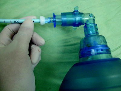
Aintree catheter with “Rapi-fit” connector.

This technique is suitable for use with a wide range of SGAs. It has a role in elective management of the patient in whom bag mask ventilation is difficult or in whom intubation difficulties are anticipated or have been encountered50,51.
Conclusion
SGAs represent a considerable advancement in the management of routine and complex airways. This chapter has devoted disproportionate attention to the LMA because it has been used more widely and more has been written about it than all the other SGAs combined; however as of this writing, over 70 varieties of SGAs are available. For some of these devices, there is limited evidence, but others such as the igel™ (Intersurgical, Wokingham UK) and air-Q™ (Mercury Medical, Clearwater FL) are generating interest and clinical support. “First generation” SGAs represented a better mask; “second generation” SGAs separated the respiratory and gastrointestinal tract, at least theoretically reducing the risk of aspiration while at the same time providing a more effective seal for PPV. All can be used as conduits for endotracheal intubation, particularly when combined with a flexible bronchoscope and an AIC.
For all their advantages, a number of issues remain. In a prospective study involving nearly three million general anesthetics in the U.K., SGAs were used in 56 % of cases and were involved in 33/133 of major airway complications resulting in death, brain injury, surgical airway, or unintended ICU admission2,52,53. These complications occurred as a result of aspiration, airway trauma, failure to establish or loss of the airway, and extubation-related complications. Improper insertion technique, inadequate confirmation, poor patient selection, inappropriate device selection with excessive reliance on first-generation SGAs, and the use of SGA in a patient with a difficult airway, without an adequate backup plan in the event of failure, were the emerging themes. The SGAs represent an advance in airway management, but patient safety will only be enhanced if the user has acquired proficiency through frequent use and critical self-assessment. In a patient with a known or presumed difficult airway, reliance upon a SGA to avoid tracheal intubation without an adequate backup plan does not advance patient safety.
References
Brain AI. The laryngeal mask—a new concept in airway management. Br J Anaesth. 1983;55(8):801–5.
Cook TM, Woodall N, Frerk C. Major complications of airway management in the UK: results of the Fourth National Audit Project of the Royal College of Anaesthetists and the Difficult Airway Society. Part 1: anaesthesia. Br J Anaesth. 2011;106(5):617–31.
Benumof JL. Laryngeal mask airway. Indications and contraindications. Anesthesiology. 1992;77(5):843–6.
Brain A. Proper technique for insertion of the laryngeal mask. Anesthesiology. 1990;73(5):1053.
Gaitini LA, Vaida SJ, Somri M, Yanovski B, Ben-David B, Hagberg CA. A Randomized controlled trial comparing the ProSeal™ laryngeal mask airway with the laryngeal tube suction in mechanically ventilated patients. Anesthesiology. 2004;101(2):316–20.
Brimacombe J, Berry A, Verghese C. The laryngeal mask airway: its uses in anesthesiology. Anesthesiology. 1994;80(3):706.
Seet E, Yousaf F, Gupta S, Subramanyam R, Wong DT, Chung F. Use of manometry for laryngeal mask airway reduces postoperative pharyngolaryngeal adverse events: a prospective, randomized trial. Anesthesiology. 2010;112(3):652–7.
Brimacombe J, Berry A. Mallampati classification and laryngeal mask airway insertion. Anaesthesia. 1993;48(4):347.
American Society of Anesthesiologists Task Force on Management of the Difficult Airway. Practice guidelines for management of the difficult airway. A report by the American Society of Anesthesiologists Task Force on Management of the Difficult Airway. Anesthesiology. 1993;78(3):597–602.
Practice Guidelines for Management of the Difficult Airway. An updated report by the American Society of Anesthesiologists Task Force on Management of the Difficult Airway. Anesthesiology. 2003;98(5):1269–77.
Benumof JL. Laryngeal mask airway and the ASA difficult airway algorithm. Anesthesiology. 1996;84(3):686–99.
Ovassapian A, Glassenberg R, Randel GI, Klock A, Mesnick PS, Klafta JM. The unexpected difficult airway and lingual tonsil hyperplasia: a case series and a review of the literature. Anesthesiology. 2002;97(1):124–32.
Asai T, Morris S. The role of the laryngeal mask for failed tracheal intubation in the patient with a “full stomach”. Anesth Analg. 1994;78(4):817–9.
Brimacombe J, Berry A, White A. An algorithm for use of the laryngeal mask airway during failed intubation in the patient with a full stomach. Anesth Analg. 1993;77(2):398–9.
Allison A, McCrory J. Tracheal placement of a gum elastic bougie using the laryngeal mask airway. Anaesthesia. 1990;45(5):419–20.
Lim SL, Tay DHB, Thomas E. A comparison of three types of tracheal tube for use in laryngeal mask assisted blind orotracheal intubation. Anaesthesia. 1994;49(3):255–7.
Benumof JL. Use of the laryngeal mask airway to facilitate fiberscope-aided tracheal intubation. Anesth Analg. 1992;74(2):313–5.
Rabb MF, Minkowitz HS, Hagberg CA. Blind intubation through the laryngeal mask airway for management of the difficult airway in infants. Anesthesiology. 1996;84(6):1510–1.
Asai T. Fiberoptic tracheal intubation through the laryngeal mask in an awake patient with cervical spine injury. Anesth Analg. 1993;77(2):404.
Parr M, Baskett GM. The intubating laryngeal mask. Anaesthesia. 1998;53:343.
Gerstein NS, Braude DA, Hung O, Sanders JC, Murphy MF. The Fastrach(TM) intubating laryngeal mask Airway(R): an overview and update. Can J Anaesth. 2010;57(6):588.
Brain AI, Verghese C, Addy EV, Kapila A. The intubating laryngeal mask. I: development of a new device for intubation of the trachea. Br J Anaesth. 1997;79(6):699–703.
Ferson DZ, Rosenblatt WH, Johansen MJ, Osborn I, Ovassapian A. Use of the intubating LMA-Fastrach in 254 patients with difficult-to-manage airways. Anesthesiology. 2001;95(5):1175–81.
Dimitriou V, Iatrou C, Brimacombe J, Voyagis GS. Intubating laryngeal mask airway in lateral position. Anesthes Analg. 2004;99(6):1877; author reply.
Combes X, Le Roux B, Suen P, Dumerat M, Motamed C, Sauvat S, et al. Unanticipated difficult airway in anesthetized patients: prospective validation of a management algorithm. Anesthesiology. 2004;100(5):1146–50.
Shung J, Avidan MS, Ing R, Klein DC, Pott L. Awake intubation of the difficult airway with the intubating laryngeal mask airway. Anaesthesia. 1998;53(7):645–9.
Frappier J, Guenoun T, Journois D, Philippe H, Aka E, Cadi P, et al. Airway management using the intubating laryngeal mask airway for the morbidly obese patient. Anesthesh Analg. 2003;96(5):1510–5, table of contents.
Henderson JJ, Popat MT, Latto IP, Pearce AC. Difficult Airway Society guidelines for management of the unanticipated difficult intubation. Anaesthesia. 2004;59(7):675–94.
Brimacombe J, Keller C, Kunzel KH, Gaber O, Boehler M, Puhringer F. Cervical spine motion during airway management: a cinefluoroscopic study of the posteriorly destabilized third cervical vertebrae in human cadavers. Anesth Analg. 2000;91(5):1274–8.
Timmermann A, Russo S, Graf BM. Evaluation of the CTrach—an intubating LMA with integrated fibreoptic system. Br J Anaesth. 2006;96(4):516–21.
Liu EH, Goy RW, Lim Y, Chen FG. Success of tracheal intubation with intubating laryngeal mask airways: a randomized trial of the LMA Fastrach and LMA CTrach. Anesthesiology. 2008;108(4):621–6.
Wender R, Goldman AJ. Awake insertion of the fibreoptic intubating LMA CTrach in three morbidly obese patients with potentially difficult airways. Anaesthesia. 2007;62(9):948–51.
Brimacombe J, Keller C. The ProSeal laryngeal mask airway: a randomized, crossover study with the standard laryngeal mask airway in paralyzed, anesthetized patients. Anesthesiology. 2000;93(1):104–9.
Keller C, Brimacombe J, Kleinsasser A, Loeckinger A. Does the ProSeal laryngeal mask airway prevent aspiration of regurgitated fluid? Anesth Analg. 2000;91(4):1017–20.
Maltby JR, Beriault MT, Watson NC. Use of the laryngeal mask is not contraindicated for laparoscopic cholecystectomy. Anaesthesia. 2001;56(8):799.
Maltby JR, Beriault MT, Watson NC, Liepert DJ, Fick GH. LMA-Classic and LMA-ProSeal are effective alternatives to endotracheal intubation for gynecologic laparoscopy. Can J Anaesth. 2003;50(1):71–7.
Cooper RM. The LMA, laparoscopic surgery and the obese patient—can vs should. Can J Anesth. 2003;50(1):5–10.
Baxter S, Brooks A, Cook T. Use of a proseal LMA for maintenance after failed intubation during a modified rapid sequence induction. Anaesthesia. 2003;58(11):1132–3.
Rosenblatt WH. The use of the LMA-ProSeal in airway resuscitation. Anesth Analg. 2003;97(6):1773–5.
O’Connor Jr JCJ, Borromeo CJ, Stix MS. Assessing ProSeal laryngeal mask positioning: the suprasternal notch test. Anesth Analg. 2002;94(5):1374–5.
Osborn IP, Behringer EC, Cooper RM, Verghese C. Detecting the etiologies of acute airway obstruction associated with the laryngeal mask airway supreme(TM). Anesthesiology. 2009;111(2):451–2. doi:10.1097/ALN.0b013e3181adf285.
Brimacombe J, Howath A, Keller C. A more ‘failsafe’ approach to difficult intubation with the gum elastic bougie. Anaesthesia. 2002;57(3):292.
Brimacombe J, Keller C. Aspiration of gastric contents during use of a ProSealTM laryngeal mask airway secondary to unidentified foldover malposition. Anesth Analg. 2003;97(4):1192–4.
Verghese C, Ramaswamy B. LMA-Supreme—a new single-use LMA with gastric access: a report on its clinical efficacy. Br J Anaesth. 2008;101(3):405–10.
Tan BH, Chen EG, Liu EH. An evaluation of the laryngeal mask airway supreme’ in 100 patients. Anaesth Intensive Care. 2010;38(3):550–4.
Seet E, Rajeev S, Firoz T, Yousaf F, Wong J, Wong DT, et al. Safety and efficacy of laryngeal mask airway Supreme versus laryngeal mask airway ProSeal: a randomized controlled trial. Eur J Anaesthesiol. 2010;27(7):602–7.
Abdi W, Dhonneur G, Amathieu R, Adhoum A, Kamoun W, Slavov V, et al. LMA supreme versus facemask ventilation performed by novices: a comparative study in morbidly obese patients showing difficult ventilation predictors. Obes Surg. 2009;19(12):1624–30.
Lopez AM, Valero R, Brimacombe J. Insertion and use of the LMA Supreme in the prone position. Anaesthesia. 2010;65(2):154–7.
Bogdanov A, Kapila A. Aintree intubating bougie. Anesth Analg. 2004;98(5):1502.
Blair EJ, Mihai R, Cook TM. Tracheal intubation via the classic and proseal laryngeal mask airways: a manikin study using the aintree intubating catheter. Anaesthesia. 2007;62(4):385–7.
Micaglio M, Ori C, Parotto M, Feltracco P. Three different approaches to fibreoptic-guided intubation via the Laryngeal Mask Airway Supreme. J Clin Anesth. 2009;21(2):153–4.
Woodall N, Frerk C, Cook TM. Can we make airway management (even) safer?—lessons from national audit. Anaesthesia. 2011;66 Suppl 2:27–33.
Woodall NM, Cook TM. National census of airway management techniques used for anaesthesia in the UK: first phase of the Fourth National Audit Project at the Royal College of Anaesthetists. Br J Anaesth. 2011;106(2):266–71.
Author information
Authors and Affiliations
Corresponding author
Editor information
Editors and Affiliations
Rights and permissions
Copyright information
© 2013 Springer Science+Business Media New York
About this chapter
Cite this chapter
Osborn, I.P. (2013). The Role of the Supraglottic Airway. In: Glick, D., Cooper, R., Ovassapian, A. (eds) The Difficult Airway. Springer, New York, NY. https://doi.org/10.1007/978-0-387-92849-4_7
Download citation
DOI: https://doi.org/10.1007/978-0-387-92849-4_7
Published:
Publisher Name: Springer, New York, NY
Print ISBN: 978-0-387-92848-7
Online ISBN: 978-0-387-92849-4
eBook Packages: MedicineMedicine (R0)