
Abstract
Evolution of locking nails has led to the introduction of interlocking nails for fractures of the distal radius as a possible effective minimally invasive surgery for instable fractures. So far locked intramedullary nailing of instable extra-articular fractures AO type A has been proven as an elegant minimally invasive method with a low complication rate and a low need of hardware removal after fracture healing. However, the indications are quite narrow compared to locked volar plating, and the indications of intramedullary locked fixation of distal radius fractures should primarily be limited to dorsally displaced extra-articular fractures, and the procedure should be avoided if the fracture cannot be reduced by closed or percutaneous means. Also special attention should be taken to avoid damage to the sensory branch of the radial nerve and to avoid locking screws penetrating into the soft tissue and into the DRUJ joint.
Access provided by Autonomous University of Puebla. Download chapter PDF
Keywords
These keywords were added by machine and not by the authors. This process is experimental and the keywords may be updated as the learning algorithm improves.
1 Summary
Evolution of locking nails has led to the introduction of interlocking nails for fractures of the distal radius as a possible effective minimally invasive surgery for instable fractures. So far locked intramedullary nailing of instable extra-articular fractures AO type A has been proven as an elegant minimally invasive method with a low complication rate and a low need of hardware removal after fracture healing. However, the indications are quite narrow compared to locked volar plating, and the indications of intramedullary locked fixation of distal radius fractures should primarily be limited to dorsally displaced extra-articular fractures, and the procedure should be avoided if the fracture cannot be reduced by closed or percutaneous means. Also special attention should be taken to avoid damage to the sensory branch of the radial nerve and to avoid locking screws penetrating into the soft tissue and into the DRUJ joint.
2 Background
Intramedullary locking nailing has been used for years in the treatment of diaphyseal fractures providing a stable fixation of fractures and rapid joint mobilization. More refined designs of the interlocking systems have improved fragment stabilization and have widened the indications to metaphyseal fractures and fractures adjacent to joints.
This evolution of locking nails has led to the introduction of interlocking nails for fractures of the distal radius as a possible effective minimally invasive surgery for instable fractures. So far two different principles have been presented.
-
An approach where the fracture is stabilized using a nail introduced in the dorsal part of the radius at Lister’s tubercle and maintaining the length of the dorsal part of the radius after reduction of dorsally angulated instable fractures without volar comminution (DNP®). As this nail only has to support the distraction of the comminuted dorsal part of the radius, the DNP nail is very thin, but still has to be locked by three screws/pegs distally and three screws proximally to stabilize the fracture and form a three-dimensional scaffold for a strong and stable construct.
-
Another approach is the insertion of an interlocking nail through the radial styloid after reduction of the fracture. This biomechanically different approach attacking the radius in a horizontal plane and the fracture at a right angle requires a more bulky nail (Targon®, Micronail®, Sonoma WRx®) with increased strength but similar number of interlocking screws to form a three-dimensional scaffold for a strong and stable construct.
The stability of these new devices have been tested and compared in experimental biomechanical studies. McCall et al. (2007) compared DNP® nails with volar locking plates and found that the DNV® volar plate provided a better stability compared to the DNP® nailplate in dorsally comminuted instable fractures. In radial styloid nails, Burkhart et al. (2010) found that a locking nail (Targon®) inserted through the radial styloid gave a more rigid fixation than volar plating. However, Capo et al. (2009) found the stability of volar plating and a locking nail inserted through the radial styloid to be equal.
3 Indications and Clinical Use
-
So far the recommended indications have been adult patients with dorsally displaced instable extra-articular fractures of the AO type A (Nishiwaki et al. 2011; van Vugt et al. 2010), but nailing may also be considered in AO type C1 with a nondisplaced intra-articular fracture line.
-
It is absolutely mandatory that the fracture is reduced before the insertion of the nail, as further reduction is almost impossible after introduction of the nail while placing the locking screws, contrary to open reduction and volar plating, where the plate may be used for further reduction of the fracture. If this is not possible, an alternative method of fracture fixation should be considered.
-
Intramedullary locking nailing is contraindicated in patients with an open epiphysis due to the nail possibly interfering with bone growth as introduction of the nail is located in the growth zone.
-
Use with care if the radius fracture is combined with a fracture of the distal ulna, because the shape of the locking nail will increase the risk of changing the rotation axis of the DRUJ (Fig. 19.1a).
Fig. 19.1 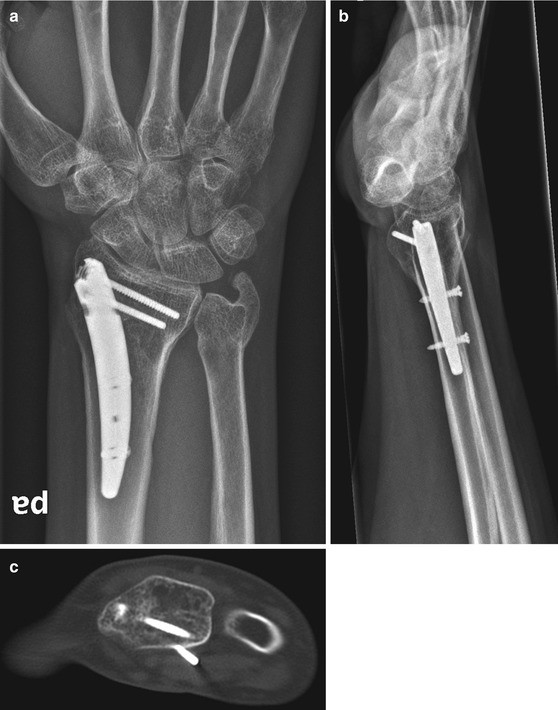
(a) An example of a Micronail® used in a distal forearm fracture resulting in tilt of the DRUJ and a change in the axis of rotation in the DRUJ due to a change in the ulnar inclination of the radius. (b) Also the volar tilt has not been restored. (c) CT scan of the same case illustrating how the distal locking screws penetrates into the soft tissue

4 Operative Technique
The operation is performed in general anaesthesia or in a regional bloc and using a tourniquet to improve visualizing the sensory nerve branches of the radial nerve and the tendons. Start by reducing the fracture by a closed technique or by introducing small elevators through stab incisions followed by a temporary fixation with K-wires. Always use a fluoroscope. Especially pay attention to restoration of volar tilt, as this may be difficult to visualize when the nail and locking screws are inserted due to hardware and instruments blocking the view on the fluoroscope. If this is not possible, an alternative method of fracture fixation should be considered. Incisions for the nail and locking screws are made with blunt dissection to avoid damage to sensory branches of the radial nerve and tendon lesions especially the EPL tendon in dorsal nailing.
Insert the distal screws first. When placing the screws in the distal part of the nail fixating the distal fragment, it is important to get a good subchondral fixation of the screws to ensure a three-dimensional scaffold with a strong and stable construct. However, pay special attention to the DRUJ in placing the screws and check carefully using fluoroscopy that they do not perforate into the joint. With the proximal locking screws, it is important too to check the correct length of the screws, as screws penetrating into the soft tissue may result in discomfort and a necessity for unplanned hardware removal.
Postoperative immobilization may be reduced to a minimum after locked nailing if a stable fixation is achieved at the operation. A splint may be used for 1–2 weeks and non-loaded wrist movements allowed after removal of the splint. A removable brace may be used as protection for another 4 weeks depending on the patient and the bone quality until fracture union permits loading of the fracture.
5 Clinical Results
-
Tan et al. (2012) compared the use of Micronail® with a simple reduction and splinting of extra-articular radius fractures and found better radiological and functional results in the initial treatment period and after 12 months when treated with Micronail® fixation.
-
Comparing Micronail® with non-bridging external fixation, Schønnemann et al. (2011) found no relevant clinical difference in the results, but the Micronail® was a more cost-effective treatment option due to a reduction in the number of postoperative follow-up visits at the hospital.
-
Safi et al. (2013) compared Micronail® with volar plating and found faster rehabilitation with Micronail®, but no other differences in radiological or clinical outcome.
-
Lerch et al. (2009) compared another radial styloid nail (Targon®) with volar plating and found no difference in clinical or radiological outcome.
-
Comparing DNP® dorsal nailing with volar plating in a randomized controlled study, Chappuis et al. (2011) found better clinical and radiological results after volar plating.
6 Complications
-
A high risk of sensory nerve injury has been reported (Schønnemann et al. 2011; Ilyas and Thoder 2008; Safi et al. 2013; Dremstrup et al. 2013) using Micronail® with up to 30 % of the patients complaining of sensory disturbances at the radial styloid after 12 months. However, Dremstrup et al. (2013) found that after 5 years the initial observed sensory disturbances were reduced to less than 10 %.
-
The closed nailing techniques also open for an increased risk of tendon damage, and especially damage to the EPL tendon in dorsal nailing should be considered (Chappuis et al. 2011; Espen et al. 2007).
-
A lack of restoration of volar tilt is important before introduction and fixation of the nail. However, both in dorsal nailing (Chappuis et al. 2011) and radial styloid nailing (Schønnemann et al 2011; Dremstrup et al. 2013), this may be technically difficult leading to an insufficient restoration of volar tilt after fixation of the fracture (Fig. 19.1b). Also lack of bony support at the volar cortex due to volar comminution may lead to transforming the dorsal angulation into a Smith type configuration, and in this case another fixation method should be considered.
-
Intramedullary nailing is primarily recommended in dorsally displaced instable extra-articular fractures of the AO type A, but nailing of AO type C1 with a nondisplaced intra-articular fracture line has also been described. This may introduce a risk of converting a nondisplaced intra-articular fracture into a displaced intra-articular fracture, and in this situation the intra-articular fracture should be carefully evaluated using the fluoroscope during the operation.
-
Stabilizing the fracture and forming a three-dimensional scaffold for a strong and stable construct with diverging locking screws make the use of fluoroscopy mandatory in placing the locking screws, as they easily penetrate into the DRUJ or into the soft tissue (Fig. 19.1c). However, the use of an oblique view on the fluoroscope may reduce the problem, and Dremstrup et al. (2013) found that hardware removal was only needed in 2 % during the first 5 years after the operation using the Micronail®, indicating that this may not be a universal problem in intramedullary nailing of distal radius fractures.
7 Conclusion
Locked intramedullary nailing of instable extra-articular fractures AO type A is an elegant minimally invasive method with a low complication rate and a low need of hardware removal after fracture healing. However, there are quite narrow indications compared to volar locked plating, and the indication for intramedullary locked fixation of distal radius fractures should primarily be limited to dorsally displaced extra-articular fractures, and the procedure should be avoided if the fracture cannot be reduced by closed or percutaneous means.
Pearls and Pitfalls
-
Keep indications to primarily dorsally displaced extra-articular fractures.
-
Avoid intra-articular fractures apart from nondisplaced AO type C1.
-
Fracture reduction has to be perfect before insertion of the nail.
-
The procedure should be avoided if the fracture cannot be reduced by closed or percutaneous means.
-
Use temporary K-wire fixation before insertion of the nail.
-
Pay attention to the radial sensory branch of the radial nerve.
-
In dorsal nailing avoid damaging the EPL tendon.
-
Avoid damage to the DRUJ when introducing locking screws.
-
In a stable fracture fixation, early mobilization is allowed resulting in rapid rehabilitation of hand and wrist function.
-
Pay attention to the correct length of the locking screws, thereby creating a fracture fixation with a very low need for implant removal.
References
Burkhart KJ, Nowak TE, Gradl G, Klitscher D, Mehling I, Mehler D, Mueller LP, Rommens PM. Intramedullary nailing vs. palmar locked plating for unstable dorsally comminuted distal radius fractures: a biomechanical study. Clin Biomech. 2010;25(8):771–5.
Capo JT, Kinchelow T, Brooks K, Tan V, Manigrasso M, Francisco K. Biomechanical stability of four fixation constructs for distal radius fractures. Hand. 2009;4(3):272–8.
Chappuis J, Bouté P, Putz P. Dorsally displaced extra-articular distal radius fractures fixation: dorsal IM nailing versus volar plating. A randomized controlled trial. Orthop Traumatol Surg Res. 2011;97(5):471–8.
Dremstrup L, Skjærbæk SS, Olesen S, Høgh A, Hansen TB. Good radiological and functional results after intramedullary nailing of distal radius fractures. J Plast Surg Hand Surg. 2013;47:286–8.
Espen D, Lauri G, Fernandez D. Stabilisation of distal radius fractures by a novel endomedullary, fixed-angle plate: first experience. Handchir Mikrochir Plast Chir. 2007;39(1):73–7.
Ilyas AM, Thoder JJ. Intramedullary fixation of displaced distal radius fractures: a preliminary report. J Hand Surg Am. 2008;33(10):1706–15.
Lerch S, Sextro HG, Wilken F, Wittenberg CE. Clinical and radiological results after distal radius fracture: intramedullary locking nail versus volar locking plate osteosynthesis. Z Orthop Unfall. 2009;147(5):547–52.
McCall TA, Conrad B, Badman B, Wright T. Volar versus dorsal fixed-angle fixation of dorsally unstable extra-articular distal radius fractures: a biomechanic study. J Hand Surg Am. 2007;32(6):806–12.
Nishiwaki M, Tazaki K, Shimizu H, Ilyas AM. Prospective study of distal radial fractures treated with an intramedullary nail. J Bone Joint Surg Am. 2011;93(15):1436–41.
Safi A, Hart R, Teknedzjan B, Kozák T. Treatment of extra-articular and simple articular distal radial fractures with intramedullary nail versus volar locking plate. J Hand Surg Eur. 2013;38:774–9.
Schønnemann JO, Hansen TB, Søballe K. Randomised study of non-bridging external fixation compared with intramedullary fixation of unstable distal radial fractures. J Plast Surg Hand Surg. 2011;45(4–5):232–7.
Tan V, Bratchenko W, Nourbakhsh A, Capo J. Comparative analysis of intramedullary nail fixation versus casting for treatment of distal radius fractures. J Hand Surg Am. 2012;37(3):460–8.
van Vugt R, Geerts RW, Werre AJ. Osteosynthesis of distal radius fractures with the Micronail. Eur J Trauma Emerg Surg. 2010;36(5):471–6.
Author information
Authors and Affiliations
Corresponding author
Editor information
Editors and Affiliations
Rights and permissions
Copyright information
© 2014 Springer-Verlag Berlin Heidelberg
About this chapter
Cite this chapter
Hansen, T.B. (2014). Closed Reduction and Intramedullary Fixation of Distal Radius Fractures. In: Hove, L., Lindau, T., Hølmer, P. (eds) Distal Radius Fractures. Springer, Berlin, Heidelberg. https://doi.org/10.1007/978-3-642-54604-4_19
Download citation
DOI: https://doi.org/10.1007/978-3-642-54604-4_19
Published:
Publisher Name: Springer, Berlin, Heidelberg
Print ISBN: 978-3-642-54603-7
Online ISBN: 978-3-642-54604-4
eBook Packages: MedicineMedicine (R0)