
Abstract
The mixed halide CH3NH3PbI3−xClx crystalline thin film has been prepared by two-step solution-processed repeated dip coating method at an ambient atmosphere. X-ray diffraction study reveals the presence of tetragonal and cubic phases in deposited film. Raman study confirms the metal halide bond in the inorganic framework and organic CH3 stretching/bending of C–H bond in CH3NH3PbI3−xClx perovskite. Scanning electron microscopy shows that cuboid and polyhedral-like crystal grains of 100 nm to 2 μm may find applications in optoelectronics. The perovskite CH3NH3PbI3−xClx thin film shows high spectral absorption coefficient of the order of 106 m−1. In optical band gap study, we found the coexistence of cubic and tetragonal perovskite phases. The energy band gap is dominated by cubic phase having Eg = 2.50 eV over tetragonal phase with band gap Eg = 1.67 eV. The room-temperature photoluminescence study confirms band edge, shallow and deep-level emissions. The temperature-dependent cathodoluminescence study shows red, green and ultraviolet emissions. The dominating green luminescence evolved for cubic phase at 2.51 eV. The red and ultraviolet emissions are also found for mixed-phase CH3NH3PbI3−xClx thin film, suitable for preparation of light-emitting devices.
Introduction
The ability of solution-processed organic–inorganic perovskites CH3NH3PbX3 (X = Br, I and Cl) to convert visible light into electricity was first discovered in 2006 using CH3NH3PbBr3 as a sensitizer on nano-porous TiO2 in a liquid electrolyte-based dye-sensitized solar cells, but the device power conversion efficiency (PCE) was found low [1]. Researchers try to boost up the PCE of solar cells using perovskite nanoparticles or thin film over conventional dyes replacing the halogen site partially but PCE still unsatisfactory. At room temperature, methyl ammonium lead tri-iodide (CH3NH3PbI3) forms a tetragonal structure of band gap ~ 1.5–1.7 eV, whereas CH3NH3PbBr3 and CH3NH3PbCl3 form cubic structure of band gap ~ 1.5–2.30 eV and ~ 3.0–3.11 eV, respectively [2, 3]. Among these perovskites, CH3NH3PbI3 and CH3NH3PbBr3 are immensely used in photovoltaic and optoelectronic applications. In comparison with these perovskites, CH3NH3PbCl3 remains overlooked to scientific community and have not investigated extensively. CH3NH3PbCl3 is a wide band gap material which exhibits high optical absorption coefficient in the UV spectral range [3]. Therefore, CH3NH3PbCl3 can be used as an visible-blind UV photodetector and hole transport layer for highly efficient organic light-emitting diodes [3, 4]. Researchers are trying to tune the band gap of perovskites in between 1.5 and 3.11 eV to generate a significant advancement for photovoltaic’s and light-emission applications. The band gap of perovskites can be tuned by different approaches. Inclusion of organic or inorganic cations can alter the band gap of perovskites [5]. Partial substitution of halogen anions such as Cl at iodide site can convert the parent halide perovskite to mixed halide perovskite (CH3NH3PbI3−xClx) and tune the band gap of parent halide perovskites. It can also modify the physical properties of parent perovskites [6, 7]. At room temperature, CH3NH3PbI3−xClx contains tetragonal crystal structure similar to CH3NH3PbI3 but exhibits exceptional optoelectronic properties, such as tunable direct band gap ranging from UV to near-IR [8], strong light absorption, high photoluminescence quantum efficiency, high charge-carrier mobility, low recombination rates etc. [9, 10]. Recently, the solar power conversion efficiency of mixed halide perovskite has been reported to 23.7% [11]. Apart from this, mixed halide perovskites are intensively investigated for light emitters in LED’s and lasers [12]. Despite progress has been achieved in terms of preparation and conversion efficiency of mixed halide perovskites, still some issues such as the presence of Cl by part at I or Br sites and its effect on structural and physical properties of CH3NH3PbI3−xClx remain still important in correlation with preparation condition and technique. Furthermore, it has been reported that thin-film growth parameters and post-treatment process such as ultrasonic substrate vibration [13, 14] and short-time annealing [15,16,17] can also play an important role for the modification of structural, morphological, tuning of band gap and luminescence properties of perovskite thin films.
Several physical and chemical deposition techniques including some new solution-processed casting methods, such as doctor blading, inkjet printing, screen printing, drop casting, slot-die coating, roll-to-roll printing, etc. [18, 19], have been employed to fabricate high-quality CH3NH3PbI3−xClx thin films. However, all these methods are a bit more expensive due to use of vacuum environment and also cause difficulties in controlling film compositions. In most cases, the precursor solution of mixed halide perovskite was prepared in a globe box at nitrogen atmosphere with a rotary evaporator which is expensive and complex process. However, in this work our main focus concentrates on cost-effective production of chlorinated perovskite film by repeated dip coating technique at air ambient and studies the effects of the partial replacement of I by Cl on structural, optical and electronic properties in order to understand photovoltaic and optoelectronic applications.
Previously, we reported multi-color emissions of CH3NH3PbI3−xClx thin film prepared by chemical dip-coating technique at ambient atmosphere [20]. In this paper, we show the coexistence of tetragonal and cubic phases in CH3NH3PbI3−xClx thin film deposited on glass substrate by repeated dip coating method at ambient atmosphere. The grown film is capable of showing high absorption coefficient, tuning the traditional band gap of perovskite and displaying strong colored emission peaks at 2.51 eV (green luminescence). The reason for the coexistence of cubic and tetragonal phases and their role on physical properties have been explained on the basis of the result of the X-ray diffraction (XRD), scanning electron microscope (SEM), Raman spectroscopy, ultraviolet (UV)-visible spectroscopy, photoluminescence (PL) and temperature-dependent cathodoluminescence (CL) experiments.
Experimental section
Preparation of perovskite solution and deposition of CH3NH3PbI3−xClx thin film by repeated dip coating technique is performed in an ambient atmosphere illustrated in Fig. 1. The details of the preparation of perovskite solutions are described in our previous work [20]. For the preparation of chlorinated perovskite film, prepared solution was heated at (80 ± 10) °C for 30 min and then a clean glass substrate was dipped into the solution for 10 s and then taken out from glass container (g) dried in air for 05 s. This process of dip coating cycle was performed for four to five times to complete the film growth process. The ash (h) color of pristine film changed from ash to dark brown (i) when heated on a hot plate at 100 °C for 5 min. Similar changes of color after heating were reported by [21, 22].

Schematics for preparation of perovskite solution and dip coating technique for the synthesis of CH3NH3PbI3−xClx thin film
X-ray diffraction (XRD) data of prepared sample were collected from a BRUKER D8 advance X-ray diffractometer in between Bragg angle of (10–50)° with Cukα radiation of wavelength λ = 0.154 nm. The surface morphology of the film was studied by a ZEISS EVO 18 Research scanning electron microscope (SEM). The phonon vibrational modes of the film were recorded by Renishaw in via Raman spectrometer using 514.5 nm line of Ar+ ion laser. Optical transmission and reflection were recorded using UV-1601PC SHIJMADZU spectrophotometer. Room-temperature photoluminescence (RTPL) measurement was performed using an F-4600, Hitachi spectroflurophotometer at an excitation wavelength, λex = 500 nm. Cathodoluminescence (CL) spectra were recorded at temperatures 80 K and 300 K using FEI Quanta 200 ESEM connected to ocean optics spectrometer. The thickness of the deposited film was measured using Newton’s rings method [23,24,25] at the middle of the film area (1 cm × 1 cm). The film thickness was found out from the difference between the diameter of the rings of the interference pattern formed on the film surface and glass substrate using the relation,
where rn is the radius of the ring with film, \( r_{n}^{'} \) is the radius of the ring without film, R is the radius of curvature of plano-convex lens used and t is the film thickness. The experiment was performed for 4 to 5 times at different places in the film area (1 cm × 1 cm) and the average film thickness was estimated to ~ 160 nm.
Results and discussion
Structural analysis
Figure 2 shows the XRD pattern of mixed halide perovskite thin film. The presence of diffraction peaks at Bragg angles 2θ ≈ 15.55°, 22.11°, 35.22° and 38.88° is corresponding to (1 0 0), (1 1 0), (2 1 0) and (2 1 1) reflections of the cubic (*) trichloride (CH3NH3PbCl3) phase [26]. The remaining reflection peaks at (1 1 0), (2 2 0), (3 1 0) and (2 2 4) corresponding to angles 14.20°, 28.55°, 31.77° and 40.55° are representing the tetragonal (#) CH3NH3PbI3−xClx phase [27, 28]. A very low intensity peak at around 12.77° corresponding to (0 0 1) plane corresponds to PbI2 (■) phase due to decomposition of CH3NH3PbI3−xClx in the air [27]. It is reported that cubic phase of CH3NH3PbCl3 is transformed into tetragonal phase when synthesized below 54 °C, whereas tetragonal to cubic phase transformation occurred when annealed at 100 °C [29]. In this work, we have prepared CH3NH3PbI3−xClx film at 80 °C and annealed at 100 °C in air ambient. From XRD pattern of annealed film (Fig. 2), it is clear that there are two major intensity peaks, one corresponding to (1 0 0) plane which is a signature of cubic CH3NH3PbCl3 phase; while other corresponding to (1 1 0) plane denotes the tetragonal CH3NH3PbI3−xClx phase. This XRD result is suggesting that cubic and tetragonal phases can coexist for the film annealed at 100 °C. Similar coexistence of cubic and tetragonal phases was reported by Luo et al. [29] for spin-coated CH3NH3PbI3 film which converted to CH3NH3PbCl3 when annealed at 100 °C for 10 min and further annealing at the same temperature for 45 min, CH3NH3PbCl3 again converted to an intermediate phase to CH3NH3PbI3 due to template effect. In general, it is established that cubic CH3NH3PbCl3 phase is originated by placing of methylammonium (MA+) cations forming C3v molecular symmetry in between the cavities of a three-dimensional array of PbCl6 octahedra. The MA+ cations perform complex rotation and orientation to satisfy the site symmetry occupying Oh sites, whereas Pb atom occupies three chlorine atoms that lie on D4h sites [30, 31] in one of each per unit cell. However, large ionic difference of Cl− and I− ions makes the formation energy for CH3NH3PbI3−xClx higher compared to cubic CH3NH3PbCl3 phase and helps to dominant CH3NH3PbCl3 over CH3NH3PbI3−xClx phase. Therefore, the structural modulation of perovskite CH3NH3PbI3 replacing I by Cl may induce the variation of electronic and optical properties which may create avenue to cater photovoltaic and optoelectronic applications [32].

XRD pattern of dipping-deposited CH3NH3PbI3−xClx thin film shows the presence of cubic (*), tetragonal (#) and PbI2 (■) phases. Inset shows the vibrational Raman spectrum excited at λex = 514.5 nm
Raman spectroscopy analysis
The vibrational mode of deposited CH3NH3PbI3−xClx film has been investigated by means of vibrational Raman spectroscopy in the range of 100–1600 cm−1, as shown in inset of Fig. 2. From this figure, it is seen that there are four distinct vibrational modes centered at 110, 174, 1032 and 1419 cm−1, respectively. The vibrational modes centered at 110 and 174 cm−1 are for tetragonal CH3NH3PbI3−xClx [33] phase corresponding to the hetero-polar ionic/covalent interactions of metal and halide bond (Pb–X bond) in the inorganic framework [34]. On the other hand, the vibrational mode of cubic CH3NH3PbCl3 phase is found at 1419 cm−1 corresponding to CH3 stretching [35] or C–H bending [36] bonds. Furthermore, the vibrational mode for organic part (CH3N \( {\text{H}}_{3}^{ + } \)) is found at 1032 cm−1 [37]. It is noted that the coexistence of cubic and tetragonal phases is evidenced by Raman spectroscopy as well.
Surface morphology
Surface morphology of solution-processed mixed halide perovskite plays a vital role in optoelectronic applications. The topology of scanning electron microscope (SEM) image of CH3NH3PbI3−xClx perovskite is shown in inset of Fig. 3. The SEM image indicates that film is composed of cuboids and polyhedral-like shapes with grain sizes 100 nm to 2 μm consistent to [38, 39] distributed all over the film surface. Such large grain size perovskites are suitable for efficient charge transfer, reduction in exciton recombination and making high-performance optoelectronic devices [40]. It is also reported that spin-coated large grain size films by ultrasonic vibration improves device performances [7].

EDS analysis of deposited CH3NH3PbI3−xClx thin film. The corresponding SEM image (inset) showing the large cuboid and polyhedral crystals
To confirm the presence of Cl in the deposited film, elemental analysis was done by energy-dispersive X-ray spectroscopy (EDS) for uncoated CH3NH3PbI3−xClx sample as shown in Fig. 3. The EDS spectrum shows peaks for C, O, Si, Na and Cl atoms using K-series X-rays and Pb atoms for M-series X-rays, respectively. The presence of O, Si and Na peaks in the spectrum originated from soda lime glass substrate. It is important to note that the EDS spectrum reveals the presence of Cl in the deposited film. The atomic % of the different elements present in the film is shown in table (inset) of Fig. 3.
Optical studies
The absorption coefficient (α) was calculated from transmission T(λ) and reflection R(λ) data using the relation [41,42,43,44]
where t is film thickness. The variation of α with photon energy for the CH3NH3PbI3−xClx thin film is shown in the inset of Fig. 4. The film displays high absorption coefficient of the order of 106 m−1 in both visible and near-IR spectral ranges. The large value of α seems to arises from s–p antibonding coupling of mixed halide perovskite [45]. It is noted that the absorption coefficient is minimum at wavelength ~ 740 nm which corresponds to traditional band gap 1.67 eV for tetragonal phase of CH3NH3PbI3−xClx. However, the sharp band edge wavelength at around 500 nm is corresponding to the band gap energy 2.50 eV of mixed-phase perovskite.

Plot of \( \left( {\alpha h\nu } \right)^{2} \) vs photon energy for direct transition in dipping-deposited CH3NH3PbI3−xClx thin film. In inset, the variation of the absorption coefficient, the optical transmittance (left scale) and reflectance (right scale) spectra with wavelength of dipping-deposited CH3NH3PbI3−xClx thin film
The optical band gap (\( {\rm E}_{\text{g}} \)) of the mixed halide perovskite film for direct transition can be expressed by the following equation [46]
where A is the band edge constant related to the effective masses associated with the valence and conduction bands and \( h\nu \) is the photon energy. The value of the optical band gap for the absorption could be estimated by extrapolating the linear portion of Eq. (2), and the intercept of the energy axis of Fig. 4 gives the value of \( {\rm E}_{\text{g}} \). The estimated band gap is found to be 2.50 eV which is lower than 3.0 eV for cubic CH3NH3PbCl3 phase but higher than 1.6 eV for traditional CH3NH3PbI3−xClx phase [1, 27, 28]. Thus, the tuning of the band gap mainly results from the interaction of cubic and tetragonal phases in which cubic phase is dominant over tetragonal phase for which we got Eg = 2.50 eV for cubic phase. This value is close to the band gap Eg = 3.0 eV of cubic perovskite. Our XRD experiment also demonstrated higher volume of cubic phase over tetragonal phase. This effect lowers the recombination process because of the replacement of excess halogen ions in heavy metal element Pb with organic framework. It is also noted that some relativistic effects such as spin–orbit coupling and higher electronegativity between the metal cation and halide anion may increase the band gap of CH3NH3PbI3−xClx film. A small peak at ~ 1.67 eV is also observed in Fig. 4 (shown by an arrow) which may arise due to the optical transitions of the free carriers for the tetragonal phase of the deposited perovskite.
Photoluminescence study
Photoluminescence (PL) is one of the useful techniques to characterize the optical properties of semiconducting materials. The room-temperature PL measurement of the CH3NH3PbI3−xClx thin film was performed at an excitation wavelength, λex= 500 nm, as shown in Fig. 5.

RTPL spectrum of dipping-deposited CH3NH3PbI3−xClx film excited at λex = 500 nm
In the spectrum, a very intense luminescence peak at 1.64 eV and two weak peaks at 2.01 eV and 2.11 eV are observed. The optical band gap estimated by optical transmittance measurement is tuned to 2.5 eV, which suggests the presence of both CH3NH3PbI3−xClx and CH3NH3PbCl3 phases. Therefore, PL emission at 1.64 eV may be due to band edge (BE) emission which attributed to the free carriers’ exciton. Besides, BE emission at 1.64 eV, two other emissions at 2.01 eV and 2.11 eV refer to either of the shallow-level defects of vacancies or interstitials of organic part, otherwise deep-level defects of halogen interstitials or anti-sites of lead and halogen as suggested by DFT calculations [47]. These defects states are related to the preparation method which allows a particular chemical structure with Cl-containing polycrystalline system having higher polarization along the C–N···Cl axis [31].
Temperature-dependent cathodoluminescence study
The cathodoluminescence (CL) is an advanced technique that detects simultaneously secondary electrons and light emissions which correlate the physical structure of the material to its local transport and optical properties of the sample. The CL measurement of CH3NH3PbI3−xClx thin film was carried out at 10 nA electron beam current with an accelerating voltage of 10 kV at temperatures 80 K and 300 K as shown in Fig. 6.

Cathodoluminescence spectra of dipping-deposited CH3NH3PbI3−xClx thin film at temperatures 80 K and 300 K
In the cathodoluminescence spectrum, a sharp emission of green luminescence (GL) peak at 2.51 eV is in between the band gap energies for tetragonal (1.6 eV) and cubic (3.0 eV) phases suggesting the interaction of tetragonal CH3NH3PbI3−xClx and cubic CH3NH3PbCl3 phases. There are few other broader emission peaks found at 1.67 eV and 1.80 eV corresponding to red luminescence (RL) and very weak peaks at 3.34 eV and 3.60 eV for ultraviolet luminescence (UVL), respectively. It should be noted that the emissions at 3.34 eV and 3.6 eV are the frequency doubling of emissions at 1.67 eV and 1.80 eV, respectively, known as harmonic emissions. However, the CL peak intensity is found lower for 300 K than the intensity at 80 K. This lowering of intensity is the direct evidence of decreasing recombination events with increasing temperature [48].
Conclusions
In summary, CH3NH3PbI3−xClx thin film has been successfully prepared by repeated dip coating method at an ambient atmosphere. Structural results provide an evidence of the formation of both tetragonal CH3NH3PbI3−xClx and cubic CH3NH3PbCl3 phases in the deposited film. Raman study confirms the metal halide bond in the inorganic framework and organic CH3 stretching/bending of C–H bond in CH3NH3PbI3−xClx perovskite. From SEM image, it is clear that the film surface is compact and composed of cuboids and polyhedral grains having high absorption coefficient of the order of 106 m−1 in both visible and near-IR spectral regions. Photoluminescence study confirms band edge accompanied with shallow and deep-level defect emissions. The green luminescence at 2.51 eV from cathodoluminescence study confirms the interaction between tetragonal CH3NH3PbI3−xClx and cubic CH3NH3PbCl3 phases. Importantly, this work presents an easy and effective method for the deposition of CH3NH3PbI3−xClx thin film with large grain size useful for high-performance perovskite optoelectronic devices.
References
Chen Q, De Marco N, Yang Y, Song TB, Chen CC, Zhao H, Hong Z, Zhou H, Yang Y (2015) Under the spotlight: the organic-inorganic hybrid halide perovskite for optoelectronic applications. Nano Today 10:355–396
Huang J, Xiang S, Yu J, Li C-Z (2019) Highly efficient prismatic perovskite solar cells. Energy Environ Sci 12:1265–1273
Nandi P, Giri C, Swain D, Manju U, Topwal D (2019) Room temperature growth of CH3NH3PbCl3 single crystals by solvent evaporation method. CrystEngComm 21:656–661
Zheng E, Yuh B, Tosado GA, Yu Q (2017) Solution-processed visible-blind UV-A photodetectors based on CH3NH3PbCl3 perovskite thin films. J Mater Chem C 5:3796–3806
Pang G, Lan X, Li R, He Z, Chen R (2019) Influence of mixed organic cations on the structural and optical properties of lead tri-iodide perovskites. Nanoscale 11:5215–5221
Ansari MIH, Qurashi A, Nazeeruddin MK (2018) Frontiers, opportunities and challenges in perovskite solar cells: a critical review. J Photochem Photobiol C 35:1–24
Xionga H, Zabihia F, Wanga H, Zhanga Q, Eslamian M (2018) Grain engineering by ultrasonic substrate vibration post treatment of wet perovskite films for annealing-free, high performance, and stable perovskite solar cells. Nanoscale 10:8526–8535
Yakunin S, Shynkarenko Y, Dirin DN, Cherniukh I, Kovalenko MV (2017) Non-dissipative internal optical filtering with solution-grown perovskite single crystals for full-colour imaging. NPG Asia Mater 9:e431
Chin S-H, Choi JW, Woo HC, Kim JH, Lee HS, Lee C-L (2019) Realizing a highly luminescent perovskite thin film by controlling the grain size and crystallinity through solvent vapour annealing. Nanoscale 11:5861–5867
Kim H, Zhao L, Price JS, Grede AJ, Roh K, Brigeman AN, Lopez M, Rand BP, Giebink NC (2018) Hybrid perovskite light emitting diodes under intense electrical excitation. Nat Commun 9:4893
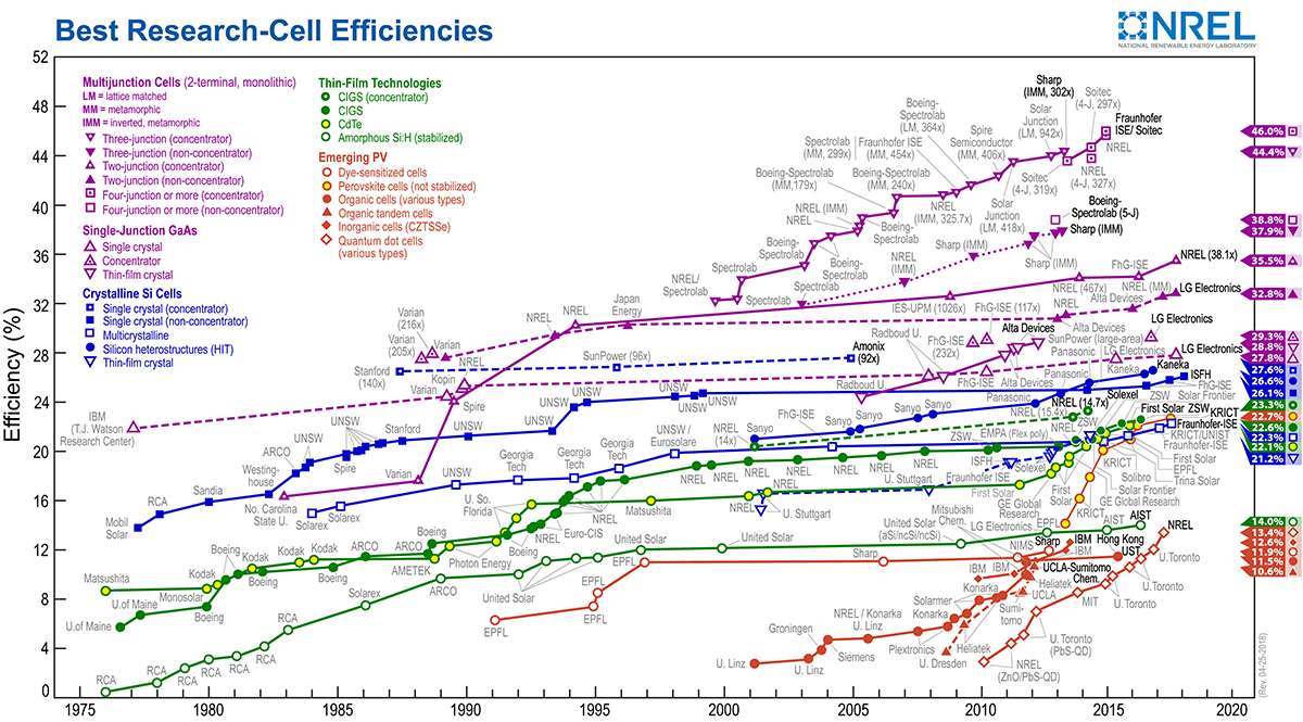
http://www.nrel.gov/pv/assets/images/efficiency-chart.png. Accessed 23 Dec 2018
Stranks SD, Hoye RLZ, Di D, Friend RH, Deschler F (2018) The physics of light emission in halide perovskite devices. Adv Mater. https://doi.org/10.1002/adma.201803336
Ahmadian-Yazdi M-R, Habibi M, Eslamian M (2018) Excitation of wet perovskite films by ultrasonic vibration improves the device performance. Appl Sci 8:308
Ahmadian-Yazdi M-R, Eslamian M (2018) Toward scale-up of perovskite solar cells: annealing-free perovskite layer by low-cost ultrasonic substrate vibration of wet films. Mater Today Commun 14:151–159
Lee JW, Yu H, Lee K, Bae S, Kim J, Han GR, Hwang D, Kim SK, Jang J (2019) Highly crystalline perovskite-based photovoltaics via two-dimensional liquid cage annealing strategy. J Am Chem Soc 141:5808–5814
Lee B, Hwang T, Lee S, Shin B, Park B (2019) Microstructural evolution of hybrid perovskites promoted by chlorine and its impact on the performance of solar cell. Sci Rep 9:4803
Sanches AWP, da Silva MAT, Cordeiro NJA, Urbano A, Lourenço SA (2019) Effect of intermediate phases on the optical properties of PbI2-rich CH3NH3PbI3 organic–inorganic hybrid perovskite. Phys Chem Chem Phys 21:5253–5261
Wang Q, Phung N, Girolamo DD, Vivo P, Abate A (2019) Enhancement in lifespan of halide perovskite solar cells. Energy Environ Sci 12:865–886
Habibi M, Rahimzadeh A, Bennouna I, Eslamian M (2017) Defect-free large-area (25 cm2) light absorbing perovskite thin films made by spray coating. Coatings 7:42
Karim AMMT, Rahman MA, Hossain MS, Khan MKR, Rahman MM, Kamruzzaman M, Ton-That C (2018) Multi-color excitonic emissions in chemical dip-coated organolead mixed-halide perovskite. Chem Sel 3:6525–6530
Adnan M, Lee JK (2018) All sequential dip-coating processed perovskite layers from an aqueous lead precursor for high efficiency perovskite solar cells. Sci Rep 8:2168
Chen J, Wan Z, Liu J, Fu S-Q, Zhang F, Yang S, Tao S, Wang M, Chen C (2018) Growth of compact CH3NH3PbI3 thin films governed by the crystallization in PbI2 matrix for efficient planar perovskite solar cells. ACS Appl Mater Interfaces 10:8649–8658
Wahl KJ, Chromik RR, Lee GY (2008) Quantitative in situ measurement of transfer film thickness by a Newton’s rings method. Wear 264:731–736
Winston AW, Baer CA, Allen LR (2013) A simple film thickness gauge utilizing Newton’s rings. In: Proceedings of the sixth national symposium on vacuum technology transactions, pp 249–254
Raveesha KH, Doddamani VH, Prasad BK (2014) On a method to employ Newton’s rings concept to determine thickness of thin films. Int Lett Chem Phys Astron 22:1–7
Maculan G, Sheikh AD, Abdelhady AL, Saidaminov MI, Haque MA, Murali B, Alarousu E, Mohammed OF, Wu T, Bakr OM (2015) CH3NH3PbCl3 Single crystals: inverse temperature crystallization and visible-blind UV-photodetector. J Phys Chem Lett 6:3781–3786
Yu H, Wang F, Xie F, Li W, Chen J, Zhao N (2014) The role of chlorine in the formation process of “CH3NH3PbI3−xClx” perovskite. Adv Funct Mater 24:7102–7108
Xu Y, Zhu L, Shi J, Lv S, Xu X, Xiao J, Dong J, Wu H, Luo Y, Li D, Meng Q (2015) Efficient hybrid mesoscopic solar cells with morphology-controlled CH3NH3PbI3−xClx derived from two-step spin coating method. ACS Appl Mater Interfaces 7:2242–2248
Luo S, Daoud WA (2016) Crystal structure formation of CH3NH3PbI3−xClx perovskite. Materials 9:123
Chi L, Swainson I, Cranswicka L, Her JH, Stephens P, Knop O (2005) The ordered phase of methylammonium lead chloride CH3ND3PbCl3. J Solid State Chem 178:1376–1385
Dimesso L, Dimamay M, Hamburger M, Jaegermann W (2014) Properties of CH3NH3PbX3 (X = I, Br, Cl) powders as precursors for organic/inorganic solar cells. Chem Mater 26:6762–6770
Sedighi R, Tajabadi F, Shahbazi S, Gholipour S, Taghavinia N (2016) Mixed-halide CH3NH3PbI3−xXx (X = Cl, Br, I) perovskites: vapor-assisted solution deposition and application as solar cell absorbers. Chem Phys Chem 17:2382–2388
Pistor P, Ruiz A, Cabot A, Roca VI (2016) Advanced Raman spectroscopy of methylammonium lead iodide: development of a non-destructive characterisation methodology. Sci Rep 6:35973
Niemann RG, Kontos AG, Palles D, Kamitsos EI, Kaltzoglou A, Brivio F, Falaras P, Cameron PJ (2016) Halogen effects on ordering and bonding of CH3NH3+ in CH3NH3PbX3 (X = Cl, Br, I) hybrid perovskites: a vibrational spectroscopic study. J Phys Chem C 120:2509–2519
Maalej A, Abid Y, Kallel A, Daoud A, Lauti A, Romain F (1997) Phase transitions and crystal dynamics in the cubic perovskite CH3NH3PbCl3. Solid State Commun 103:279–284
Brivio F, Frost JM, Skelton JM, Jackson AJ, Weber OJ, Weller MT, Goni AR, Leguy AMA, Barnes PRF, Walsh A (2015) Lattice dynamics and vibrational spectra of the orthorhombic, tetragonal, and cubic phases of methylammonium lead iodide. Phys Rev B 92:144308
Glaser T, Muller C, Sendner M, Krekeler C, Semonin OE, Hull TD, Yaffe O, Owen JS, Kowalsky W, Pucci A, Lovrincic R (2015) Infrared spectroscopic study of vibrational modes in methylammonium lead halide perovskites. J Phys Chem Lett 6:2913–2918
Dong Q, Yuan Y, Shao Y, Fang Y, Wang Q, Huang J (2015) Abnormal crystal growth in CH3NH3PbI3−xClx using a multi-cycle solution coating process. Energy Environ Sci 8:2464–2470
Xiao L, Xu J, Luan J, Yu X, Zhang B, Dai S, Yao J (2018) Preparation of CH3NH3PbCl3 film with a large grain size using PbI2 as Pb source and its application in photodetector. Mater Lett 220:108–111
Gedamu D, Asuo IM, Benetti D, Basti M, Ka I, Cloutier SG, Rosei F, Nechache R (2018) Solvent-antisolvent ambient processed large grain size perovskite thin films for high-performance solar cells. Sci Rep 8:12885
Moss TS (1959) Optical properties of semiconductor. Academic Press, New York
Karim AMMT, Khan MKR, Rahman MM (2015) Structural and opto-electrical properties of pyrolized ZnO–CdO crystalline thin films. J Semicond 36:053001
Ashaduzzman M, Khan MKR, Karim AMMT, Rahman MM (2018) Influence of chromium on structural, non-linear optical constants and transport properties of CdO thin films. Surf Interfaces 12:135–144
Islam MA, Karim AMMT, Julkarnain M, Badrul AKM, Khan MKR, Khan KA (2017) Opto-transport properties of e-beam evaporated annealed CuInSe2 thin films. Surf Interfaces 8:170–175
Song TB, Chen Q, Zhou H, Jiang C, Wang HH, Yang YM, Liu Y, Youab J, Yang Y (2015) Perovskite solar cells: film formation and properties. J Mater Chem A 3:9032–9050
Tauc J (1974) Amorphous and liquid semiconductors. Plenum Press, New York
Buin A, Pietsch P, Voznyy O, Comin R, Ip AH, Sargent EH, Xu B (2014) Materials processing routes to trap-free halide perovskites. Nano Lett 14:6281–6286
Jiang DS, Jung H, Ploog K (1988) Temperature dependence of photoluminescence from GaAs single and multiple quantum-well heterostructures grown by molecular-beam epitaxy. J Appl Phys 64:1371
Acknowledgements
Authors are thankful to the department of Glass and Ceramic Engineering, Rajshahi University of Engineering & Technology, Bangladesh, for providing XRD and SEM facilities. The authors are also grateful to the Department of Materials Science and Engineering, City University of Hong Kong, for providing Raman spectroscopy measurement. One of the authors M. Azizar Rahman acknowledges the financial support of Australian Government through the Research Training Program Scholarship.
Author information
Authors and Affiliations
Corresponding author
Additional information
Publisher's Note
Springer Nature remains neutral with regard to jurisdictional claims in published maps and institutional affiliations.
Rights and permissions
About this article

Cite this article
Karim, A.M.M.T., Hossain, M.S., Khan, M.K.R. et al. Solution-processed mixed halide CH3NH3PbI3−xClx thin films prepared by repeated dip coating. J Mater Sci 54, 11818–11826 (2019). https://doi.org/10.1007/s10853-019-03740-0
Received:
Accepted:
Published:
Issue Date:
DOI: https://doi.org/10.1007/s10853-019-03740-0