
Abstract
Background
Over the last 30 years there has been interest in the use of autologous fat transplantation for breast reconstructive and cosmetic purposes. Up until now injection of adipose tissue into the breast has been subject to two limiting factors. First, fat injection into the breast could result in fat necrosis, cyst formation, and indurations that could be mistaken as cancerous calcifications. Second, the degree of reabsorption of the injected adipose tissue is unpredictable.
Methods
Patients included in the study were candidates for either breast reconstruction after tumor resection or breast augmentation and were divided into three groups. Group I included patients with asymmetry after mastectomy and breast reconstruction; Group II consisted of patients with congenital breast asymmetry; and Group III included patients requesting bilateral breast augmentation. All patients signed a consent form acknowledging potential complications of infiltrating fat into the breast.
Results
A total of 820 consecutive female patients were operated on between 1983 and 2007. The age distribution of the patients ranged from 19 to 78 years, with a mean of 45.6 years. There were 381 patients in Group I, 54 in Group II, and 385 in Group III. Complications included ecchymosis in 76 patients, striae in 36 patients, 12 hematomas, and 5 infections. Long-term breast asymmetry was observed in 34 cases. Six hundred seventy patients have undergone mammography and ultrasonography 6 months and 1 year after their first intervention under our care. The majority of complications resulting from lipofilling of the breast have been seen in this series during the first 6 months after each session. Breast lesions, including calcifications, cysts, and cancer, that are not apparent in the first year after the final procedure of lipofilling we believe may not be directly associated with the autologous fat grafting to the breast. This has been confirmed by the long-term follow-up of 230 patients (range = 2–25 years, mean = 11.3 years) who have been followed up yearly with mammographic examination.
Conclusion
In the last 25 years the results of autologous fat transplantation have been predictable and satisfying on the condition that the treatment is performed in stages with small quantities of adipose tissue fat injected in each treatment session. To prevent major complications the final expected result should not be the aim of a single procedure. Mammary lipografting is a procedure that can be offered to patients for breast reconstructive and cosmetic purposes.
Over the last 30 years there has been continuous interest in breast augmentation using autologous fat transplantation for reconstructive and cosmetic purposes [1]. The senior author presented his technique of autologous fat transplantation to the breast in 1983 [2], and in 1987 Bircoll [3] reported his experience using fat removed by liposuction and transplanted by transcutaneous injection to the breast. A 1987 American Society of Plastic and Reconstructive Surgeons position paper predicted that fat grafting would compromise breast cancer detection and should therefore be prohibited [4].
Until now adipose tissue injection into the breast or mammary lipoaugmentation has been subject to two limiting factors. First, fat injection in and around the breast could result in cyst formation, indurations, and fat necrosis that could be mistaken as cancerous calcifications. Second, the degree of reabsorption of the injected adipose tissue is unpredictable. Fat grafting remains shrouded in the stigma of the variable results experienced by most plastic surgeons when they first grafted fat.
In 2005, Spear et al. [5] reported that autologous fat transplantation is a very safe technique that can improve or correct significant contour deformities after breast reconstruction which otherwise would require more complicated and riskier procedures to improve. There are centers around the world where autologous fat transfer for breast reconstruction has become a routine procedure due to its simplicity, safety, and reproducibility [6]. It is systematically offered to all of their patients as the final, perfecting procedure of breast reconstruction, irrespective of the technique used for the initial reconstructive procedure, and also for the repair of certain conservative treatments [7]. In recent years autologous fat grafting to the breast has been reported to be a useful procedure for cosmetic breast enhancement in many patients who desire such a procedure, although there is still skepticism about this procedure [8].
We present the senior author’s technique and report on our 25 years of experience in performing this specific procedure with emphasis on the “pearls,” “pitfalls,” and complications.
Materials and Methods
All patients who were included in the study were candidates for either breast augmentation or breast reconstruction after tumor resection. The patients were divided into three groups. Group I included patients with asymmetry after mastectomy and breast reconstruction; Group II consisted of patients with congenital breast asymmetry; and Group III included patients requesting bilateral breast augmentation. All patients included in the study had preoperative mammography and ultrasonography examinations. Only patients with the American College of Radiology Breast Imaging Reporting and Data System (ACR BI-RADS) category 1 or 2 were included. Group I patients were disease free for at least 1 year after breast reconstruction. All patients had to sign a consent form that presented potential complications of infiltrating fat into the breast and also agree to undergo routine postoperative mammography and ultrasonography.
Technique
-
1.
Marking of the areas for liposuction and fat grafting was done while the patient was standing (Fig. 1).
Fig. 1 
Marking of the areas for liposuction and fat grafting was done while the patient was standing
-
2.
Preoperative sedation in the surgical suite is administered.
-
3.
After injection of normal saline wetting solution containing 1:500,000 of adrenaline using a small-bore cannula and waiting 15 min, a 60-cc syringe attached to a 4-mm blunt cannula is inserted through a small incision in the selected area to be lipoaspirated.
-
4.
Fat is aspirated using the syringe method. The donor sites could include the abdominal, flank, thigh, and knee fat areas [9, 10].
-
5.
The aspirated fatty tissue is treated in the following manner: With the syringe held vertically with the open end down, the fat is allowed to decant (Fig. 2). After 10-15 min, the fat becomes nearly yellow. The preparation of 10-ml syringes for injection into the breast is shown in Fig. 3.
Fig. 2 
Decanting of fat in preparation for grafting
Fig. 3 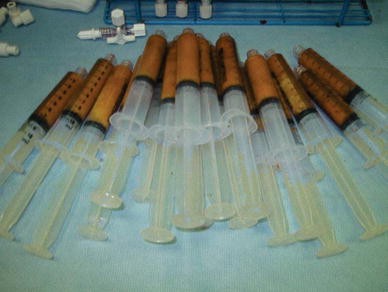
Preparation of 10-ml syringes for injection into the breast
-
6.
The breast is divided into four cosmetic units (Fig. 4). Fat is woven into the subcutaneous and intraglandular spaces of the breast using a 2.5-mm cannula attached to a 10-ml syringe with multiple passes, injecting only a small amount with each pass as the cannula is withdrawn in order to obtain the most reliable clinical outcome (Fig. 5). The entire breast is addressed by filling one cosmetic unit at a time.
Fig. 4 
Schematic division of breast into four cosmetic units
Fig. 5 
Schematic representation of the injection planes of the breast
-
7.
A light dressing is used on the breasts postoperatively in order not to exert pressure on the breasts.


Results
A total of 820 consecutive female patients were operated on between 1983 and 2007. The age of the patients ranged from 19 to 78 years (mean = 45.6 years). Twenty-five to 180 ml of fat was grafted into each breast in each session (mean = 145 ml). The number of sessions needed to achieve the desired result ranged from one to five (mean = 3 sessions). The total amount of fat transplanted in each breast ranged from 25 to 900 ml (mean = 540 ml). There were 381 patients classified into Group I. Lipografting was performed for correction of breast asymmetry and deformities after mastectomy and breast reconstruction. Autologous fat grafting with removal of silicone implants was performed in 253 patients who had undergone unilateral placement of implants for reconstruction of the breast. In 98 cases lipofilling was performed following unilateral breast reconstruction with a myocutaneous flap transfer. In 30 cases unilateral lipotransfer was done to correct breast deformities after local tumor resection. There were 54 cases classified into Group II. In 43 cases congenital breast asymmetry was corrected by lipotransfer to the breast, and in 11 cases of Poland syndrome, unilateral autologous fat transplantation was performed. Group III included 385 cases of bilateral fat transfer for breast augmentation (Fig. 6).

Schematic representation of the patient group classifications
Complications included small areas of ecchymosis in 76 patients, the development of striae in 36 patients, 12 hematomas, and 5 infections. The hematomas were resolved without any intervention and antibiotics were given in the cases of infection (Fig. 7). Long-term breast asymmetry was seen in 34 cases. Thirteen of the 34 patients underwent one session of lipofilling for correction of breast asymmetry. In Group III there were 36 patients who requested additional breast volume and had bilateral breast augmentation by the insertion of breast implants. The majority of the women had a significant improvement in their breast size and/or shape postoperatively. Six hundred seventy patients have undergone mammography a 6 months and 1 year after their first intervention under our care. Postoperative mammograms after autologous fat transplantation to the breast identify changes one would expect after a breast reduction surgical intervention (Fig. 8).

Schematic representation of complications observed in this series of patients

Mammographic exam 12 months after performing autologous fat transplantation to the breast
Patient 1
A 43-year-old female was referred to our clinic after she had been operated on for breast cancer in the right breast. Subcutaneous mastectomy had been performed and a silicone implant inserted (Group I). On examination the patient had a significant deformity with retraction of the skin on the right breast and ptosis of the left breast (Fig. 9a, b). The implant was removed and then the patient underwent three sessions of autologous fat transplantation, with 3 months between each one (session 1 = 92 ml, session 2 = 108 ml, and session 3 = 89 ml). The total volume of fat grafted was 289 ml. Six months after the last session the patient underwent mastopexy of the left breast and nipple-areola reconstruction on the right. The patient is shown in Fig. 9c and d 4 years after the last procedure with no complications and a satisfactory aesthetic result.

a, b Preoperative views of a 43-year-old female patient after performing right subcutaneous mastectomy and insertion of a breast silicone implant. c, d Postoperative views after removal of the right breast’s silicone implant, three sessions of autologous fat transplantation, left breast mastopexy, and nipple-areola reconstruction on the right breast 4 years after the last intervention
Patient 2
A 18-year-old female with Poland syndrome was referred to our clinic (Group II). She expressed the desire to undergo the least invasive procedure available to achieve an aesthetically pleasing result (Fig. 10a). The patient underwent five sessions, with 3 months between each one, of autologous fat transplantation (session 1 = 127 ml, session 2 = 209 ml, session 3 = 82 ml, session 4 = 206 ml, session 5 = 185 ml). The total volume of fat grafted was 809 ml on the left breast. Six months after the last session the patient underwent nipple-areola reconstruction on the left breast. The patient is shown in Fig. 10b 7 years after the last procedure with no complications and a satisfactory aesthetic result. The CT scan shows the left breast lipofilling 18 months after the last session (Fig. 11).

a Preoperative view of a 18-year-old female patient with Polands’ syndrome on the left side. b Postoperative view after five sessions of breast lipofilling and nipple-areola reconstruction on the left breast 7 years after the last procedure

CT scan shows the left breast’s lipofilling 18 months after the last session
Patient 3
A 27-year-old female came to our clinic requesting breast augmentation but she did not want to undergo insertion of implants (Group III) (Fig. 12a, b). There were two sessions, 3 months apart, of bilateral autologous fat transplantation (session 1 = 105 ml, session 2 = 135 ml). The total volume of fat grafted was 240 ml in each breast. The patient is shown in Fig. 12c, D 11 years after the last procedure with no complications and a satisfactory aesthetic result.

a, b Preoperative view of a 27-year-old female patient requesting breast augmentation. c, d Postoperative view after two sessions of autologous fat transplantation
Discussion
Autologous fat grafting to the breast is not a simple procedure and should be performed only by well-trained and skilled surgeons. A recent study confirms that this procedure is being performed incorrectly by untrained and untutored physicians and could result in major complications [11]. An extensive literature review indicated that the major complications observed after lipografting of the breast were related mainly to technical errors and to the wrong anatomic site of harvesting and implantation of the fat [12]. The primary complication of breast lipografting is the formation of liponecrotic cysts which have characteristically benign appearances in sonography, mammography, or magnetic resonance imaging (MRI) [13, 14]. Calcifications in breast parenchyma can also be expected after breast fat injection, and according to a recent study lipofilling for breast augmentation should not be performed in patients with a family history of breast cancer [15]. Fat necrosis, cyst formation, and indurations can be seen as in any other surgical manipulation of the breast [16, 17]. A range of mammographic findings such as parenchymal asymmetrical densities, radiolucent cyst, heterogeneity of the subcutaneous tissues, and benign-looking calcifications can be expected after autologous fat transplantation to the breast. Ultrasonographic features like anechoic lesions with posterior acoustic enhancement or shadowing, cystic lesions with internal echo, and increased echogenicity of the subcutaneous tissues can also be expected after breast lipofilling [18, 19]. The ACR BI-RADS classification of the 670 patients who underwent mammography 6 months and 1 year after their first intervention under our care is given in Table 1.
In recent years, advanced radiologic screening techniques have made it easier for radiologists to distinguish between the changes associated with benign necrosis of breast tissue and changes associated with cancer. Knowledge of the appearance of the breast on mammography and ultrasonography and the evolution of patterns of fat necrosis in patients who have undergone breast fat injection is mandatory in the evaluation of post-lipofilling breast lesions. Figure 13 shows the mammographic appearance of a breast before autologous fat transplantation and Fig. 14 shows the breast 12 months after the final session. In selected cases, MRI may be more accurate in evaluating breast findings. Transplanted fat in the breast has a smaller MRI T1 signal compared with native breast fat and the T2 signal of the transplanted fat is higher than that of native fat. This could probably be due to a slightly lower fat content and/or fibrosis of the injected areas or due to higher perfusion [20]. In Group III patients who requested breast augmentation by placement of silicone implants using the periareolar incision, lipoma-like tissue was observed. Similar lipoma-like tissue has been observed after autologous fat transplantation to the face and gluteal area [21, 22].

Mammographic appearance of a breast with no surgical intervention

Mammographic appearance of a breast 12 months after the final session of autologous fat transplantation
Numerous studies support the idea that radiologists have a high level of confidence in differentiating between fat necrosis calcifications after breast surgery and those related to breast cancer [23]. This view can be supported by another study that affirms that the lipomodeling technique does not affect the postoperative follow-up of patients with breast cancer and an imaging-controlled biopsy is possible in case of any doubt [24]. Figure 15 shows the ultrasonographic appearance of a breast before autologous fat transplantation and Fig. 16 shows the breast 12 months after the last lipofilling session. Grafted fat has many of the attributes of an ideal soft tissue filler, but the results, like those of any procedure, are technique dependent. Quantitative evidence of clinical fat survivability and predictability of volume restoration does not exist, yet reports of patient satisfaction with this procedure are plenty [25–29]. The need to standardize the autologous fat grafting technique is critical [30]. The success of autologous fat grafting in the breast depends on many factors: the techniques and instruments used to harvest the fat tissue, the fat processing, the volume of fat implanted, the levels of fat placement, and even the individual patient.

Ultrasonographic appearance of a breast with no surgical intervention

Ultrasonographic appearance of a breast 12 months after the final session of autologous fat transplantation
There are several principles that have guided our lipofilling practice in the last 25 years. The first principle is the avoidance of retroglandular injection of fat. It has been shown that the retroglandular plane does not have enough vascularization for autologous fat grafts to survive [31]. The ideal plane for breast autologous fat injection is the subcutaneous tissue, which is rich in adipose tissue and subsequently has a rich blood supply. The intraglandular adipose tissue graft is another option for lipofilling because the mammary tissue is also well vascularized. By injecting fat into areas with a rich blood supply there is an increased chance of adipocyte survival and integration with the surrounding tissue. The second principle is that the injection method has to be in a radial retrograde fashion, using the “drop-to-drop” technique and injecting in different planes. By injecting small “pearls” of adipose tissue in a drop-to-drop manner, there is an increased chance of adipocyte survival and avoidance of creating encapsulated tumors caused by fat bolus injection which subsequently become calcified (Figs. 17, 18). The third principle is that the final expected result of breast fat grafting should not be the aim of a single procedure but rather that of a multisession procedure in order to prevent major complications. When the patient desires voluptuous breasts, those can be obtained by repeated intramammary and intraglandular adipose tissue grafting. The fourth principle is the division of the breast into four cosmetic units and systematic lipofilling of those cosmetic units in each treatment session. This prevents miscalculations and under- or overtreatment of each breast cosmetic unit. The fifth principle is that a 3-month interval between treatment sessions is needed in order to perform successful fat grafting in the breast. During his 25 years of experience, the senior author has dedicated himself to producing modest breast augmentation results in order to prevent complications that could result after injection of an excessive amount of fat.

Mammographic appearance of an encapsulated breast tumor 6 months after bolus injection of fat in a 34-year-old patient

Mammographic appearance of calcified breast tumor 12 months after bolus injection of fat in a 34-year-old patient
Even though the aforementioned principles have been followed closely, there were still complications, as stated above. Most of those complications were observed in the early postoperative period and were resolved. Fat graft reabsorption was observed in our series [29]. Although quantitative measurements for fat reabsorption exist and fat volume survival can be predicted by the use of serial MRI, such measures were not performed as a standard method of evaluation [32, 33]. In some cases in which MRI was performed to evaluate breast lesions following lipofilling, the grafted fat was distinguished from the native fat but there was no preoperative and no immediate postoperative MRI control to accurately quantify the percentage of fat survival. Therefore, we could not objectively measure the rate of fat reabsorption following our technique. The multiple-session autologous fat grafting that was performed in the majority of the cases in this series of patients could have counterbalanced the possible partial fat reabsorption. A major disadvantage of this treatment plan is that the patients need to undergo several sessions before the final result can be achieved, a factor that sometimes discourages patients. Although 670 patients out of 820 have had mammography and ultrasonography 6 months and 1 year after their first session of breast lipofilling under our care, almost 20% of patients failed to have those exams. This was because some of the patients underwent only one treatment session and did not return to our clinic, were out-of-town patients who were followed up by their home physician, or simply failed to show up for the mammography. All the patients were informed and signed a consent form that stated that lipomodeling of the breast could be associated with a risk of calcification and multiple cyst formation and screening imaging is advised thereafter during their lifetime.
In all patients with a susceptible breast lesion less than 1 year after the final intervention, the opinion of an experienced radiologist and further evaluation may be needed because the susceptible lesion could be a primary breast cancer or a locoregional recurrence of breast cancer. Nevertheless, we have observed in our series that the majority of breast lesions resulting from lipofilling of the breast, such as fat necrosis, cyst formation, and indurations, are seen during the first 6 months after each session. Any breast lesions, including calcifications, cysts, tumor locoregional occurrence, or primary breast cancer, that are not apparent in the first year after the final procedure of lipofilling we believe are not directly associated with the autologous fat grafting to the breast. This has been confirmed by long-term follow-up of 230 patients (range = 2–25 years, mean = 11.3 years) who have been followed up yearly by mammographic and ultrasonographic examinations. Although lipofilling of the breast in Group III patients has produced aesthetically pleasant results, there were 36 patients who were not satisfied by the breast volume achieved. This is a limitation of the technique and at the same time one of its principles in order to avoid major complications. Patients should be advised that insertion of breast implants may be needed in order to achieve the final desired breast volume in some cases.
We believe that the introduction of regenerative cell-based strategies in our practice, such as those encompassing the use of stem cells, can hold tremendous promise for augmentation of the breast. In cell-assisted lipotransfer (CAL), autologous adipose-derived stem cells (ADSCs) are used in combination with lipoinjection. A stromal vascular fraction (SVF) containing ADSCs is freshly isolated from half of the aspirated fat and recombined with the other half. This process converts relatively ADSC-poor aspirated fat to ADSC-rich fat. The preliminary results suggest that CAL is effective and safe for soft tissue augmentation and superior to conventional lipoinjection [34]. Another study has confirmed that the CAL fat can survive better (35% on average) than non-CAL fat, and microvasculature can be detected more prominently in CAL fat, especially in the outer layers of the fat transfer [35].
Conclusion
In the last 25 years the results of autologous fat transplantation have been satisfying and stable when the treatment is done in multiple sessions, each consisting of injection of small quantities of adipose tissue fat. The final expected result should not be the aim of a single procedure in order to prevent major complications. Mammary lipografting is a procedure that can be offered to patients for breast reconstructive and cosmetic purposes.
References
Pulagam SR, Poulton T, Mamounas EP (2006) Long-term clinical and radiologic results with autologous fat transplantation for breast augmentation: case reports and review of the literature. Breast J 12(1):63–65
Illouz YG (1983) Body contouring by lipolysis: a 5-year experience with over 3000 cases. Plast Reconstr Surg 72(5):591–597
Bircoll M (1987) Cosmetic breast augmentation utilizing autologous fat and liposuction techniques. Plast Reconstr Surg 79(2):267–271
American Society of Plastic and Reconstructive Surgeons (ASPRS) (1987) Ad-Hoc Committee on New Procedures. Report on autologous fat transplantation, September 30, 1987. Plast Surg Nurs 7(4):140–141
Spear SL, Wilson HB, Lockwood MD (2005) Fat injection to correct contour deformities in the reconstructed breast. Plast Reconstr Surg 116(5):1300–1305
Delay E, Delpierre J, Sinna R, Chekaroua K (2005) How to improve breast implant reconstructions. Ann Chir Plast Esthet 50(5):582–594
Missana MC, Laurent I, Barreau L, Balleyguier C (2007) Autologous fat transfer in reconstructive breast surgery: indications, technique and results. Eur J Surg Oncol 33(6):685–690
Zheng DN, Li QF, Lei H, Zheng SW, Xie YZ, Xu QH, Yun X, Pu LL (2008) Autologous fat grafting to the breast for cosmetic enhancement: experience in 66 patients with long-term follow up. J Plast Reconstr Aesthet Surg 61(7):792–798
Ullmann Y, Shoshani O, Fodor A, Ramon Y, Carmi N, Eldor L, Gilhar A (2005) Searching for the favorable donor site for fat injection: in vivo study using the nude mice model. Dermatol Surg 31(10):1304–1307
Rohrich RJ, Sorokin ES, Brown SA (2004) In search of improved fat transfer viability: a quantitative analysis of the role of centrifugation and harvest site. Plast Reconstr Surg 113(1):391–395; discussion 396–397
Hyakusoku H, Ogawa R, Ono S, Ishii N, Hirakawa K (2009) Complications after autologous fat injection to the breast. Plast Reconstr Surg 123(1):360–370; discussion 371–372
Zocchi ML, Zuliani F (2008) Bicompartmental breast lipostructuring. Aesthetic Plast Surg 32(2):313–328
Castelló JR, Barros J, Vázquez R (1999) Giant liponecrotic pseudocyst after breast augmentation by fat injection. Plast Reconstr Surg 103(1):291–293
Hyakusoku H, Ogawa R, Ono S, Ishii N, Hirakawa K (2009) Complications after autologous fat injection to the breast. Plast Reconstr Surg 123(1):360–370; discussion 371–372
Carvajal J, Patiño JH (2008) Mammographic findings after breast augmentation with autologous fat injection. Aesthet Surg J 28(2):153–162
Leibman AJ, Styblo TM, Bostwick JIII (1997) Mammography of the postreconstruction breast. Plast Reconstr Surg 99:698
Danikas D, Theodorou SJ, Kokkalis G, Vasiou K, Kyriakopoulou K (2001) Mammographic findings following reduction mammaplasty. Aesthetic Plast Surg 25:283
Bilgen IG, Ustun EE, Memis A (2001) Fat necrosis of the breast: clinical, mammographic and sonographic features. Eur J Radiol 39(2):92–99
Taboada JL, Stephens TW, Krishnamurthy S, Brandt KR, Whitman GJ (2009) The many faces of fat necrosis in the breast. AJR Am J Roentgenol 192(3):815–825
Goehde SC, Kuehl H, Ladd ME (2005) Magnetic resonance imaging of autologous fat grafting. Eur Radiol 15(12):2423–2426
Pereira LH, Sterodimas A (2009) Long-term fate of transplanted autologous fat in the face. J Plast Reconstr Aesthet Surg [Epub ahead of print]
Pereira LH, Sterodimas A (2009) Macroscopic and microscopic proof of long-term survival of gluteal fat transplantation. Plast Reconstr Surg 123(4):162e–163e
Kneeshaw PJ, Lowry M, Manton D, Hubbard A, Drew PJ, Turnbull LW (2006) Differentiation of benign from malignant breast disease associated with screening detected micro calcifications using dynamic contrast enhanced MRI. Breast 15(1):29–38
Pierrefeu-Lagrange AC, Delay E, Guerin N, Chekaroua K, Delaporte T (2005) Radiological evaluation of breasts reconstructed with lipomodeling. Ann Chir Plast Esthet 51(1):18–28
Illouz YG (1986) The fat cell “graft”: a new technique to fill depressions. Plast Reconstr Surg 78(1):122–123
Pereira LH, Sterodimas A (2008) Free fat transplantation for the aesthetic correction of mild pectus excavatum. Aesthetic Plast Surg 32(2):393–396
Coleman SR (1995) Long-term survival of fat transplants: controlled demonstrations. Aesthetic Plast Surg 19(5):421–425
Guerrerosantos J (1996) Autologous fat grafting for body contouring. Clin Plast Surg 23(4):619–631
Kaufman MR, Miller TA, Huang C, Roostaien J, Wasson KL, Ashley RK, Bradley JP (2007) Autologous fat transfer for facial recontouring: is there science behind the art? Plast Reconstr Surg 119(7):2287–2296
Sterodimas A, Huanquipaco JC, de Souza Filho S, Bornia FA, Pitanguy I (2009) Autologous fat transplantation for the treatment of Parry-Romberg syndrome. J Plast Reconstr Aesthet Surg [Epub ahead of print]
Illouz YG (1989) Body sculpturing by lipoplasty. London, Churchill Livingstone, pp 390–394
Murillo WL (2004) Buttock augmentation: case studies of fat injection monitored by magnetic resonance imaging. Plast Reconstr Surg 114(6):1606–1614; discussion 1615–1616
Wolf GA, Gallego S, Patrón AS, Ramírez F, de Delgado JA, Echeverri A, García MM (2006) Magnetic resonance imaging assessment of gluteal fat grafts. Aesthetic Plast Surg 30(4):460–468
Sterodimas A, de Faria J, Correa WE, Pitanguy I (2009) Tissue engineering in plastic surgery: an up-to-date review of the current literature. Ann Plast Surg 62(1):97–103
Yoshimura K, Sato K, Aoi N, Kurita M, Hirohi T, Harii K (2008) Cell-assisted lipotransfer for cosmetic breast augmentation: supportive use of adipose-derived stem/stromal cells. Aesthetic Plast Surg 32(1):48–55; discussion 56–-57
Author information
Authors and Affiliations
Corresponding author
Rights and permissions
About this article
Cite this article
Illouz, Y.G., Sterodimas, A. Autologous Fat Transplantation to the Breast: A Personal Technique with 25 Years of Experience. Aesth Plast Surg 33, 706–715 (2009). https://doi.org/10.1007/s00266-009-9377-1
Received:
Accepted:
Published:
Issue Date:
DOI: https://doi.org/10.1007/s00266-009-9377-1