
Abstract
Humans consider themselves as “crown of the creation,” since they feel to be guided by a breeze of the “divine spirit” that enables some insights into the scene of life and into basics of earth. However, with respect to their relationships on earth, humans are just members of the animal kingdom and thus endangered by the same agents of disease that threaten the life of animals. Since humans as predators are also members of the food chain on earth, they may also become infected with agents of diseases that are on or inside of animals that belong to the daily human food. Diseases due to such animal-based or animal-transmitted pathogens are called zoonosis. Such pathogens may belong to the groups of prions, viruses, fungi, bacteria, and animal parasites, which may interact in a broad spectrum of pathways. The main topics of this book—Blastocystis species—belong to these pathogens. Thus many pathways of transmission of the numerous above-cited pathogens will run identically or are at least very similar. Therefore it is worthwhile to throw a glimpse onto the, in general, already available transmission pathways of the agents of zoonotic diseases while giving definitions and showing important examples.
Access provided by Autonomous University of Puebla. Download chapter PDF
Keywords
These keywords were added by machine and not by the authors. This process is experimental and the keywords may be updated as the learning algorithm improves.
10.1 Introduction
Since about 20,000–100,000 years, when larger settlements were founded, the specimens of the Homo sapiens started to live in close neighborhood to farmed animals and/or animals (such as rats, mice, dogs, flies, etc.), which seek shelter or food inside or close to human dwellings. This was the chance for several, primarily strictly host-specific agents of diseases or of parasites of animals to begin a phase of coevolution inside animals and humans and thus to enlarge their host spectrum considerably and as consequence to obtain better chances for a long-term survival on earth. Such processes of adaptations were found in viral, bacterial, fungal, and parasitic infections, which after a slow beginning then occurred more and more often simultaneously in the constantly growing human population. Many of these natural cross-infections surely failed, while others probably ended fatal for those hosts—animals or humans—which were infected as a second host after the agent of disease had been propagated for a long time in its first hosts, which therefore had the chance to become adapted. Thus then these first hosts suffered only from low-graded symptoms of disease or even showed no disturbances after an infection. Therefore there are mostly much heavier symptoms of disease in those cases, where agents of diseases hit a host population, which had not yet been in contact with such aggressors. For example, mankind probably suffered longer from infections with Plasmodium vivax than with P. falciparum, which demands today a much higher death toll per year than all other malaria types together. Similarly opportunistic parasites such as Giardia lamblia introduce practically no symptoms of disease in immunocompetent hosts, while they lead often to severe, slimy diarrheas in naïve hosts (=those without any previous contact to the parasite) or in immunocompromised persons (e.g., in AIDS-patients). Another factor supporting the development of zoonotic diseases was given when mankind started farming of animals close to their housings and enlarged it in our days of mass production of animals in overcrowded stables. This close neighborhood of animals and humans (often at short distances to towns with huge populations) increases the chances for ping-pong transmissions of agents of diseases between animals and humans. This is especially the case, when flies that are attracted by feces, get the chance to become vectors by taking up pathogens from feces of some hosts and transport them to the food of other hosts. The potential importance of this often-neglected transportation system was recently shown in a study lasting 3 years, which proved the occurrence of more than 100 important parasites and bacteria on the surface and in the intestine of different fly species which were caught close to stables of horses, pigs, cattle, chicken, or rabbits respectively close to dog ponds or common human recreation sites (Gestmann et al. 2012; Förster et al. 2012). These examinations showed that severe agents of diseases such as EHEC (Enterohemorrhagic Escherichia coli bacteria), Staphylococcus aureus, Campylobacter sp., etc. and/or several common parasites might be easily transmitted. Thus they may introduce severe outbreaks of diseases among crowded populations of humans and animals.
10.2 Which Are the Definitions of Zoonosis?
This term has its origin in the Greek words zoon = animal and nosos = disease that originally meant “disease of animals.” The present use of many Greek terms as termini technici for diseases is based on the facts that this early human high culture left many written documents on diseases and potential medications and that their physicians also used the medicinal knowledge of previous human high cultures such as the Assyrians, Babylonians, Sumerers, or Egyptians. Table 10.1 summarizes some important landmarks in the discovery of zoonotic agents of disease from the early beginning to the early twenties of the last century that was full of discoveries, that are still today of high importance (Mehlhorn 2012a, b, c; Grüntzig and Mehlhorn 2010a, b).
In the more recent past the original sense of the word “zoonosis” as “disease of animals” was erroneously changed by scientists who apparently were not familiar with the “old Greek” language. They started to use the word “zoonosis” to describe a peculiar disease, the agents of which were constantly transmitted in a cycle between animals and humans. However, since there are significant variations in the ways, modes, and directions of the transmission pathways, many international groups of researchers described several subgroups of zoonosis. However, there is no final agreement in the use of the terms among the international community of scientists. In general up to five different types of zoonosis were used in literature (see Mehlhorn 2008, 2012a, b, c).
-
1.
Direct zoonosis: This term is often used to characterize the transmission of agents of disease, the shape of which remains identical in animals and later in infected humans. Examples of such direct zoonosis occur during the transmission of the viruses of the rabies disease (Lyssa-virus), of the bacteria of the brucellosis (Brucella species), of the parasitic protozoans introducing the entamoebiasis (Entamoeba histolytica) or the giardiasis (Giardia duodenalis). Furthermore the top targets of this book—the Blastocystis species—would also fit into the category of a direct zoonosis, since the fecally excreted cyst stages are taken up orally and then may colonize the intestinal tract of humans.
-
2.
Cyclic zoonosis: In these cases a successful transmission from one host to another one may only occur after a morphologic transformation of the agent of disease has occurred in an animal or in a human. This example is the case in the life cycles of tapeworms such as Echinococcus granulosus or Taenia solium, both of which are found as adult worms in the intestine of the final host (=predators, carnivores) and as larval stages in the muscles and/or other tissues of intermediate hosts (prey animals, herbivores or omnivores).
-
3.
Metazoonosis: This term is used to describe transmission pathways that include arthropods as distributors (=vectors), which in some cases ingest an agent of disease, allow its reproduction in their bodies or not, but transmit these pathogens to another host during a bite or by fecal contamination. Examples for this category would be the transmission of plague bacteria (Yersinia pestis) by fleas, the viruses of the Dengue or Yellow fever by Aedes mosquitoes, or the mechanical transmission of bacteria and parasites that are attached at the feet, mouthparts of flies, or that are included in the feces of any other insect (e.g., cockroaches).
-
4.
Saprozoonosis: This group contains agents of disease that occur in nature on the soil or on degenerating contaminated food of animals which thus become infected. These pathogens may be transmitted, when humans eat raw meat or come into contact with fecally contaminated plants or waste waters. Examples for this group would be the transmission of the bacterial agents of listeriosis, of the spores of fungi of mycosis, or cysts of amoebic dysenteria.
-
5.
Other authors grouped zoonotic transmission procedures according to the main direction that is taken by an agent of disease.
-
(a)
Zooanthroponosis: In these cases the infectious agents occur mainly in animals and may be transmitted from there to humans. Examples for this category would be the transmission of the Trichinella spiralis larvae in muscles of wild boars to humans when eating undercooked meat. Also the transmission occurs when Balantidium coli cysts are swallowed by humans.
-
(b)
Anthropozoonosis: This term describes the pathway of transmission, when a pathogen of an animal has its origin in humans and leads to disease in animals. This may be the case when e.g., wild bears or wolves feed on humans infected with Trichinella muscle larvae or when pigs or cattle swallow human feces containing the sporocysts and/or the oocysts of Sarcocystis species. Of course pathways of transmission starting from humans are much more rare than those with a source in animals reaching humans from there.
-
(a)
When looking at the great variations of transmission pathways that have been established by zoonotic agents between humans and animals during coevolution, an absolutely correct classification according to the categories described above is mostly very difficult. Therefore the proposal to use exclusively the term zoonosis to characterize any of the numerous ping-pong infections between humans and animals seems most reasonable. In this case, however, it makes sense to differentiate according to the main pathways of transmission into:
-
(a)
Food-borne zoonotic diseases
-
(b)
Vector-borne zoonosis
-
(c)
Cyclic zoonosis
-
(d)
Contamination-borne zoonosis
10.3 Examples of Zoonotic Pathways in the Transmission of Pathogens Between Humans and the Animals and from Their Surroundings
Except for prions and viruses which depend on living cells and are reproduced by them all other members of the living society on earth—plants, fungi, animals (including humans)—have to fight for their survival, i.e., they have to defend their living space (=biotope) against competitors. Only those, which had been the winners in this daily survival fight, are our contemporaries. All members of the living society on earth belong to a giant food chain—a pyramid with predators at top sites and preys and producers at the lower levels. Therefore it is not astonishing that there had been established correlations among the different members of the food chain and that man and animals are attacked by various organisms seeking shelter and propagation inside their bodies. Such invasions may occur occasionally or might have been established since long and thus run often according to extremely fixed pathways of transmission. Tables 10.2, 10.3, 10.4, 10.5, 10.6 in the following Sects. 10.3.1–10.3.5 list some of the most important human diseases, which have their sources in organisms that exist longer on earth and thus in the human surroundings of our days than mankind. This is not only true for the rather recent Homo sapiens with his only ~100,000-years-old history but also for its precursors such as specimens of the genera Ardepithecus or Australopithecus, etc. Since man still exists although there are these huge amounts of hostile agents of diseases around him, there is good hope that humans will have a future on earth.
10.3.1 Prions with a Zoonotic Potential
Prions—an abbreviation of the description as proteinaceous infectious particles—are no true living organisms but wrongly folded intracellular proteins inducing other proteins to do the same under peculiar not yet well-understood conditions (Prusiner 1998; Rabenau 2009). These wrongly folded proteins, which e.g., in the case of the BSE-epidemiology (Bovine Spongious Encephalopathy) in Great Britain asked a death toll of more than 200 persons and left hundres other victims with severe brain damages besides a loss of more than 500,000 cattle, which had been killed either due to acute infections or due to preventive measurements. Since there was a clear proof for transmission from cattle to humans these diseases were also called transmissible encephalopathies. Similar degenerative symptoms may also occur due to hereditarily transmitted destructions in the genome of some families, but are rare compared to the wave of infections that rolled on after the feeding of animal meet powder (produced from BSE-infected animals) to uninfected ones. The reason was that no one had expected that such powdered animal meat should have been heated up to at least 141 °C in order to destroy these aggressive wrongly folded proteins, while normal cell proteins degenerate mostly already at 60 °C.
In nature there exists a broad potential of transmission of such infectious proteins from one host to the other. This was shown by the experiments of our group (Post et al. 1999). During these experiments Sarcophaga carnaria fly larvae were fed with brains of Scrapie-infected hamsters (see Table 10.2). One to seven days later these maggots were squeezed and the minced material was orally inoculated to uninfected hamsters. The same was done with pupae and adult flies (Fig. 10.1) obtained from the originally infected fly larvae. In all cases the hamsters showed 3–5 months after the experimental infection the full range of symptoms of BSE respectively Scrapie-disease including the full loss of motility control. Furthermore they were all prion-positive as was shown by molecular biological tests.

Macrophoto of a pupa and an adult of the gray flesh fly Sarcophaga carnaria
These experiments strongly supported the supposition that apparently these wrongly folded proteins are parts of nature being transmitted since long (probably without producing broad outbreaks but only single fatal cases). Such a transmission scenario would be given e.g., in cases when fly larvae feed on the remnant placenta of an infected animal and were later engorged by other grass feeding ruminants. Such a transmission will also explain the astonishing findings during the BSE-outbreak in Europe that some cows that had definitively not been fed with animal meat powder were positively tested for BSE.
However, evaluating all these aspects together, it can be stated that prions (Table 10.2) will not be a considerable threat for human health. Under natural conditions (i.e., when ruminants are fed by normal grass or plants) the natural infection rate of those animals with prions will never lead to such a significant increase of the amounts of prions inside the ruminant populations, that this later could lead to a significant infection potential.
10.3.2 Virus-Borne Zoonotic Diseases
The term virus has its origin in the Latin word virus, which means mucus, poison, moisture. Viruses themselves are no living organisms but are obligatory based as intracellular pathogens on the reproduction machinery of their host cells, which is situated in the nucleus and along the ribosomes. Therefore successful viruses had developed methods to become firmly attached to potential host cells, to enter such cells, and to find their way to the steering system, where their genetic material—RNA or DNA (in species specifically varying arrangements)—might be reproduced and finally released from the invaded cell. Thus for viruses it is most important to reach the surfaces of such cells otherwise they will burst or become digested by cells of the defense systems of attacked hosts. Therefore the viruses have developed some major pathways of transmission (Table 10.3):
-
Transmission by vector bites (Fig. 10.3)
-
Transmission by feces of vectors (Figs. 10.1, 10.2, 10.3, 10.9a, b)
Fig. 10.2 
Macrophoto of the adult green flesh fly Lucilia sericata
Fig. 10.3 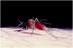
Macrophoto of an Aedes mosquito sucking blood on human skin
-
Transmission by feces of infected hosts
-
Transmission by contact or by inhaling of excretions or raw meat of infected hosts


While it is much easier for humans to avoid the last three transmission pathways using normal hygienic preventive methods, the contact to vectors occurs practically unlimited—at least in low numbers. Therefore the transmission and propagation of the pathogens of the so-called emerging diseases (Mehlhorn 2012a) increases constantly under the recent worldwide accelerating conditions of globalization and global warming (i.e., local change of the climate and huge migration and traveling activities worldwide; Dobler and Aspöck 2008, 2010; Neumeister et al. 2009).
Table 10.3 summarizes important viral diseases of humans which are discussed in more detail in Sects. 10.4–10.7 (Aspöck 2010; Dobler and Aspöck 2008, 2010; Darai et al. 2009; Löscher and Burchard 2010; Neumeister et al. 2009). The existence of viruses and their high potential to create new combinations, however, endangers life on earth, since they are easily transferred by genetic variations into highly dangerous pathogens. Due to their reproduction inside host cells there are only very restricted possibilities of chemotherapy apart from vaccination trials, which, however, are very often not successful, since many viruses often change their surfaces that have to be recognized by vaccines.
10.3.3 Bacteria of Zoonotic Importance
The present life on earth would not be possible without bacteria. They are found everywhere in nature, inside body cavities, and they even may settle inside cells. Since long some of these bacteria have undergone (as our mitochondria) a close symbiosis with all living organisms. However, a broad spectrum of these organisms that are invisible with naked eyes, may also introduce severe diseases, which in times of worldwide epidemics and pandemics of plaque or cholera reduced mankind significantly in many regions. For example, in Europe the population was reduced during the so-called 30 years-war (1618–1648) down to 15 % mainly by plaque epidemics. Even today the bacteria of tuberculosis or cholera request millions of victims every year.
It is not long ago when both “popes of bacteriology,” the German Robert Koch (1843–1910) and the French Louis Pasteur (1822–1895) and their famous scholars started to enlighten the hidden world of “good and bad” bacteria and to develop preventive measurements, which later were supported by the invention of the sulfonamides by the German Bayer Company scientist Gerhard Domagk (1895–1964) and the penicillin by the Scottish scientist Alexander Fleming (1881–1955). Both got (1939: Domagk and 1945: Fleming) the Nobel Prize for Medicine. However, the bacteria struggled back by a constant and often very quick development of resistances, so that today many bacterial diseases are far from being under control (and even may start a new career as “emerging diseases.”
Table 10.4 summarizes a selected spectrum of bacterial diseases, which are spread between humans and animals in their surroundings. The peculiar pathways of transmission are discussed in the Sects. 10.4–10.7 of this article (Alam and Zurek 2004; Bielaszewska et al. 2011; De Jesus et al. 2004; Ekdahl et al. 2005; Emerson et al. 2004; Fischer et al. 2001; Fotedar 2001; Fotedar et al. 1992; Grübel et al. 1999; Hold et al. 2007; Karch 2005; Kobayashi et al. 1999; Mian and Jacal 2002; Nichols 2005; Olsen and Hammack 2000; Rasko et al. 2011; Sasaki et al. 2000; Szalanski et al. 2004). Again the vector-transmitted pathogens of this group are most difficult to control, since the bites of the mostly lonely acting blood suckers (ticks, mites, mosquitoes, tabanids) or contact to skin lickers (flies) can only hardly be avoided.
10.3.4 Fungi with an Anthropophilic Zoonotic Potential
Fungi, the name of which has its origin in the Latin word: fungus = mushroom are heterotrophic organisms, which are covered by a chitin containing outer layer consisting of a network of fibrils. Most species of the fungi belong to the free nature, settle and feed at moist places. Several species of the fungi (French: champignons) are eatable.
However, some of the fungi species (see Table 10.5) invade body cavities of humans or animals respectively spread on their skin—mostly at places, where a high humidity is guaranteed as it is in the case at the hair-covered scalp, below finger or toe nails, in the groins, or in the shoulder-hollows. These places will be invaded especially in patients with immune suppression and can be treated only with difficulties (Figs. 10.4 and 10.5). This is due to the hidden places of the infection, the poor penetration of chemical substances through the surface of the fungi and due to a general lack of really potent fungicides with low side effects. The main transmission pathways are contacts to contaminated surfaces, direct contacts to contaminated human skin or to hair of infected animals. Other possibilities of transmission of fungi from human or animal feces or from other excretions are given via contaminations of food or materials (Abbot 2002; Banjo et al. 2005; Darai et al. 2009; Neumeister et al. 2009).

Macrophoto of a culture of Aspergillus fumigatus which may enter into the lung of mammals after injection of three different concentrations of the neem-seed derived extract MiteStop (Fa. Alpha-Biocare). Note the clear, doses dependent spots of clearance

Macrophoto of a culture of Aspergillus fumigatus which may enter into the lung of mammals after injection of three different concentrations of the neem-seed derived extract MiteStop (Fa. Alpha-Biocare). Note the clear, doses dependent spots of clearance
10.3.5 Parasites with a Zoonotic Life Cycle
Parasites are per definition in a strict sense animals that live on costs of other animals. Of course there also exist plants that stay as parasites on other plants, but they are here not considered, since they do not harm the health of humans. The name “parasites” has its origin in the Greek word “parasitos,” which described “employees” at noble courts that had to taste the food for poisons and thus were nourished on foreign costs. The parasites attack animals and humans. However, although many parasitic species rely on many hosts (e.g., Toxoplasma gondii), some had become specialists in the last 10,000–20,000 years and have adapted themselves as specialists (e.g., Ascaris lumbricoides) to the “late runner” on earth, i.e., the Homo sapiens. Parasites occur in many phyla of the Zoological Kingdom. Thus they are found among the unicellular organisms (Protozoa), among animals with a reduced number of cells (Myxosporida), among the various groups of worms (Plathyhelminthes, Nemathelminthes, Acanthocephala, Pentastomida, leeches), and among the billion-headed crowds of ticks, mites, and insects. The members of the latter three groups may also act as vectors of different pathogens (prions, viruses, bacteria, fungi, and parasites; see Tables 10.2, 10.3, 10.4, 10.5, 10.6). This transmission might be done just mechanically by means of contaminated mouthparts, by body contacts, and/or by placing pathogen-contaminated feces onto food, skin, or wounds. However, it may also occur as result of a defined developmental cycle with a reproduction of the pathogen inside the vector. Very often such reproduced pathogens even enter the eggs inside the female ovary or uterus so that the next vector generation is already carrier of the once ingested pathogen (i.e., this occurs inside ticks when the viruses of the spring-summer-meningoencephalitis or the kinetes (=motile zygotes) of the protozoan Babesia species enter the not yet fertilized eggs. Having entered the egg the parasites are firmly included therein and the thick eggshell, which is formed after fertilization and thickened during the laying process by adding a layer of wax, protects the growing tick larva and the therein included pathogens from desiccation. Examples of important zoonotic life cycles are compiled in Table 10.6) (Aspöck 2010; Darai et al. 2009; Getachew et al. 2007; Greenberg 1973; Kollaritsch and Paulke-Korinek 2010; Mehlhorn 2008; Mehlhorn et al. 2011; Mehlhorn 2012a, b, c; Oyerinde 1976; Szostakowska et al. 2004).
10.4 Food-Borne Zoonosis
When taking into consideration a very wide definition of the term food-borne zoonosis any pathogen that occurs inside or on meat of animals (inclusive fish) respectively is found within milk would fit into this group. However, in this case the differentiation from contamination-borne zoonosis would be rather difficult. Therefore it seems more reasonable to draw the limitations much stronger. This, however, leads to the fact that only a few groups of pathogens could be placed into this category. The prions—especially those of the bovine spongious encephalopathy (BSE)-group—would surely find their place here, since they introduce disease as well in animals as in humans after eating meat (even cooked one) of infected animals (Table 10.2). The viral pathogens with a zoonotic potential are mainly transmitted by bites of arthropods (mites, ticks, insects) or by fecal contamination, and only rarely—if at all—by infected food of humans and/or animals (Table 10.3). The explanation is that viruses need for their reproduction the very well-adapted DNA/RNA machinery of a living cell, which has problems to survive the passage of the stomach and intestinal system of humans (Altekrues et al. 1997; Darai et al. 2009; Neumeister et al. 2009; Steinmueller et al. 2006; WHO 2002a, b).
Likewise most bacteria prefer pathways of transmission other than the direct inclusion inside organs of warm- or cold-blooded animals respectively, fish. Thus there are not many bacteria like those of the Brucella-, Listeria- or Salmonella-groups that are frequently found not only on contaminated meat but also in milk (see Table 10.4).
Fungi with a zoonotic potential (Table 10.5) have no direct food-borne transmission pathways apart from fecal contaminations, that are common pathways in the species of the Blastocystis -group (if these species are really accepted as a fungus—see the other chapters).
On the other hand food-borne transmissions are very common in parasites (Table 10.6). There are protozoans such as several Sarcocystis species (Fig. 10.6) and Toxoplasma gondii which develop intracellular stages that later are transmitted, if this infected tissue is swallowed by a predator (inclusive humans). Then it starts a further developmental phase there. Man might act as final host (where the sexual process is running in his intestine, e.g., in Sarcocystis species) or as intermediate host with an exclusively asexual reproduction of the parasite in his tissues (e. g. in Toxoplasma gondii).

Life cycle of Sarcocystis suihominis with two obligatory hosts. 1 Motile sporozoites hatch from the ingested sporocysts inside the intestine of the intermediate host, i.e., swine. 2 Two generations of schizonts are formed (5–6 and 12–17 days after infection) inside endothelial cells of blood vessels, giving rise to 50–100 merozoites by endodyogeny. 3 Free motile merozoites; first-generation merozoites enter other endothelial cells and form schizonts, whereas merozoites of the second generation induce formation of tissue cysts. 4 Cyst formation inside typical cells (muscle fibers, brain cells); within these cysts the parasites are reproduced by repeated endodyogeny leading to thousands of cyst merozoites which are situated inside chamber-like hollows. 5 When the final host man has eaten cyst-containing raw or insufficiently cooked meat, the cyst merozoites are set free and enter cells of the lamina propria. 6 Formation of female (macrogametes, 6.3) via gamonts (6.1, 6.2) within 14 h of infection. 7 Fusion of gametes. 8 Formation of the oocyst wall around the zygote. 9–11 Formation of two sporocysts (containing four sporozoites each) inside the host cell. The smooth oocyst wall often becomes disrupted. Thus, fully sporulated oocysts are found in the feces (11). DM developing merozoites; DR disrupted oocyst wall; HC host cell; N nucleus; NH nucleus of the host cell; OW oocyst wall; PC primary cyst wall; PV parasitophorous vacuole; RB residual body; S sporocyst; SP sporozoite; WB wall-forming bodies
Also among the trematodes (phylum Plathyhelminthes) there are many examples of food-borne parasitosis in humans. In general humans are final hosts, since the adult worms live in their intestine (e.g., Fasciolopsis buski), in their lung (Paragonimus species), or in their liver (Clonorchis sinensis, Opisthorchis species, Fasciola hepatica, etc.; Table 10.6). The infection of humans with stages (metacercariae) of trematodes occurs by eating infected raw or undercooked intermediate hosts (e.g., fish, crustaceans) or plants with metacercariae that had been attached there by intermediate hosts (e.g., snails; Mehlhorn 2008).
For tapeworms (Cestodes; phylum Plathyhelminthes) humans might be final or intermediate hosts. In the case of the large human tapeworms Taenia saginata, T. solium, T. asiaticum and Diphyllobothrium latum, humans act as final hosts and shelter in their intestine the several meter long, hermaphroditic adult worms after being infected by ingestion of larval stages (cysticercus in the case of Taenia species respectively plerocercoids of D. latum) inside undercooked meat or fish. In the case of T. solium humans can be both final and intermediate hosts, since the cysticercus larvae may also develop inside their muscles and/or brain after they had orally ingested worm eggs from human feces (Mehlhorn 2008, 2012c).
On the other hand, however, in the cases of the tiny dog, respectively, fox tapeworms Echinococcus granulosus and E. multilocularis humans remain exclusively intermediate hosts, within which large cysts develop. Transmission of tapeworms of dog or fox to humans does only occur via the fecal contamination pathway when ingesting worm eggs from feces of dogs or foxes. However with respect to the general life cycle of these tapeworms humans are “dead ends,” since in general they are not eaten by the final hosts (dogs, foxes).
Among the nematodes there are rather few examples for food-borne worm infections in humans with respect to the huge number of nematode species. However, one prominent example is given by the Trichinella species, of which up to now seven had been described. These worms live as adult, rather tiny worms (♀, ♂) of only 1–3 mm in length inside the intestine of predators (inclusive man) (Fig. 10.7). The females produce larvae; however, these stages do not leave this infected individual (i.e., acting here as final host), but they penetrate into the muscle cells, where they stay until another predator feeds such infected meat. Thus these predators are final and intermediate hosts at the same time and free stages do not occur outside a body (Mehlhorn 2008).

Light micrograph of a muscle fiber of a rat containing three larvae of Trichinella spiralis
In the case of the species of the genera Anisakis, Porrocaecum, Contracaecum (Fig. 10.8), and related worms humans are intermediate hosts. Man, however, is not relevant for the transmission. He is a “dead end,” since the adult worms live in the intestinal tractus of large marine mammals after they had ingested the larvae inside fish. Humans do the same: they eat raw fish containing these worm larvae, which live for a short time (1–3 weeks) in the human intestinal system before they die. Similar infections are also possible, when humans eat raw meat of reptiles; then they may ingest also larval pentastomids which finally may grow up to considerable size in humans (Mehlhorn 2008, 2012c).

Light micrograph of the muscles of a fish containing many Anisakis larvae
10.5 Vector-Borne Zoonosis
Vectors in the sense of parasitological terms are animals that are able to transport pathogens from one host to another, while these vectors search their food on such hosts (Abbot 2002; Aspöck 2010; Hald et al. 2008; Holt et al. 2007; Löscher and Burchard 2010; Neumeister et al. 2009). The simplest way of such a transfer is the transport of pathogens that glue at mouthparts, at feet, or at the body surface of such vectors (Figs. 10.1, 10.2, 10.3, 10.9, 10.10, 10.11, 10.12), which are mostly mites, ticks, mosquitoes, flies, midges, simuliids, tabanids, leeches, etc. This type of transmission is described as mechanical, since the pathogens are not multiplicated on the vectors. For example, if 800 bacteria of a special species glue at the fine hair of a fly, the transmission maximum would be 800, but in general the finally transmitted amount is much less. Nevertheless this method might be still highly effective, if it is considered that many bacteria (such as e.g., toxic or hemorrhagic strains of Escherichia coli (EHEC, ETEC) need only an initial dose of 20–30 specimens to start severe infections. Recent studies examined various fly species of the genera Musca, Calliphora, Sarcophaga, Lucilia, etc. for their “normal load” of bacteria and parasites (protozoans and worm eggs). These flies had been caught close to stables of pigs, horses, cattle, rabbits, chickens, dog ponds respectively close to human recreation sites inside and outside of towns. Astonishing masses and varieties of pathogens were found on the examined flies. These studies of Förster et al. (2012) and Gestmann et al. (2012) confirmed earlier ones (Förster 2009; Förster et al. 2007, 2009) showing that more than 100 important bacteria species and more than 15 infectious stages of parasites might be transmitted via flies as vectors.

Photograph of a petri dish filled with bacterial culture medium. Onto the surface of this petri dish a Musca fly was placed for 30 s (a). The photo at the right (b) shows the growth of bacteria 2 days after the exposition. This proves that the feet of this fly had been highly contaminated with bacteria

Two adult Ixodes ticks laying eggs, which already may contain the viruses of the spring-summer meningoencephalitis

(a, b) Photograph of a tabanid of the genus Chrysops and a wandering worm (Loa loa) in an eye (b, right)

Scanning electron micrograph of a black fly (Simulium morsitans), the vector of Onchocerca volvulus worms
Additional experimental transmission experiments showed that practically all types of bacteria were also transferred to bacterial culture plates, if the flies had contact for only 20–30 s to bacteria on a culture plate. In another series of experiments single flies were placed onto a fluid containing a definite number of Toxocara canis eggs. Then these flies were washed and squeezed (to get the intestinal fluid). When counting the obtained eggs from these flies the astonishing result was noted that merely two-thirds of the original number was found again.
These experiments clearly show that the importance of licking flies as mechanical vectors is apparently underestimated.
The same should be true for biting and/or blood sucking mites, ticks, mosquitoes, flies, fleas, lice, tabanids, etc., since most of them are able to switch quickly from one host to another, so that many tiny blood droplets remain liquid thus keeping the pathogens in an infectious status.
The situation becomes even worse in those cases, where a regular reproduction of the pathogens has been developed inside the vectors during evolution. Such reproductive processes occur regularly in vectors bearing special species of viruses, bacteria, and parasites (see Tables 10.3, 10.4, 10.6). Since these vectors even are able to transmit the pathogens into their eggs as e.g., the spring–summer–meningoencephalitis virus in ticks (Fig. 10.10), the distribution intensity is enormously enlarged not only by the flying activity of the vectors but also by the transmission into their progeny.
Therefore flying vectors that give pathogens a chance to reproduce inside their bodies represent a hidden risk, since they may help to spread severe epidemics. The consequences for mankind would have been terrific, if mosquitoes, tabanids, or ticks would be able to transmit the AIDS viruses as easy as they do it with other viruses.
Nevertheless the various vector-borne zoonosis demand even today a high death toll in many regions of the world. Considering the recent enormous increase of the Dengue-virus propagation by Aedes mosquitoes, the progress of the West-Nile-Virus into many countries as well as the still not yet solved malaria situation or filarial worm problems new and concentrated control measurements are highly needed. Especially in times of global warming and intense globalization epidemics may quickly become pandemics, which will not stop at the borders of countries, where the population believe even today to live on “safe grounds.”
10.6 Cyclic Zoonosis
This term comprises pathways of transmission of agents of diseases that run repeatedly in a fixed direction and are based on at least two different hosts, where always defined developmental processes take place (Mehlhorn 2008). With respect to these conditions the transmission pathways of the malarial, babesial, and sarcosporidian parasites would fit in this category, because in these life cycles asexual processes like merogony (schizogony) start in a first host (human tissues) and are followed by gamogony and sporogony in a second host, which belongs—depending on the species—to the group of vectors (mosquitoes, ticks) or to mammalian species (as it is the case in the Sarcocystis species; Table 10.6, Fig. 10.6). Although the life cycle of the trematodes fits into this category, since, for example, the adult schistosomal worms live in the blood system of humans and other mammalians, while the asexual reproduction (via sporocysts and the finally infectious cercariae) takes places inside the tissues of water snails (Table 10.6). The life cycles of tapeworms belong to this group, too. Only if humans eat the cysticercus-larva with raw meat or the plerocercoid-larva in raw fish, the adult worms can develop from these ingested larval stages inside the human intestine (Table 10.6).
Among the nematodes examples of cyclic zoonosis are much more rare than in trematodes and cestodes (Aspöck 2010; Mehlhorn 2008). However, especially the filarial worms have developed very sophisticated developmental cycles, which need a very tight coordination in the different biotopes, if the transmission of the parasite from one host to the other should be successful. In the case of the filarial worms Wuchereria bancrofti, Brugia malayi, and Loa loa the adults live either in the lymph nodes or in the subcutaneous tissues (Loa loa), while their sheathed larvae (=microfilariae enveloped by the egg shell) stay in the blood. However, since these three worm species use different vectors with different behaviors and activity periods, the larvae of these worms are concentrated at different day times in the peripheral blood vessels. Wuchereria bancrofti and B. malayi are transmitted by nightly active mosquitoes, thus their larvae are found in the peripheral blood mainly at 10 p.m. On the other hand Loa loa is transmitted by day time active tabanids (Chrysops species; Fig. 10.11). Thus their larvae are found in day-time from 1 p.m. to 3 p.m. in the peripheral blood vessels. The adult worms of Onchocerca volvulus (accumulations of their dead larvae in human eyes induce the so-called river blindness) stay in humans inside the subcutaneous tissues, where the females are often found in groups inside nodules. The infectious (unsheathed) larvae stay all day long in the lymph fluid of the skin system. There they are taken up by the simuliids (black flies; Fig. 10.12) which do not have as fine mouthparts as the mosquitoes, but scissor like ones, which lead to the formation of small “lacunes” of blood and lymph in the skin. Such an all-day-presence of the larvae in the skin makes sense for Onchocerca larvae, since the simuliids only suck blood during day time.
Besides all differences in their behavior inside humans the first larvae (=microfilariae) of all four species described above proceed a nearly identical development inside their vectors. Having reached the insect’s gut the larva 1 enters the body cavity of the new host, grows up there (while molting twice), and the larva 3 enters finally the head of the vector. If this infected insect starts a new blood sucking action, the relatively thick larva 3 perforates the connecting membranes of the different mouthparts, creeps outside down to the biting site at the human skin and enters there the body of the new host.
Although this cyclic development seems very complicated, it is so perfect, that even today more than hundred fifty million humans in the tropics suffer from diseases due to these worms, since available means of chemotherapy are still rather poor.
10.7 Contamination-Borne Zoonosis
The diseases categorized inside this group are characterized by the fact that both humans and animals carry these pathogens, but their cross transmission occurs more or less occasional and the direction of the transmission is not fixed. This means that humans may get the pathogens from animals and that also the opposite way is possible (Tables 10.2, 10.3, 10.4, 10.5, 10.6). Examples for such contamination-borne zoonosis can be found in all groups of pathogens, which in addition also may use other pathways of transmission (WHO 2002a, b; Mehlhorn 2008).
Rabies viruses can be transmitted directly by saliva during bites of infected animals, by blood transfusion, or by contact to virus-contaminated saliva. Many bacteria change the host if there is contact either to human or animal feces or to contaminated food. The same is true for several species belonging to the group of fungi and thus also for the different Blastocystis species. Among the non cyclically transmitted parasites the species of the genera Giardia and Cryptosporidium as well as the different microsporidian species (Table 10.6) belong to the group of contamination-borne zoonosis. In all these cases fecal stages (cysts, oocysts, respectively, spores) might contaminate food by direct contact with feces or by help of the transportation activity of licking flies (Table 10.6). But even in cyclically transmitted parasites such as Trichinella spiralis, Plasmodium species, Trypanosoma species, Babesia species, and Leishmania species mechanical transmission of the different pathogens may occur by contaminated mouthparts of large sized blood suckers (tabanids, leeches, blood licking bats) or even more frequently by transfusion of blood obtained from semi-immune donors with a low and thus only hardly detectible parasitemia.
Of course all these contamination-borne transmissions cannot become completely avoided—especially not in rural regions, however, strict hygienic conditions—as far as reasonable—will limit large outbreaks, as they had occurred in the USA or Australia when hundred thousands of humans had used drinking water that was contaminated with oocysts of Cryptosporidium species respectively with cysts of G. lamblia (duodenalis) (Fig. 10.13a, b).

Light micrographs of cysts of Cryptosporidium sp. oocysts (a) and a four-nucleated cyst of Giardia duodenalis. Such cysts had been excreted by infected animals in lakes in Milwaukee respectively Sydney. Humans became infected after drinking contaminated water and mass epidemics occurred
Therefore preparation of safe drinking water will be worldwide one of the most prominent tasks of the future.
The outbreak of a kidney destroying disease due to hemorrhagic strains of Escherichia coli (EHEC, EPEC, etc.), which in 2011 took its start somewhere in Germany, spread due to travelers into many countries within weeks, and killed more than 300 persons, showed that the populations in crowded cities are highly vulnerable. Even today (i.e., 1½ years after the start of an intense search for the source of this epidemic), it is not completely finished and it remains unsolved, whether the finally detected contaminated soja sprouts had been the only source.
10.8 Myiasis
Myiasis, the name of which has its origin in the Greek term myia = fly, midge, is a very peculiar form of zoonosis (Mehlhorn 2008). Around the world there exists a group of flies, which do not proceed their larval development in debris of any kind or even not in dead bodies as it is done by many fly species, but which place their eggs or freshly hatched larvae onto the skin of animals and humans. These larvae bore—anterior end with the two mouth-hooks forward—into the skin. Exclusively the terminal end of the penetrated larva with its two terminal plates of the tracheal = oxygen uptaking system has contact to the surface. In order to avoid falling out unwillingly from their feeding hollow, these larva developed often very stiff circularly arranged hooks along their different segments. This peculiar appearance led to the description of these larvae as “screw worms.” Some of these fly larvae are specialists and attack exclusively animals (e.g., Haematobia bovis). Others enter both humans and animals (e.g., Oestrus species), while again others have developed a peculiar preference for humans (Dermatobia hominis; Fig. 10.14).

Skin hollow (a) and the therefrom extracted second stage larva of Dermatobia hominis (b).
Especially D. hominis has developed an unbelievable way of superinfection. The adult females catch while flying, a female mosquito of the blood sucking group and attach a bundle of own eggs on the body of this insect. As soon as this mosquito has a touch down on a host, the Dermatobia larvae hatch immediately during the sucking phase of their transporter mosquito and enter the skin—anterior end forward. After two molts the larva 3 finally leaves the inflamed hole in the skin, drops down to earth, and pupates there within a short time. Depending on the outside temperature the development of the adult stage inside the pupa takes only a few days, so that propagation can go on. In this case a broad spectrum of animals and the Homo sapiens are involved in a very effective, sophisticated manner. Thus these flies use mosquitoes and motile mammals (humans) to enlarge the spectrum of their biotopes.
10.9 Conclusions
The health of humans and their animals are threatened by a big bunch of pathogens, which have established during coevolution with their hosts a broad spectrum of pathways of transmissions. They all together belong to the group of zoonosis, where ping-pong infections occur between humans and animals. These infections might occur directly via contact to pathogen-containing feces, contaminated drinking water, or infected food. The indirect methods of transmission are based on the activity of so-called vectors, which transmit pathogens during blood sucking, while licking on skin and/or lips of humans or by contamination on human food.
With respect to these well-established pathways, the use and amelioration of measurements to avoid such transmissions is of high importance especially in increasing populations of animals and humans. Thus the following measurements have high priority in the near future:
-
Preparation of safe, noncontaminated drinking water for humans and animals
-
Quick clearance of human and animal feces
-
To fight against possible vectors of pathogens in human houses and in stables of animals
-
To use, wherever possible, vaccines and to develop new ones
-
To establish a regular international control system to detect (at the very early moment) possible transmissions of pathogens and thus avoid outbreaks of epidemics and pandemics
-
To keep masses of farmed animals in closed systems under most effective hygienic conditions
-
To inform regularly farmers, leaders of institutions, veterinarians, physicians, and also the public on recent protection methods
-
To intensify at the search for new compounds and new methods to control those pathogens, where treatment is not yet possible or where pathogens have developed resistances
References
Abbot SP (2002) Insects and other arthropods as agents of vector-dispersal in fungi. http://www.thermapure.com./pdf/AbbottInsectdispersal.pdf, 1–5 pp
Alam MJ, Zurek L (2004) Association of Escherichia coli O157:H17 with houseflies on a cattle farm. Appl Environ Microbiol 70:7578–7580
Altekrues SF, Cohen MT, Swerdlow DI (1997) Emerging foodborne diseases. Emerg Infect Dis 3:285–293
Aspöck H (ed) (2010) Sick through arthropods, vol 30, Denisia. Austrian Museal Publisher, Vienna
Banjo AD, Lawal OA, Adedujji OO (2005) Bacteria and fungi isolated from house fly (Musca domestica L.) larvae. Afr J Biotechnol 4:780–784
Bielaszewska M, Mellmann A, Zhand W, Köck R, Fruth A, Bauwens A, Peters G, Karch H (2011) Characterization of the E. coli strain associated with an outbreak of haemolytic uremic syndrome in Germany. The Lancet 11(9):671–676. doi:10.1016/S1473-3099(11)70165-7
Darai G, Handermann M, Sonntag HG, Tidona CA, Zöller L (2009) Lexikon der Infektionskrankheiten des Menschen, 3rd edn. Springer, Heidelberg
De Jesus AJ, Olsen AR, Bryce JR, Whiting RC (2004) Quantitative contamination and transfer of Escherichia coli from food by houseflies, Musca domestica L. (Diptera: Muscidae). Int J Food Microbiol 93:259–262
Dobler G, Aspöck H (2008) Virus diseases. In: Mehlhorn H (ed) Encyclopedia of parasitology. Springer, New York
Dobler G, Aspöck H (2010) Krankheiten durch Viren. In: Aspöck H (ed) (2010) Sick through arthropods. Denisia 30:388 pp
Ekdahl K, Norman B, Andersson Y (2005) Could flies explain the elusive epidemiology of campylobacteriosis? BMC Infect Dis 5:1–11
Emerson PM, Lindsay SW, Alexander N, Bah M, Dibba SM, Faal HB, Lowe KO, McAdam KP, Ratcliffe AA, Walraven GE, Bailey RL (2004) Role of flies and provision of latrines in trachoma control: cluster-randomized controlled trial. Lancet 363:1093–1098
Fischer O, Mátlová L, Dvorská L, Svástová P, Bartl J, Melichárek I, Weston RT, Pavlik I (2001) Diptera as vectors of mycobacterial infections in cattle and pigs. Med Vet Entomol 15:208–211
Förster M (2009) Gefährliche Plagegeister—Rundschau Fleischyg. Lebensmittelüberwach 7:4–6
Förster M, Klimpel S, Sievert K (2009) The house fly (Musca domestica) as a potential vector of metazoan parasites caught in a pig-pen in Germany. Vet Parasitol 160:163–167
Förster M, Klimpel S, Mehlhorn H, Sievert K, Messler S, Pfeffer K (2007) Pilot study on synanthropic flies (e. g. Musca, Sarcophaga, Calliphora, Fannia, Lucilia, Stomoxys) as vectors of pathogenic microorganisms. Parasitol Res 101:243–246
Förster M, Gestmann F, Mehlhorn H, Sievert K, Messler S, Neuhausen N, Petersdorf S, Pfeffer K (2012) In: Mehlhorn H (ed) Flies as vectors of parasites potentially inducing severe diseases, vol 3, Parasitol research monographs. Springer, Heidelberg, New York
Fotedar R (2001) Vector potential of houseflies (Musca domestica) in the transmission of Vibrio cholera in India. Acta Trop 78:31–34
Fotedar R, Banerjee U, Samantray JC, Shirniwa S (1992) Vector potential of hospital houseflies with special reference to Klebsiella species. Epidemiol Infect 109:143–147
Gestmann F, Förster M, Mehlhorn H, Messler S, Neuhausen N, Pfeffer K (2012) Flies as vectors of parasites potentially inducing severe diseases in humans and animals. In: Mehlhorn H (ed) Arthropod as vectors of emerging diseases, vol 3, Parasitology research monographs. Springer, Heidelberg, New York
Getachew S, Gebre-Michael T, Erko B, Balkew M, Medhin G (2007) Non-biting cyclorrhaphan flies (Diptera) as carriers of intestinal human parasites in slum areas of Addis Ababa, Ethiopia. Acta Trop 103:186–194
Greenberg B (1973) Flies and disease, vol 2. Princeton University Press, Princeton, NJ, 447 pp
Grüntzig I, Mehlhorn H (2010) Expeditions into the empire of plaques. Düsseldorf University Press, Düsseldorf
Grüntzig J, Mehlhorn H (2010b) Robert Koch: Seuchenjäger und Nobelpreisträger. Spektrum Akadem Verlag, Heidelberg
Grübel P, Hoffmann JS, Chong FK, Burstein NA, Mepani C, Cave DR (1999) Vector potential of houseflies (Musca domestica) for Helicobacter pylori. J Clin Microbiol 35:1300–1303
Hald B, Skovgard H, Pedersen K, Bunkenborg H (2008) Influx insects as vectors for Campylobacter jejuni and Campylobacter coli in Danish broiler houses. Poult Sci 87:1428–1434
Holt PS, Geden CJ, Moore RW, Gast RK (2007) Isolation of Salmonella enteritica serovar enteritis from houseflies (Musca domestica) found in rooms containing Salmonella serovar enteritis-challenged hens. Appl Environ Microbiol 73:6030–6035
Karch H (2005) Enterohaemorrhagic Escherichia coli in human medicine. Int J Med Microbiol 295:405–418
Kobayashi M, Sasaki T, Saito N, Tamura K, Suzuki K, Watanabe H, Agui N (1999) House flies: not simple mechanical vectors of enterohaemorrhagic Escherichia coli O157:H7. Am J Trop Med Hyg 61:625–629
Kollaritsch H, Paulke-Korinek M (2010) Durchfallerkrankungen. In: Löscher T, Burchard KT (eds) Reisekrankheiten. Thieme, Stuttgart
Löscher T, Burchard GD (eds) (2010) Tropenmedizin in Klinik und Praxis. Thieme, Stuttgart
Mehlhorn B, Mehlhorn H, Walldorf V (2011) Schach den Blutsaugern und Schädlingen. Düsseldorf University Press, Düsseldorf
Mehlhorn H (ed) (2008) Encyclopaedia of parasitology, 3rd edn. Springer, Heidelberg, New York
Mehlhorn H (ed) (2012a) Arthropods as vectors of emerging diseases, 3rd edn, Parasitology research monographs. Springer, Heidelberg, New York
Mehlhorn H (2012b) Parasites of animals, 7th edn. Springer, Spektrum Akadem Verlag, Heidelberg
Mehlhorn H (2012c) Parasites of humans, 7th edn. Springer, Spektrum Akadem Verlag, Heidelberg
Mian LSH, Jacal JV (2002) Isolation of salmonellas from muscoid flies at commercial animal establishments in a Bernardino country, California. J Vector Ecol 27:82–85
Neumeister B, Geiss HK, Braun RW, Kimmig P (eds) (2009) Mikrobielle Diagnostik, 2nd edn. Thieme, Stuttgart
Nichols GL (2005) Fly transmission of Campylobacter. Emerg Infect Dis 11:361–364
Olsen AR, Hammack TS (2000) Isolation of Salmonella spp. from the housefly Musca domestica L. and the dump fly Hydrotaea aenescens (Wiedemann) (Diptera: Muscidae), at cage-layer houses. J Food Prot 63:958–960
Oyerinde JPO (1976) The role of the housefly (Musca domestica) in the dissemination of hookworm. Ann Trop Med Parasitol 70:455–462
Post K, Riesner D, Walldorf V, Mehlhorn H (1999) Fly larvae and pupae can serve as vectors for scrapie transmission. Lancet 84:1969–1970
Prusiner SB (1998) Prions. Proc Natl Acad Sci USA 95:13363–13383
Rabenau HF (2009) Diagnostik prionenbedingter Erkrankungen. In: Neumeister B et al (eds) Mikrobiologische Diagnostik. Thieme, Stuttgart
Rasko DA, Webster DR, Sahl IW et al (2011) Origins of the E. coli strain causing an outbreak of haemolytic uremic syndrome in Germany. New Engl J Med 365:709–717. doi:10.105/NEJMoa11069LO
Sasaki T, Kobayashi M, Agui N (2000) Epidemiological potential of excretion and regurgitation by Musca domestica (Diptera: Muscidae) in the dissemination of Escherichia coli O157:H/to food. J Med Entomol 37:945–949
Steinmueller N, Demma L, Bender JB, Eidson M, Angulo FJ (2006) Outbreaks of enteric disease associated with animal contact: not just a foodborne problem anymore. Emerg Infect Dis 43:1596–1602
Szalanski AL, Owens CB, McKay T, Steelman CD (2004) Detection of Campylobacter and Escherichia coli O157:H7 from filth flies by polymerase chain reaction. Med Vet Entomol 18:241–246
Szostakowska B, Kruminis-Lozowska W, Racewicz M, Knight R, Tamang L, Myjak P, Graczyk TK (2004) Cryptosporidium parvum and Giardia lamblia recovered from flies on a cattle farm and in a landfill. Appl Environ Microbiol 70:3742–3744
WHO (2002) Food safety and foodborne illness. World Health Organization (Fact sheet No. 237, Geneva)
WHO (2002) Foodborne diseases, emerging. World Health Organization, Geneva (Fact sheet No. 124)
Author information
Authors and Affiliations
Corresponding author
Editor information
Editors and Affiliations
Rights and permissions
Copyright information
© 2012 Springer-Verlag Berlin Heidelberg
About this chapter
Cite this chapter
Mehlhorn, H. (2012). What Are Zoonotic Diseases?. In: Mehlhorn, H., Tan, K., Yoshikawa, H. (eds) Blastocystis: Pathogen or Passenger?. Parasitology Research Monographs, vol 4. Springer, Berlin, Heidelberg. https://doi.org/10.1007/978-3-642-32738-4_10
Download citation
DOI: https://doi.org/10.1007/978-3-642-32738-4_10
Published:
Publisher Name: Springer, Berlin, Heidelberg
Print ISBN: 978-3-642-32737-7
Online ISBN: 978-3-642-32738-4
eBook Packages: Biomedical and Life SciencesBiomedical and Life Sciences (R0)