
Abstract
This chapter provides the general workflow to simply entrap nucleic acids (in the specific case DNA) onto a Nylon membrane by dot-blot technique. The described method is a simple and fast procedure to transfer a known amount of sample onto an inherent support, such as a Nylon membrane. The quantity of the specific target could be then determined by probe hybridization.
The protocol is based on a sample denaturation by temperature (i.e., PCR products or crude extracts) and the direct transfer to the membrane by the use of a dot-blot or slot-blot apparatus, without any previous electrophoretic separation.
Access provided by Autonomous University of Puebla. Download chapter PDF
Keywords
These keywords were added by machine and not by the authors. This process is experimental and the keywords may be updated as the learning algorithm improves.
1 Introduction and Purpose
This protocol provides the general workflow to simply entrap nucleic acids (in this case DNA) onto a Nylon membrane [1–3]. Dot-blot is generally a simple, fast and sensitive technique that enables to transfer a known amount of sample onto an inherent support, such as a Nylon membrane. The quantity of the specific target is then determined by probe hybridization.
Denatured samples (i.e., PCR products or crude extracts) are directly applied to the membrane with the dot-blot apparatus, without electrophoretic separation. Since the specificity of the probe for the target sequence cannot be determined by this method, generally the specificity of the hybridization signal is preliminarily tested in a Southern blot experiment by running the target sequence on a polyacrilamide gel (see Chap. 22).
The time required for the whole procedure is half an hour.
2 Protocol
2.1 Reagents
-
Nylon membrane
-
20X SSC: 3 M NaCl, 0.3 M Na3 citrate.2H2O. Adjust pH to seven with 1 M HCl
-
Dye for Dot-Blot: 0.25% bromophenol blue, 2.5% ficoll in water. Store at 4°C
2.2 Equipment
-
Disinfected Footnote 1 adjustable pipettes, range: 2–20 μl, 20–200 μl
-
Pipette tips
-
1.5 ml tubes (autoclaved)
-
Centrifuge suitable for centrifugation of 1.5 ml tubes at 13,200 or 14,000 rpm
-
Dot-blot apparatus
-
Vacuum system
-
UV Crosslinker
2.3 Method
-
For each sample to be blotted, transfer 20 μl of PCR product or DNA in a 1.5 ml tube.
-
Denature at 95°C for 10 minFootnote 2 and chill on ice.
-
Spin down briefly to remove the condensate from the tube walls.
-
For 20 μl of PCR product add 30 μl of 20X SSC supplemented with dye.Footnote 3
-
In the meantime equilibrate the membrane for 5–10 min in 10X SSC.
-
Load the membrane on the dot-blot apparatus, switch on the vacuum pump and load the samples on the membrane.Footnote 4
-
When the transfer is finished, remember first of all to detach the vacuum alimentation and then to switch off the pump, otherwise you could have a reflux in the apparatus.
-
Mark the membrane using a needle or scissors (for example, you can cut every time the same corner of the membrane).
-
Dry the membrane in oven at max 65°C for 15–20′.
-
Crosslink two times face up in the UV cross linker (each cross linking takes about 1′ and 18² and goes from 0.25 to 0 J).
-
For the hybridization procedure refer to Chap. 22 (Fig. 23.1)
Fig. 23.1 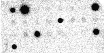
Autoradiography of a dot blot membrane hybridized with a radiolabeled probe

2.4 Troubleshooting
-
If the dots are irregularly shaped, check the vacuum system and repeat the transfer.
Notes
- 1.
Clean the pipette with alcohol or another disinfectant and leave them under the UV lamp for at least 10 min. Alternatively it is possible to autoclave the pipette depending on the provider instructions.
- 2.
It is possible to use a thermoblock, the Thermomixer or the thermocycler. If the latter has been chosen, load the samples directly in the PCR tubes.
- 3.
Add approximately 500 μl of dye for dot blot in 20 ml of SSC 20x. The use of this dye gives the possibility to evaluate the shape of the dot and consequently the transfer.
- 4.
Sometimes it could be convenient to use the dot-blot apparatus without the upper part (the cover) to check the transfer of the samples. In this way, when the vacuum is applied, the membrane adheres directly to the wells.
References
Cseke L, Kaufman P, Podila G, Tsai C (2004) Handbook of molecular and cellular methods in biology and medicine, 2nd edn. CRC, Boca Raton
Sambrook J, Russel DW (2001) Molecular cloning. A laboratory manual, 3rd edn. Cold Spring Harbour Laboratory Press, New York
Stanta G, Bonin S, Utrera R (1998) RNA quantitative analysis from fixed and paraffin-embedded tissues. Methods Mol Biol 86:113–119
Author information
Authors and Affiliations
Editor information
Editors and Affiliations
Rights and permissions
Copyright information
© 2011 Springer-Verlag Berlin Heidelberg
About this chapter
Cite this chapter
Bonin, S., Dotti, I. (2011). General Protocol for Dot-Blot. In: Stanta, G. (eds) Guidelines for Molecular Analysis in Archive Tissues. Springer, Berlin, Heidelberg. https://doi.org/10.1007/978-3-642-17890-0_23
Download citation
DOI: https://doi.org/10.1007/978-3-642-17890-0_23
Published:
Publisher Name: Springer, Berlin, Heidelberg
Print ISBN: 978-3-642-17889-4
Online ISBN: 978-3-642-17890-0
eBook Packages: MedicineMedicine (R0)