
Abstract
Cleft lip and cleft palate are major public health problems that should receive a comprehensive treatment (Freitas et al. 2012). These defects arise on intrauterine development of the face and may have long-standing implications on dental arch morphology and impair facial growth as well. Cleft correction itself may also harm facial growth potential, even if performed properly (Mølsted et al. 2005). Anatomical and physiological cleft-related problems can have implications on speech, eating, and aesthetic, sometimes leading to deep psychological consequences. Proper dental care from birth to adulthood is necessary to overcome these conditions while avoiding further harm. In this setting, the orthodontist plays an important role in the prevention, correction, and reduction of the consequences of cleft lip and cleft palate (Long et al. 2000).
Access provided by CONRICYT-eBooks. Download chapter PDF
18.1 Introduction
Cleft lip and cleft palate are major public health problems that should receive a comprehensive treatment (Freitas et al. 2012). These defects arise on intrauterine development of the face and may have long-standing implications on dental arch morphology and impair facial growth as well. Cleft correction itself may also harm facial growth potential, even if performed properly (Mølsted et al. 2005). Anatomical and physiological cleft-related problems can have implications on speech, eating, and aesthetic, sometimes leading to deep psychological consequences. Proper dental care from birth to adulthood is necessary to overcome these conditions while avoiding further harm. In this setting, the orthodontist plays an important role in the prevention, correction, and reduction of the consequences of cleft lip and cleft palate (Long et al. 2000).
Orthodontic treatment within the interdisciplinary team that takes care of children with cleft lip and cleft palate has a role to counteract the morphological impact on transverse, vertical, and anteroposterior maxillary dimensions imposed by reconstructive surgeries or by underdevelopment intrinsic to the pathology. Treatment as a whole should do a care protocol in order to harmonize the face and improve dental positioning.
One of the sought-after results for these patients is a good relation between upper and lower dental arches. In some cases, minor orthodontic treatment is sufficient to provide good occlusion. However, specially in patients operated too early or in more severe cases, good occlusion achievement may require quite complex treatment by the orthodontist, extensive orthopedics, or even surgical repositioning of the jaws through orthognathic surgery. Previous cephalometric studies have shown that primary surgery tends to affect the facial growth and dental development (Capelozza Filho et al. 1996). Therefore, a close follow-up by the orthodontist is of paramount importance to achieve a satisfactory outcome.
There are mainly three phases in which interventions in this area may take place. First one should be even before tooth eruption, when maxillary orthopedics can be applied in order to minimize deformities on alveolar bone ridge. This phase will not be covered in this chapter and its classical approach is nasoalveolar molding (Grayson et al. 1999). A second time window is during early mixed dentition, when orthopedics and orthodontics are used mainly to provide space for adequate permanent dentition. Finally on the end of facial growth, orthodontics may be necessary for final compensation or for decompensation in preparation for orthognathic surgery. At any time, the main goal is to maximize final esthetics and function. Nevertheless, successful treatment depends on the degree of skeletal and dental commitment that the patient presents.
18.2 Classification
In order to improve results in cleft management, it is important to apply periodic protocols and evaluations of the treatments employed. Some interventions will take many years to show their consequences. Therefore, classification methods and evaluation parameters were developed to compare intervention protocols and prognosis regarding facial skeletal growth. When comparing results from different approaches, used for instance by different centers, it is important to be sure that one is comparing patients of the same severity (Mølsted et al. 2005). Outcome studies based on classifications can provide information that clinicians may use in order to preview treatment difficulties and limitations of each case (Gray and Mossey 2005).
For unilateral clefts, which comprise the majority of cases, the most commonly used index is the Goslon yardstick, which analyzes the occlusal relationship through plaster models and clinical analysis (Mars et al. 1987). More recently, virtual tools based on dentofacial scanning were added. In bilateral clefts, the analysis proposed by Ozawa and colleagues in 2005 is the method most commonly used (Ozawa et al. 2011). These indexes classify patients according to features such as sagittal, transverse, and vertical relations between dental arches into categories with different prognosis.
Goslon yardstick was originally designed to classify patients during mixed and early permanent dentition (Mars et al. 1987). Later, adaptations for patients around 5 years were published (Atack et al. 1997; Mars et al. 2006). These systems divide patients into five groups:
-
Group 1: Positive overjet with average inclined or retroclined incisors with no crossbite or open bite. Long-term outcome: excellent.
-
Group 2: Positive overjet with average inclined or proclined incisors with unilateral crossbite or crossbite tendency with or without open-bite tendency around cleft site. Long-term outcome: good (Fig. 18.1).
Fig. 18.1 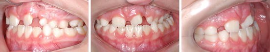
Patient classified as group 2 according to Goslon yardstick. Maxilla is sagittally well positioned in relation to mandible. Despite the need for transverse expansion to correct unilateral posterior crossbite, prognosis is good. Probably, orthognathic surgery will not be needed in the future; therefore dental compensation can be used if necessary in order to correct occlusion
-
Group 3: Edge-to-edge bite with average inclined or proclined incisors or reverse overjet with retroclined incisors. Unilateral crossbite with or without open-bite tendency around cleft site. Long-term outcome: fair.
-
Group 4: Reverse overjet with average inclined or proclined incisors. Unilateral crossbite with or without bilateral crossbite tendency with or without open-bite tendency around cleft site. Long-term outcome: poor (Fig. 18.2).
Fig. 18.2 
Patient classified as group 4 according to Goslon yardstick. Maxilla retracted in relation to mandible. Prognosis is fair, but probably orthognathic surgery will be advisable in the future. Maxillary expansion, leveling, and alignment must be performed, having in mind that there is a high chance that this maxilla will be brought forward during orthognathic operation and therefore decompensation would be needed as preparation
-
Group 5: Reverse overjet with proclined incisors, bilateral crossbite, and poor maxillary arch form and palatal vault anatomy. Long-term outcome: very poor.


Ozawa et al. published the Bauru index, with the same purpose of Goslon yardstick, but designed for patients with bilateral complete clefts (Ozawa et al. 2011). It’s interesting to note that this index changes little from the mixed to permanent dentition. It also consists of a scale of 1–5 with increasing severity degree, considering interarch relationship, shape of the upper dental arch, and inclination of upper incisors:
18.3 Dental Peculiarities
The development of primary dentition around the cleft region may be delayed. Teeth in this area may also show abnormalities in shape, structure, number, and position (Haque and Alam 2015; Galante et al. 2005). Usually, the more extensive the cleft, the more frequent these abnormalities are. Due to some of these irregularities, proper oral hygiene maintenance may be impaired, leading to cavities and early teeth loss. Preservation of teeth next to the cleft is very important, as their presence helps to maintain bony structure in the area.
Supernumerary teeth may be present in unilateral or bilateral cleft regions. Primary tooth eruption is delayed (Kobayashi et al. 2010). On the other hand, in patients with cleft lip and palate, natal and neonatal teeth occur more often and, because of their typical extreme mobility, extraction is indicated (Cabete et al. 2000).
Eruption of permanent teeth is also delayed by 6 months in average (de Carvalho Carrara et al. 2004). Permanent lateral and central incisors may have alterations in enamel structure (Gomes et al. 2009). Permanent lateral incisors are the most frequently absent teeth in patients with complete unilateral cleft (da Silva et al. 2008). Great care with oral hygiene is advised in order to prevent further teeth decay (Freitas et al. 2012).
18.4 Orthodontic Treatment
The goal of orthodontic treatment in cleft patients should be to counter the dental problems and incorrect relationships between alveolar bone bases. Orthodontic treatment in these children has a complexity related to the type and size of the cleft. Teeth may be analyzed according to their intra-arch and interarch relationships. In unilateral clefts, there may be a midline shift towards the cleft, often leading to the need for asymmetric extractions for correction. Extractions may also be necessary in order to correct crowding, which is a common feature on the maxilla due to poor sagittal and transverse growth (Capelozza Filho et al. 1996). When the cleft involves the alveolar ridge, the neighboring teeth show changes in their mesiodistal angulation added to abnormalities previously described. Central incisors are especially prone to present giroversion.
One great improvement on cleft lip and palate treatment was the introduction of secondary alveolar bone grafting. This procedure rebuilds bone anatomy of the alveolar cleft, allowing tooth movement in the region of the lateral incisors and making room for eruption of permanent canines (Bergland et al. 1986).
Over the years several studies have reported that patients with complete unilateral cleft had progressive restriction of anteroposterior maxillary growth, mainly due to consequences of primary surgery. The tension exerted by a rebuilt lip and the scar can be caused by cheiloplasty restricting growth and anterior maxillary development. Early palatoplasty also seems to have a restrictive influence on sagittal growth of the maxilla; thereby, in both unilateral and bilateral clefts, we often observe an anterior crossbite as a consequence of these constraining factors (Nollet et al. 2005; Liao and Mars 2006). Due to the restraining action of palatoplasty and the absence of midpalatal suture, there is a lack of maxillary development in the transversal direction. This maxillary atresia leads to a posterior crossbite, making maxillary expansion procedures a routine therapy in cleft patients (Capelozza Filho et al. 1996; Liao and Mars 2006) (Figs. 18.3–18.4).

Examples of different palatal expansion devices that may be employed, depending on factors like rate and vector of expansion

From left to right: Patient with bilateral cleft, with posterior crossbite. Hyrax expander in place. Postexpansion transversal gain. Device in place to promote transversal gain in anterior region
Diagnosis and treatment plan for cleft patients are based on the same diagnostic methods used for noncleft patients, meaning facial analysis, plater models, and radiologic as tomographic analysis. Classification of the case according to Goslon yardstick for unilateral clefts and Bauru method for bilaterals can help on prediction of the outcome.
Treatment may involve the steps described in the following protocol (Freitas et al. 2012):
-
1.
Orthodontics before alveolar bone grafting
-
2.
Secondary alveolar bone grafting
-
3.
Orthodontics after alveolar bone grafting
-
4.
Orthognathic surgery
-
5.
Finalization and containment
Pre-alveolar bone graft orthodontic treatment aims to promote maxillary transverse gain in order to align the teeth and the alveolar bone ridge. As a side result, there is a widening of the cleft, where the bone graft will be placed. The ideal age for secondary alveolar grafting is about 8–12 years old, on a moment just previous to canine eruption, as controlled by radiographic means. Surgery at this age also proves convenient because vertical and anteroposterior growth of the maxilla may be quite stabilized by then. Commencement of pre-grafting orthodontics must be planned having this time frame in mind. Orthodontic appliances are used, such as Hyrax expanders, Haas, or quad-helix. After expansion, a fixed containment device is provided in order to minimize relapse. Expanders allow an improvement in maxillary transverse deficiency but sagittal deficiency should be treated by means of devices that provide stimulus in this direction. Protraction masks can be used with this intention, but should only be applied to cases where there is a palatal inclination of the alveolar process. Fixed orthodontic appliances may be used in this step, but care should be taken on the periodontal limitations mainly in complete bilateral clefts. Repositioning of the premaxilla may also be necessary, in which case it should be performed at this phase.
In orthodontics after alveolar bone grafting, a quantitative and qualitative assessment of the grafted bone through clinical and radiographic examination of the area should be conducted while monitoring of the nonerupted canine; if the canine has already erupted, one must wait for 60–90 days after bone grafting surgery, before preforming orthodontic movement (Freitas et al. 2012).
Orthodontic treatment of patients that will not require orthognathic surgery involves the elimination of problems in the cleft region. If lateral incisors are present and have appropriate root and crown length, they must be correctly positioned. If they are missing, one must decide if the space will be closed by mesial movement of canine or if the space will be maintained for future prosthetic rehabilitation. This decision is based on the position canine eruption, on intermaxillary relationship, and on tooth size discrepancy. In patients with unilateral cleft, asymmetric extractions of premolars or laterals may be necessary for correction of deviated midline (Freitas et al. 2012).
Patients with complete bilateral clefts or unilateral clefts classified as Bauru or Goslon 3–5, by the end of facial growth, will probably present anterior crossbite and require orthognathic surgery. Orthodontic preparation on these patients involves alignment and leveling of both dental arches. Incisor decompensation is not necessary on the maxilla since superior incisors are usually already vertical, due to the restraining force of operated superior lip. Inferior incisors must be decompensated from their lingual inclination, provided that periodontal tissue is healthy and allows for the movement. Early classification, during childhood, is important to keep the orthodontist from compensating cases like Goslon 4–5 that will require orthognathic surgery in the future. After alignment, leveling, and decompensation, model cast analysis is performed to simulate final intercuspation. When this analysis shows that surgery is already viable, orthodontist and surgeon can decide on the magnitude and direction of movements of the jaws at the operation, always involving maxillary advancement. After postsurgical bone consolidation has occurred, orthodontic finalization can take place (Figs. 18.5–18.7).

At the end of facial growth, this unilateral cleft patient shows posterior crossbite and dental crowding due to maxillary transverse deficiency along with reduced maxillary dimensions as a whole. This Angle class III occlusion must be corrected surgically. Orthodontics in preparation for orthognathic surgery must involve extraction of superior malpositioned premolar, transverse expansion, alignment, leveling, and decompensation, which is performed mainly for correction of lingual inclination of inferior incisors

Case shown in Fig. 18.5 just before maxillary advancement. Negative overjet after decompensation reflects sagittal malposition. Arches are leveled and aligned. Residual posterior crossbite will be corrected by the advancement itself

Case shown in Fig. 18.5 before and after orthodontic and surgical treatment. Canine in position of lateral must receive esthetic treatment in order to mimic lateral shape
As on any orthodontic treatment, appliance removal must be done when esthetic and functional goals are achieved. Nevertheless, some adaptations may be necessary. If a canine had to be moved into lateral incisor position, the protection provided by canine contact during lateral excursion is lost. In these cases, contacts of posterior teeth in group function must be able to provide protection on lateral excursion.
Orthodontic relapse is a concern in cleft patients. Therefore, usage of containing devices is of paramount importance. Upper containment device (Hawley plate) must be used 24 h a day for 1 year. After this period, the removable device can be used during the night. Inferior fixed lingual container from canine to canine should be placed when fixed appliance is removed and must be left in place indefinitely. Prosthodontics and periodontal care may be necessary and the patient must be educated about the need for continuation of oral hygiene for maintenance of oral health.
References
Atack NE, Hathorn IS, Semb G, Dowell T, Sandy JR. A new index for assessing surgical outcome in unilateral cleft lip and palate subjects aged five: reproducibility and validity. Cleft Palate Craniofac J. 1997;34:242–6.
Bergland O, Semb G, Abyholm FE. Elimination of the residual alveolar cleft by secondary bone grafting and subsequent orthodontic treatment. Cleft Palate J. 1986;23:175–205.
Cabete HF, Gomide MR, Costa B. Evaluation of primary dentition in cleft lip and palate children with and without natal/neonatal teeth. Cleft Palate Craniofac J. 2000;37:406–9.
Capelozza Filho L, Normando AD, da Silva Filho OG. Isolated influences of lip and palate surgery on facial growth: comparison of operated and unoperated male adults with UCLP. Cleft Palate Craniofac J. 1996;33:51–6.
de Carvalho Carrara CF, de Oliveira Lima JE, Carrara CE, Gonzalez VB. Chronology and sequence of eruption of the permanent teeth in patients with complete unilateral cleft lip and palate. Cleft Palate Craniofac J. 2004;41:642–5.
Freitas JA, Garib DG, Oliveira M, et al. Rehabilitative treatment of cleft lip and palate: experience of the Hospital for Rehabilitation of craniofacial anomalies-USP (HRAC-USP)--part 2: pediatric dentistry and orthodontics. J Appl Oral Sci. 2012;20:268–81.
Galante JM, Costa B, de Carvalho Carrara CF, Gomide MR. Prevalence of enamel hypoplasia in deciduous canines of patients with complete cleft lip and palate. Cleft Palate Craniofac J. 2005;42:675–8.
Gomes AC, Neves LT, Gomide MR. Enamel defects in maxillary central incisors of infants with unilateral cleft lip. Cleft Palate Craniofac J. 2009;46:420–4.
Gray D, Mossey PA. Evaluation of a modified Huddart/Bodenham scoring system for assessment of maxillary arch constriction in unilateral cleft lip and palate subjects. Eur J Orthod. 2005;27:507–11.
Grayson BH, Santiago PE, Brecht LE, Cutting CB. Presurgical nasoalveolar molding in infants with cleft lip and palate. Cleft Palate Craniofac J. 1999;36:486–98.
Haque S, Alam MK. Common dental anomalies in cleft lip and palate patients. Malays J Med Sci. 2015;22:55–60.
Kobayashi TY, Gomide MR, Carrara CF. Timing and sequence of primary tooth eruption in children with cleft lip and palate. J Appl Oral Sci. 2010;18:220–4.
Liao YF, Mars M. Hard palate repair timing and facial growth in cleft lip and palate: a systematic review. Cleft Palate Craniofac J. 2006;43:563–70.
Long RE, Semb G, Shaw WC. Orthodontic treatment of the patient with complete clefts of lip, alveolus, and palate: lessons of the past 60 years. Cleft Palate Craniofac J. 2000;37:533–42.
Mars M, Plint DA, Houston WJ, Bergland O, Semb G. The Goslon yardstick: a new system of assessing dental arch relationships in children with unilateral clefts of the lip and palate. Cleft Palate Craniofac J. 1987;24:314–22.
Mars M, Batra P, Worrell E. Complete unilateral cleft lip and palate: validity of the five-year index and the Goslon yardstick in predicting long-term dental arch relationships. Cleft Palate Craniofac J. 2006;43:557–62.
Mølsted K, Brattström V, Prahl-Andersen B, Shaw WC, Semb G. The Eurocleft study: intercenter study of treatment outcome in patients with complete cleft lip and palate. Part 3: dental arch relationships. Cleft Palate Craniofac J. 2005;42:78–82.
Nollet PJ, Katsaros C, Van’t Hof MA, Kuijpers-Jagtman AM. Treatment outcome in unilateral cleft lip and palate evaluated with the GOSLON yardstick: a meta-analysis of 1236 patients. Plast Reconstr Surg. 2005;116:1255–62.
Ozawa TO, Shaw WC, Katsaros C, et al. A new yardstick for rating dental arch relationship in patients with complete bilateral cleft lip and palate. Cleft Palate Craniofac J. 2011;48:167–72.
da Silva AP, Costa B, de Carvalho Carrara CF. Dental anomalies of number in the permanent dentition of patients with bilateral cleft lip: radiographic study. Cleft Palate Craniofac J. 2008;45:473–6.
Author information
Authors and Affiliations
Corresponding author
Editor information
Editors and Affiliations
Rights and permissions
Copyright information
© 2018 Springer International Publishing AG
About this chapter
Cite this chapter
Camara, P., Bastos, E.O., Curi, D., Alonso, N. (2018). Orthodontic Treatment of Patients with Orofacial Cleft. In: Alonso, N., Raposo-Amaral, C. (eds) Cleft Lip and Palate Treatment. Springer, Cham. https://doi.org/10.1007/978-3-319-63290-2_18
Download citation
DOI: https://doi.org/10.1007/978-3-319-63290-2_18
Published:
Publisher Name: Springer, Cham
Print ISBN: 978-3-319-63289-6
Online ISBN: 978-3-319-63290-2
eBook Packages: MedicineMedicine (R0)