
Abstract
Rickettsial diseases, caused by a variety of obligate intracellular, Gram-negative bacteria from the genera Rickettsia, Orientia, Ehrlichia, Neorickettsia, Neoehrlichia, and Anaplasma, belong to the class Alphaproteobacteria, are considered some of the most covert emerging and re-emerging diseases and are being increasingly recognized. Rickettsial infections are generally incapacitating and difficult to diagnose; untreated cases have case fatality rates as high as 30–45 % with multiple organ dysfunction, if not promptly diagnosed and appropriately treated especially in the developing and under-developed countries. The vast variability and non-specific presentation of this infection have often made it difficult to diagnose clinically. Prompt antibiotic therapy shortens the course of the disease, lowers the risk of complications, and in turn reduces morbidity and mortality due to rickettsial diseases. There is a distinct need for physicians and health care workers at all levels of care to be aware of the clinical features, available diagnostic tests and their interpretation, and the therapy of these infections.
Access provided by Autonomous University of Puebla. Download chapter PDF
Keywords
1 Introduction
Rickettsial infections are caused by a variety of obligate intracellular, Gram-negative bacteria of the genera Rickettsia, Orientia, Ehrlichia, Neorickettsia, and Anaplasma, belonging to the class Alphaproteobacteria. Bacteria of the genus Rickettsia are the well-known members of the order Rickettsiales. Rickettsia are divided into the typhus group and spotted fever group (SFG) . Orientia spp. makes up the scrub typhus group (http://www.cdc.gov/). Rickettsial diseases are zoonoses where human beings are accidentally involved in a chain of transmission between trombiculid mites (chiggers ), ticks or fleas, and animals (most commonly rodents).
Classification of the Rickettsial diseases :
Diseases | Rickettsial agent | Insect vectors | Mammalian reservoirs |
|---|---|---|---|
Typhus group | |||
(a) Epidemic typhus | R. prowazekii | Louse | Humans |
(b) Murine typhus | R. typhi | Flea | Rodents |
(c) Scrub typhus | O. tsutsugamushi | Mite | Rodents |
Spotted fever group | |||
(a) Indian tick typhus | R. conorii | Tick | Rodents, dogs |
(b) Rocky Mountain spotted fever | R. rickettsii | Tick | Rodents, dogs |
(c) Rickettsial pox | R. akari | Mite | Mice |
Among the major groups of rickettsioses diseases commonly reported in India are scrub typhus , murine flea-borne typhus , and Indian tick typhus .
2 Presenting Manifestations
Rickettsial infections are generally incapacitating and difficult to diagnose; untreated cases have case fatality rates as high as 30–45 % with multiple organ dysfunction, if not promptly diagnosed and appropriately treated (Batra 2007). The vast variability and non-specific presentation of this infection have often made it difficult to diagnose clinically. Given below are some of the presenting symptoms and signs of rickettsial infections:
Acute fever is the most common presenting symptom often associated with breathlessness, cough, nausea, vomiting, myalgia, and headache (Mathai et al. 2003; Vivekanandan et al. 2010).
An eschar
at the site of chigger
bite can be seen in early disease and is a useful diagnostic clue in scrub typhus
, though its frequency varies from 7 to 97 % (Paris et al. 2013). Eschars are painless, ulcers upto 1 cm in size, with a black necrotic centre (resembling the mark of a cigarette burn) (Figs. 6.1 and 6.2). Usually a single eschar is found on the neck, axillae, chest, abdomen and groin, but multiple eschars have also been documented (Kaushik et al. 2014). Eschar on moist intertriginous surfaces (axilla, scrotum, perianal region) may be missed if not looked into carefully because they may lack the black scab and appear as shallow yellow-based ulcers without surrounding hyperemia
.

Eschar at neck region

Eschar with the typical central black scab with a cigarette burn like appearance on the skin
Rash (in fair skinned people)
Though rash is considered a hallmark of rickettsial disease, it is neither seen at presentation nor in all patients. Presence of rash is common in spotted fever and extremely rare in scrub typhus . Rash usually becomes apparent after 3–5 days of the onset of symptoms . Initially, rash is in the form of pink, blanching, discrete maculae, which subsequently becomes maculopapular, petechial, or hemorrhagic (Rathi and Rathi 2010).
None of the clinical symptoms and signs including eschar are diagnostic of rickettsial disease. Therefore, epidemiological factors pertaining to geographical area, habitat, occupation, movement of the subject (vocational or recreational) could assist in reaching a diagnosis of rickettsial disease with certainty and initiating treatment on time.
The complications of scrub typhus usually develop after the first week of illness. Jaundice, renal failure, pneumonitis, acute respiratory distress syndrome (ARDS) , septic shock, myocarditis, and meningoencephalitis are various complications known with this disease (Mahajan 2005).
Pneumonia is one of the most frequent complications of scrub typhus that manifests as a non-productive cough and breathlessness and leads to ARDS which could be life-threatening. Severe complications besides acute respiratory distress syndrome (ARDS) include hepatitis, renal failure, meningo-encephalitis and myocarditis; shock may occur in varying proportions of patients (Mahajan 2005).
Prompt antibiotic therapy , even based on suspicion, shortens the course of the disease, lowers the risk of complications , and in turn reduces morbidity and mortality due to rickettsial diseases. Currently, doxycycline is regarded as the drug of choice (Liu and Panpanich 2002).
There is a distinct need for physicians and health care workers at all levels of care in India to be aware of the clinical features, available diagnostic tests and their interpretation, and the therapy of these infections. Therefore, these guidelines are developed to help physicians towards correct diagnosis and treatment. Lack of awareness in physicians and community workers, as well as diagnostic delays, results in patients presenting to tertiary care centers with ARDS with severe complications resulting in a higher risk of mortality .
3 Guidelines for Management
3.1 Case Definition
3.1.1 Definition of Suspected/Clinical Case
Acute undifferentiated febrile illness of 5 days or more with or without eschar should be suspected as a case of Rickettsial infection. (If eschar is present, fever of less than 5 days duration should be considered as scrub typhus .) Other presenting features may be headache and rash (rash more often seen in fair persons), lymphadenopathy, multi-organ involvement like liver, lung, and kidney involvement. The differential diagnosis of dengue, malaria, pneumonia, leptospirosis , and typhoid should be kept in mind.
3.1.2 Definition of Probable Case
A suspected clinical case showing titres of 1:80 or above in OX2, OX19, and OXK antigens by Weil Felix test and an optical density (OD) > 0.5 for IgM by ELISA are considered positive for typhus and spotted fever groups of Rickettsiae .
3.1.3 Definition of Confirmed Case
A Confirmed case is the one in which:
Rickettsial DNA is detected in eschar samples or whole blood by PCR.
Or
Rising antibody titers on acute and convalescent sera detected by indirect Immunofluorescence assay (IFA) or indirect immunoperoxidase assay (IPA) .
3.2 Laboratory Criteria
There are various laboratory tests available for diagnosis of rickettsial diseases. Indirect immunoperoxidase assay (IPA) and immunofluorescence assay (IFA) are considered serological gold standards, but are available at laboratories with higher level of facilities and expertise. Molecular diagnosis by PCR is available only at few centres in India. However, ELISA-based tests , particularly immunoglobulin M (IgM) capture assays, can be made available at secondary level and tertiary levels of health care like district hospitals and medical colleges.
Weil-Felix test , which is helpful in establishing presumptive diagnosis in diseases caused by members of typhus and spotted fever groups of Rickettsiae, can be considered at primary level of health care as they can be easily set up with moderate level of infrastructure and expertise at least in areas affected by scrub typhus .
3.2.1 Specific Investigations
Weil Felix: The sharing of the antigens between Rickettsia and Proteus is the basis of this heterophile antibody test. It demonstrates agglutinins to Proteus vulgaris strain OX19, OX2, and Proteus mirabilis OXK. Though this test lacks high sensitivity and specificity, it still serves as a useful and inexpensive diagnostic tool for laboratory diagnosis of rickettsial disease. This test should be carried out only after 5–7 days of onset of fever. Titer of 1:80 is to be considered possible infection. However, baseline titers need to be standardized for each region.
IgM and IgG ELISA: ELISA techniques , particularly immunoglobulin M (IgM) capture assays for serum, are probably the most of sensitive tests available for rickettsial diagnosis and the presence of IgM antibodies and indicate comparatively recent infection with rickettsial disease. In cases of infection with O. tsutsugamushi, a significant IgM antibody titer is observed at the end of the first week, whereas IgG antibodies appear at the end of second week. The cut off value is Optical Density of 0.5. Baseline titres need to be established keeping in view the regional variations.
Polymerase Chain Reaction (PCR) : It is a rapid and specific test for diagnosis. It can be used to detect rickettsial DNA in whole blood and eschar samples. The PCR is targeted at the gene encoding the major 56 kDa and/or 47 kDa surface antigen gene. The results are best within first week for blood samples because of the presence of rickettsemia (O. tsutsugamushi, R. rickettsii, R. typhi and R. prowazekii) in the first 7–10 days.
Immunofluorescence Assay (IFA): This is a reference serological method for diagnosis of Rickettsial diseases and is considered the serological ‘gold standard’; however, cost and requirement of technical expertise limit its wide use. Therefore, it is recommended only for research and in areas where sero-prevalence of rickettsial diseases has been established and a reference facility is already available which has the necessary expertise required to conduct these tests.
Indirect Immunoperoxidase Assay (IPA) : It gives comparable result as IFA, but requires special instrumentation and experienced personnel for interpretation of the test. Currently, we do not recommend any rapid test for diagnosis of scrub typhus as they need additional evaluation.
3.2.2 Supportive Laboratory Investigations
These are required as additional diagnostic clues and sometimes can indicate severity and development of complications. These investigations can assist in deciding upon appropriate management of patients.
-
(a)
Hematology
-
Total Leucocytes Count during early course of the disease may be normal, but later in the course of the disease, WBC count may become elevated to more than 11,000/cu.mm.
-
Thrombocytopenia (i.e. <1,00,000/cu.mm) is seen in majority of patients.
-
-
(b)
Biochemistry
-
Raised Transaminase levels are commonly observed.
-
3.2.3 Imaging
-
Chest X-Ray showing infiltrates , mostly bilateral (Figs. 6.3, 6.4 and 6.5).
Fig. 6.3 
X-ray of a patient with scrub typhus showing bilateral lower lobe interstitial infiltrates
Fig. 6.4 
X-Ray of a 30-year old woman presenting with fever for 10 days, non-productive cough for 5 days, and complaining of breathlessness. X-ray shows bilateral reticulonodular (interstitial) opacities in the lower lobes before treatment
Fig. 6.5 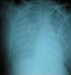
X-ray of the same patient taken 2 days after admission to a tertiary care center with severe breathlessness. X-ray now shows bilateral extensive air-space consolidation suggestive of an acute respiratory distress syndrome


3.3 Treatment
There is paucity of evidence based on randomized controlled trials for the management of rickettsial diseases including scrub typhus .
These guidelines for treatment cover the most common infection, the scrub typhus , murine typhus , and Indian Tick typhus .
Without waiting for laboratory confirmation of the Rickettsial infection, antibiotic therapy should be instituted when rickettsial disease is suspected.
3.3.1 At Primary Level
The Healthcare provider needs to do the following:
-
(a)
Recognition of disease severity. If the patients come with complications to primary health facility and the treating physician considers it as rickettsial infection, treatment with doxycycline should be initiated before referring the patient.
-
(b)
Referral to secondary or tertiary centre in case of complications like ARDS, acute renal failure, meningo-encephalitis, multi-organ dysfunction. In addition to recommended management of community-acquired pneumonia, doxycycline is to be initiated when scrub typhus is considered likely.
-
(c)
Patients with fever of more than 5 days where malaria, dengue, and typhoid have been ruled out; the following drugs should be administered when scrub typhus is considered likely—
Adults
-
(a)
Doxycycline 200 mg/day in two divided doses for individuals above 45 kg for duration of 7 days. Patients should be advised to swallow capsules with plenty of fluid during meals while sitting or standing.
Or
-
(b)
Azithromycin 500 mg in a single oral dose for 5 days.
Children
-
(a)
Doxycycline in the dose of 4.5 mg/kg body weight/day in two divided doses for children below 45 kg.
Or
-
(b)
Azithromycin in the single dose of 10 mg/kg body weight for 5 days.
Pregnant women
-
(a)
Azithromycin 500 mg in a single dose for 5 days.
-
(b)
Azithromycin is the drug of choice in pregnant women, as doxycycline is contraindicated.
3.3.2 At Secondary and Tertiary Care
-
(a)
The treatment as specified above in uncomplicated cases.
-
(b)
In complicated cases the following treatment is to be initiated—
-
Intravenous doxycycline (wherever available) 100 mg twice daily in 100 mL normal saline to be administered as infusion over half an hour initially followed by oral therapy to complete 7–15 days of therapy.
Or
-
Intravenous Azithromycin in the dose of 500 mg IV in 250 mL normal saline over 1 h once daily for 1–2 days followed by oral therapy to complete 5 days of therapy (Jang et al. 2014).
Or
-
Intravenous chloramphenicol 50–100 mg/kg/day 6 hourly doses to be administered as infusion over 1 h initially followed by oral therapy to complete 7–15 days of therapy.
-
Management of the individual complications should be done as per the existing practices.
-
Doxycycline and/or Chloramphenicol-resistant strains have been observed in South-East Asia. These strains are sensitive to Azithromycin (Kim et al. 2007).
Disclaimer: These guidelines on diagnostic and treatment of rickettsial infections are based on a review of the currently available evidence and best practices and may be revised in light of future developments in the field .
References
http://wwwnc.cdc.gov/travel/yellowbook/2014/chapter-3-infectious-diseases-elated-to-travel/rickettsial-spotted-and-typhus-fevers-and-related-infections-anaplasmosis-and-ehrlichiosis (as accessed on 21st August, 2014).
Sharma P, Kakkar R, Kaore S, Yadav V, Sharma R. Geographical Distribution, Effect of season & Life Cycle of Scrub Typhus. JK Science. 2010; 12 (2): 63–64.
Mittal V, Gupta N, Bhattacharya D, Kumar K, Ichhpujani RL, Singh S, et al. Serological evidence of rickettsial infections in Delhi. Indian J Med Res. 2012;135(4):538–41.
Yuehong Wei, Yong Huang, Lei Luo, Xincai Xiao, Lan Liu, and Zhicong Yang. Rapid Increase of Scrub Typhus: An Epidemiology and Spatial-Temporal Cluster Analysis in Guangzhou City, Southern China, 2006–2012. 2014 PLoS One. 2014; 9(7): e101976.
Batra HV. Spotted fevers and typhus fevers in Tamil Nadu. Indian J Med Res. 2007; 126:101–103.
Mathai E, Rolain JM, Verghese GM, Abraham OC, Mathai D, Mathai M, Raoult D. Outbreak of scrub typhus in southern India during cooler moths. Ann N Y Acad Sci. 2003; 990:359–364.
Vivekanandan M, Mani A, Priya YS, Singh AP, Jayakumar S, Purty S. Outbreak of scrub typhus in Pondicherry. J assoc Physicians India. 2010; 58:24–28.
Paris D H, Shelite T R, Day N P and Walker D H. Review Article: Unresolved Problems related to scrub typhus: A seriously neglected life-threatening disease. Am J Trop. Med. Hyg. 2013; 89 (2): 301–307.
Kaushik R M, Kaushik R, Bhargava A. Multiple eschars in scrub typhus. Tropical Medicine and Health [serial on the Internet]. 2014 [cited 2014 7th May]: Available from: http://jlc.jst.go.jp/DN/JST.JSTAGE/tmh/2013-33.
Rathi N and Rathi A. Rickettsial Infections: Indian Perspective. Indian Pediatrics 2010;47: 157–164.
Mahajan S K.Scrub typhus. J Assoc Physicians India 2005; 53: 954–958.
Liu Q, Panpanich R. Anitbiotics for treating scrub typhus. Cochrane Database of Systematic Reviews 2002; Issue 3 Art No. CD002159.
Mi-Ok Jang, Hee-Chang Jang, Uh Jin Kim, Joon Hwan Ahn, Seung-Ji Kang, Sook-In Jung, Hee-Young Shin and Kyung-Hwa Park 2014.Outcome of intravenous azithromycin therapy in patients with complicated scrub typhus compared with that of doxycycline therapy using propensity-matched analysis. Antimicrob Agents Chemother 58 (3): 1488–1493.
Kim D-M, Yu K D, Lee J H, Kim H K, Lee S H. Controlled trial of a 5-Day Course of Telithromycin versus Doxycycline for treatment of mild to moderate scrub typhus. Antimicrobial agents and chemotherapy 2007; 51 (6): 2011–15.
Author information
Authors and Affiliations
Corresponding author
Editor information
Editors and Affiliations
Rights and permissions
Copyright information
© 2016 Springer International Publishing AG
About this chapter
Cite this chapter
Rahi, M., Gupte, M.D., Bhargava, A., Varghese, G.M., Arora, R. (2016). DHR-ICMR Guidelines for Diagnosis and Management of Rickettsial Diseases in India. In: Thomas, S. (eds) Rickettsiales. Springer, Cham. https://doi.org/10.1007/978-3-319-46859-4_6
Download citation
DOI: https://doi.org/10.1007/978-3-319-46859-4_6
Published:
Publisher Name: Springer, Cham
Print ISBN: 978-3-319-46857-0
Online ISBN: 978-3-319-46859-4
eBook Packages: Biomedical and Life SciencesBiomedical and Life Sciences (R0)