
Abstract
The hypoosmotic swelling (HOS) test evaluates the functional integrity of the sperm’s plasma membrane and also serves as a useful indicator of its fertility potential. The functional integrity can be demonstrated by allowing sperm to react in a hypoosmotic medium. The hypoosmotic swelling test presumes that only cells with intact membranes (live cells) will swell in hypotonic solutions. The results of the HOS test correlate closely with the hamster egg penetration test. Spermatozoa with intact membranes swell within 5 min in hypoosmotic medium, and all flagellar shapes are stabilized in 30 min.
Access provided by Autonomous University of Puebla. Download chapter PDF
Keywords
1 Introduction
The hypoosmotic swelling (HOS) test evaluates the functional integrity of the sperm plasma membrane and also serves as a useful indicator of its fertility potential. The functional integrity can be demonstrated by allowing sperm to react in a hypoosmotic medium. The hypoosmotic swelling test presumes that only cells with intact membranes (live cells) will swell in hypotonic solutions. The results of the HOS test correlate closely with the hamster egg penetration test. Spermatozoa with intact membranes swell within 5 min in hypoosmotic medium and all flagellar shapes are stabilized by 30 min [1–3].
1.1 Specimen Collection
The physician instructs the patient on proper collection technique. The patient collects the specimen into a sterile container and brings it to the laboratory within 1 h, keeping the sample at body temperature. Sperm vitality should be assessed as soon as possible after liquefaction of the semen sample, preferably at 30 min, but within 1 h of ejaculation to prevent the deleterious effects of dehydration, or large changes in temperature, on vitality.
1.2 Equipment and Materials
-
A.
Hypoosmotic solution—prepare monthly (or as needed) as follows:
Combine the following in 100 mL of distilled water:
-
1.
0.735 g sodium citrate dihydrate (Fig. 12.1)
Fig. 12.1 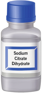
Sodium citrate dihydrate [Reprinted with permission, Cleveland Clinic Center for Medical Art & Photography © 2015. All Rights Reserved]
-
2.
1.351 g fructose (Fig. 12.2)
Fig. 12.2 
d-(−)-Fructose [Reprinted with permission, Cleveland Clinic Center for Medical Art & Photography © 2015. All Rights Reserved]
-
1.
-
B.
Microcentrifuge tubes
-
C.
3″ × 1″ glass slides (plain)
-
D.
22 × 22 mm coverslips
-
E.
Pipette tips (100 μL, 1000 μL)


1.3 Quality Control
A positive control using a donor or patient specimen should be run monthly or when fresh reagents are prepared with a result of 58 % or greater vitality to verify the integrity of the media. Record initial results on testing control sheet the day when the control is run.
1.4 Procedure
Note: Specimens to be tested should be liquefied and no more than 1 h old.
-
A.
Combine 0.1 mL well-mixed semen with 1 mL hypoosmotic solution (HOS) (Fig. 12.3).
Fig. 12.3 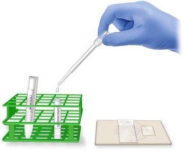
Adding well-mixed semen sample to HOS solution [Reprinted with permission, Cleveland Clinic Center for Medical Art & Photography © 2015. All Rights Reserved]
-
B.
Mix gently by drawing sample in and out of the pipette.
-
C.
Incubate at 37 °C for 30–60 min.
-
D.
After incubation, place one drop of the semen mixture on a glass slide and top with a coverslip (Fig. 12.4).
Fig. 12.4 
Droplet of semen sample and HOS mixture placed on slide [Reprinted with permission, Cleveland Clinic Center for Medical Art & Photography © 2015. All Rights Reserved]
-
E.
Observe for tail swelling (Fig. 12.5) under 40× phase contrast lens.
Fig. 12.5 
Degrees of hypoosmotic swelling of sperm tails [Reprinted with permission, Cleveland Clinic Center for Medical Art & Photography © 2015. All Rights Reserved]
-
F.
Using a two-channel cell counter, differentiate 100 sperm by swelling vs. non-swelling tails.
Example:
$$ \frac{55\ \left(\mathrm{swollen}\ \mathrm{tails}\right)}{100\ \mathrm{total}\ \mathrm{sperm}\ \mathrm{counted}} \times 100 = 55\% $$


1.5 Results
-
Reference range: ≥58 % (CI: 55–63 %) Tail swelling
-
HOS test values approximate those of the eosin test [4]. The lower reference limit for vitality (membrane-intact spermatozoa) is 58 % (fifth centile, 95 % CI 55–63).
References
Hossain AM, Selukar R, Barik S. Differential effect of common laboratory treatments on hypoosmotic swelling responses of human spermatozoa. J Assist Reprod Genet. 1999;16(1):30–4.
Jeyendran RS, Van der Ven HH, Perez-Pelaez M, Crabo BG, Zaneveld LJ. Development of an assay to assess the functional integrity of the human sperm membrane and its relationship to other semen characteristics. J Reprod Fertil. 1984;70:219–28.
WHO Manual. Laboratory manual for the examination of human semen and semen-cervical mucus interaction. 5th edition, Geneva. World Health Organization Manual. Switzerland, 2010.
Ramirez JP, Carreras A, Mendoza C. Sperm plasma membrane integrity in fertile and infertile men. Andrologia. 1992;24(3):141–4.
Author information
Authors and Affiliations
Corresponding author
Editor information
Editors and Affiliations
Cleveland Clinic
Cleveland Clinic

Rights and permissions
Copyright information
© 2016 Springer International Publishing Switzerland
About this chapter
Cite this chapter
Agarwal, A., Gupta, S., Sharma, R. (2016). Hypoosmotic Swelling Test (HOS). In: Agarwal, A., Gupta, S., Sharma, R. (eds) Andrological Evaluation of Male Infertility. Springer, Cham. https://doi.org/10.1007/978-3-319-26797-5_12
Download citation
DOI: https://doi.org/10.1007/978-3-319-26797-5_12
Published:
Publisher Name: Springer, Cham
Print ISBN: 978-3-319-26795-1
Online ISBN: 978-3-319-26797-5
eBook Packages: MedicineMedicine (R0)