
Abstract
Rapid and precise assessment of sperm viability is vital in evaluating seminal quality. The viability (%) is determined by identifying sperm with an intact cell membrane, either by carrying out dye exclusion tests or by hypo-osmotic swelling. In this chapter, we have attempted to cover some important assays with the underlying principles including assay based on the hypotonic swelling and its variations as an indicator of the biochemical integrity of the membrane. This chapter contains procedures related to hypo-osmotic swelling and application of supravital dye and various fluorescent dyes such as Hoechst 33342, DAPI, PI and SYBR-14 including their combination for determining spermatozoa viability. A comparison of the assay vis-à-vis their merit has been provided.
Access provided by CONRICYT-eBooks. Download chapter PDF
Keywords
6.1 Introduction
Analysis of semen quality is a multistage process: mostly the motility evaluation is the first step followed by determining the proportions of live/dead sperm [1] and evaluation of other quality parameters. Since functional activity and the intactness of sperm membrane are of fundamental importance in fertilization process, most of the assays are focused on membrane integrity. Functional activity is based on the property of cell membrane to permit the transport of molecules selectively. Hypo-osmotic swelling assay exploits this property of the sperm membrane and is a useful assay in determining viability and functional integrity of the plasma membrane.
Generally, while analysing semen for routine purposes or in much research-oriented experimentation, it is crucial to discriminate dead from live spermatozoa. Identification of live sperm is by observing their motility, while the ability to distinguish live but immotile spermatozoa from dead ones requires application of other tests. For this purpose, many dye exclusion techniques have evolved. The term ‘viable spermatozoon’ is often linked to a cell with intact plasma membrane, since the plasma lemma is pivotal for sperm function and interactions with other cells, including their environment. Keeping this in mind, therefore, most viability assays just assess integrity of the plasma lemma, either by using impermeable probes (cannot penetrate an intact membrane) or alternatively membrane-permeable (penetrating) probes (depict spermatozoon with eroded plasma lemma) or many a times a combination, a matter that shall be described in detail later in the chapter. Since sperm ‘viability' can be measured by numerous methods, some of them have been provided here. We have also included lipid peroxidation assay, which reflect the membrane’s function (Fig. 6.1).

Assessment of sperm membrane damages: (1) by the use of membrane-permeable (MP) or membrane (non)-impermeable dyes, (2) by biochemical integrity of the plasma membrane (hypo-osmotic swelling of the sperm tail) and (3) by assessing distortion in the lipid bilayer, either by fluorescent staining with chlortetracycline (CTC), C8NBD, Merocyanine 540 and annexin V or by assessing peroxidation of fatty acid moieties (C11BODIPY and thiobarbiturate test)
6.2 Comparison of Different Staining Procedures for Viability Evaluation
Eosin-nigrosin stain for the evaluation of bull sperm was introduced for the first time by Blom in 1950 [2]: the test was carried out by adding eosin-opal blue to bull semen followed by mixing with a nigrosin solution. Thereafter, the simplified one-step technique was introduced [3] and evaluated for various mammalian sperm [4]. In 1954, Mixner and Saroff [5] reported that eosin-nigrosin stain for sperm viability is not suitable for analysing frozen-thawed sperm because of interference with glycerol with the stains [5]. In 1986, Garner et al. used CFDA in combination with PI for evaluation of viability of sperm from dogs, horses, boars, bulls, mice and humans [6]. They showed that motile sperm were stained with the CFDA fluorochrome, stain spreading throughout the sperm cytoplasm, at the same time not staining with PI. Since the CFDA is an enzyme-based stain (enzyme substrate conversion to a fluorescent product), time dependency is a major problem [7]. Subsequently, Garner et al. and Ensico et al. [7, 8] used SYBR-14 along with PI to evaluate sperm viability in bovine sperm. Former stain is also a green fluorescent molecule that labels green the nucleus of sperm with intact membrane. Garner et al. [9] demonstrated that the combination of SYBR-14 and P1 not only was useful in staining freshly ejaculated sperm but was ineffective in assessing the viability of thawed, cryopreserved sperm. In comparison, nucleic acid stains are considered less variable than enzyme-based stains for viability evaluation. This is more so because sperm DNA is considered a more appropriate target for probes for its stainability as well as uniform staining [10]. To date, combination of SYBR-14+PI has been shown to be a reliable and accurate way of evaluating the percent number of viable and dead sperm [11] (Table 6.1).
6.3 Staining for Vitality Determination
6.3.1 Supravital Stains
6.3.1.1 Eosin-Nigrosin Stain
Principle
For faster determination of viability of bull sperm, eosin is the stain of choice [19]. Initially, a two-step staining technique using eosin was originally employed for the examination of bull sperm [2]. Thereafter, the simplified one-step technique was introduced [3] and evaluated for various mammalian sperm [4]. The objective of a differential stain is to stain the spermatozoa in such a way that two populations (live and dead) can be distinguished from each other and also remain evident from the background.
The eosin-nigrosin staining technique is based on the principle that eosin penetrates the membrane of dead sperm due to increased permeability which appear pink in colour, while eosin cannot penetrate the membrane of live sperm cells because of their selective permeability and hence live cells appear colourless. The nigrosin stain provides a dark background which helps to differentiate live and dead spermatozoa. The objective of the staining is to evaluate the structural plasma integrity.
Materials
Hancock’s Stain (Hancock 1951) [20]
-
1.67 g eosin Y.
-
10 g nigrosin.
-
Dilute to 100 mL DW.
Blom’s Stain (Blom 1950) [2]
-
5 g eosin B.
-
10 g nigrosin.
-
Dilute to 500 mL DW.
To prepare both stains, add the nigrosin to the water while stirring and heating until all is dissolved. Thereafter, add the eosin to the nigrosin solution. Boiling should be avoided throughout the procedure. However, it is difficult to dissolve stain at this concentration and is quite hypotonic.
Modified Eosin-Nigrosin Formula [21]
-
3.3 g eosin Y.
-
20.0 g nigrosin.
-
1.5 g sodium citrate.
-
Dilute to 300 mL DW.
Preparation of the Stain
-
(a)
Dissolve the ingredients by stirring and heating as described above.
-
(b)
Adjust the pH to 6.8–7.0 if necessary.
-
(c)
Allow the stain to stand a few days and then filter it.
-
(d)
Since Hancock’s and Blom’s stains are slightly hypotonic, a small amount of sodium citrate is added to increase the osmolarity. This prevents formation of bent tail, which is a common problem with the above two stains.
-
(e)
Stock solution of eosin-nigrosin can be stored for several years if refrigerated. At RT these stains may be kept without harm up to 6 months. However, many times bacteria and fungi may multiply in the stains at RT.
Procedure
-
(a)
Place large drops of warm eosin-nigrosin stain near the one end of a warm slide.
-
(b)
Place a semen drop (size varying with the density of the sample: large drop for frozen- thawed semen and small drop for fresh neat semen) near the stain and quickly mix the two on the slide.
-
(c)
Leave it for 1 min.
-
(d)
Draw a smear (thick smear for diluted frozen-thawed semen and thin smear for undiluted fresh semen) on a clean, grease-free slide.
-
(e)
Dry the smear quickly by blowing air across it or by placing on a warming plate.
-
(f)
Examine the slide under microscope.
-
(g)
First, focus at low magnification (40x objective lens) in order to get an overview.
-
(h)
Now, place a drop of immersion oil at the area of actual observation.
-
(i)
Turn the revolving nosepiece to engage the immersion objective, and then focus with the help of coarse and fine adjustment knob.
-
(j)
Count approximately 200 sperm randomly from different fields and calculate % of live and dead sperm.
Observation
Live spermatozoa remain unstained, while dead spermatozoa take pink or red stain against blue-black dark background. Count partially stained sperm as dead ones.
Calculation
\( {\displaystyle \begin{array}{l}\mathrm{Percent}\ \mathrm{live}\ \mathrm{sperm}=\hfill \\ {}\kern0.50em \ \kern0.125em \frac{\mathrm{Number}\ \mathrm{of}\ \mathrm{live}\ \mathrm{sperm}\times 100\ }{\mathrm{Total}\ \mathrm{number}\ \mathrm{of}\ \mathrm{live}\ \mathrm{and}\ \mathrm{dead}\ \mathrm{sperm}}\hfill \end{array}} \)
Interpretation
-
The live sperm count in normal bull should be above 80%.
-
Discard semen sample containing more than 30% dead sperm.
-
A minimum of 40% viable sperm in post-thaw semen is considered good.
-
Structural defects in the sperm flagellum are indicated by the presence of a large proportion of viable but immotile cells [22].
-
Epididymal pathology is indicated by a high percentage of immotile and non-viable cells (necrozoospermia) [23, 24].
Points to Ponder
-
(a)
When EN-stained slides are allowed to dry slowly, some of the sperm may die and be stained before the drying process is complete, thus giving a false result.
-
(b)
When examining slides under oil immersion, make sure that the oil used is free of air bubbles. Turn the revolving nosepiece of the microscope to move the oil immersion objective back and forth few times to dislodge bubbles.
-
(c)
Following every use, clean the oil from the objective front lens by gently wiping it with gauze slightly moistened with a mixture of cleaning solution (ether, 70%+ alcohol, 30%) (Fig. 6.2).
Fig. 6.2 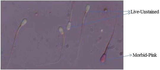
Microphotograph of spermatozoa stained with eosin and nigrosin. Spermatozoa that are white coloured are live, while one stained red/pink is dead

6.3.2 Fluorescent Probes for Sperm Viability
The sperm viability is also detected by the fluorescent dyes. Such fluorescent probes can be classified into two categories [25]: firstly the membrane-impermeable dyes are able to penetrate the damaged plasma membrane of dead sperm, stain the nucleus and are seen directly under a fluorescence microscope, e.g. Hoechst 3358, YO-PRO-1 and PI, whereas the second group consists of acylated membrane probes (have no fluorescence of its own, but turn into fluorescent substances while they pass the intact plasma membrane after gaining entry into the living sperm, e.g. CFDA, CAM, SYTO-1 and SYBR-14). Some of the fluorescent probes for determining sperm cell viability by staining DNA are:
-
Carboxyfluorescein diacetate (CFDA)
-
4′,6-Diamidino-2-phenylindole (DAPI)
-
Hoechst stains
-
Propidium iodide (PI) (Table 6.2)
Table 6.2 The excitation, emission and other characteristics of common fluorescent dyes
6.3.2.1 Sperm Viability by Hoechst 33342 (Viadent) Stain
With the development of CASA, new criteria for sperm evaluation were available for many species. Hamilton Thorne Research (Beverly, Massachusetts) produced an integrated visual optical system (IVOS) equipped with UV illumination to be used with the DNA-specific Hoechst 33342 stain for CASA [12].
Hoechst stains are bisbenzimides. These compounds were initially developed by a German company named Hoechst AG numbering all their compounds serially, meaning the dye Hoechst (H) 33342 is the 33342nd compound made by the company. The three related Hoechst stains are H 33258, H 33342 and H 34580. Of the three, the most commonly used stains are H 33258 and H 33342; these are excited by UV light at around 350 nm and emit blue-cyan fluorescent light (Em around 461 nm). Between these two, H 33342 exhibits a tenfold more cell permeability than H 33258.
Principle
The non-viable sperm heads are rapidly penetrated by the Viadent stains. Following staining with the stain, same fields of the slides are examined alternately under standard and fluorescent illumination, and by comparing the cells visible under those two views, the analyser easily discriminates between the live and dead cells. The researcher must identify and track first the sperm cells under visible illumination as with standard analysis and thereafter apply fluorescence illumination for one additional frame to identify dead sperm.
Materials
Hoechst stains are supplied as Viadent stain (40 μg Hoechst 33258 stain in each tube).
Procedure
-
(a)
Pipette 1000 μL of sperm cell suspension into the Viadent stain tube (a stain concentration of 40 μg/mL is achieved).
-
(b)
Slowly vortex for 5 s.
-
(c)
Add 1 mL of stain solution to 3 mL of sperm cell suspension (a stain concentration of 10 μg/mL is achieved). Slowly vortex for 5 s and keep at 37 °C.
-
(d)
Prepare a carefully diluted sample at a concentration of 20–60 million/mL.
-
(e)
Add 500 μL of stain solution to 500 μL of diluted semen sample (creates the final stain concentration of 5μg/mL).
-
(f)
Incubate the stained sample at 37 °C for 2 min.
-
(g)
The sample is now ready for analysis with CASA.
Points to Ponder
-
(a)
If sperm are not visualized on the Playback screen (sperm image is focused but too faint), reconsider the staining level which may be too low. Increase the Viadent stain concentration so as to get improved staining level.
-
(b)
Differences in individual samples or types of extender may necessitate adjustments in staining concentration and/or staining time.
6.3.2.2 DAPI/ PI Staining
DAPI (4′,6-diamidino-2-phenylindole) was first produced in 1971 in the laboratory of Otto Dann [26] who was in search of drugs to treat trypanosomiasis. Though it failed to meet its intended purpose of a drug, further investigation showed its strong binding affinity to AT-rich regions of DNA, becoming more fluorescent thereafter. Because of the ability of the DAPI to pass through an intact sperm membrane, it is useful to stain both live and dead cells. After binding to double-stranded DNA, DAPI shows an absorption maximum of 358 nm (UV), and emission maximum is at 461 nm (blue). Thus, this probe is excited with UV light and is detected through a blue/cyan filter.
Propidium iodide (PI), with a molecular mass of 668.4 Da, is a fluorescent molecule used to stain cells. Upon binding of PI to nucleic acids, the fluorescence ex/em maximum is 535/617 nm. After binding to DNA, fluorescence of PI is enhanced from 20- to 30-fold. PI is intact membrane impermeant and therefore cannot penetrate viable cells. This makes PI suitable for identifying dead cells in a population.
We have outlined the procedure of Makarevich et al. [27] for evaluating the vitality of sperm cells.
Principle
DAPI/PI staining is based on the labelling of dead cells by PI (red fluorescence) and total sperm by DAPI on analysis of a smear-dried preparation. Sperm viability is calculated as the PI-stained sperm to total sperm count ratio. The total sperm count is made by counting sperm labelled by a DAPI (blue fluorescent dye).
Protocol
-
(a)
Centrifuge semen at 600 g for 10 min and resuspend the sperm pellets in HBS medium (HEPES-buffered saline added with 1% BSA, pH 7.4) to the original volume.
-
(b)
Add PI stock solution (0.5 mg/mL) to a final concentration 5 μg/mL and incubate for 15 min at 37 °C.
-
(c)
Mix 3 μL of the sperm suspension with 1 μL of 1% glutaraldehyde solution and smear over the microslide.
-
(d)
Remove excessive liquid by drying (2 min) in air and cover the sample with VECTASHIELD anti-fade medium containing DAPI fluorochrome (H-1200, Vector Laboratories Inc.) and cover by a coverslip.
-
(e)
Analyse the sperm samples under fluorescent microscope equipped with specific wavelength filters for TRITC or DAPI channels using a 40x magnification.
-
(f)
Calculate sperm viability by subtracting PI-stained cells from total cell counts from DAPI-stained cell counts.
6.3.2.3 Carboxyfluorescein Diacetate and Propidium Iodide Staining
Carboxyfluorescein diacetate (CFDA) is an esterified fluorogenic substrate that has been widely used for assessing esterase activity in cells. Ester, after passing through viable cell membranes, is hydrolysed by cellular esterases into a negatively charged molecule under physiological conditions. This property makes it a suitable functional group for staining viable cells.
Principle
Carboxyfluorescein diacetate fluorescent probe, along with PI, is used to assess the sperm viability [28]. It is cell permeant and upon hydrolysis converted into fluorescent carboxyfluorescein with green fluorescence by cytoplasmic esterase enzymes. Once esterase converts it into fluorescent products, CFDA is retained by sperm cells because of their negative charges. These esterase substrates make them suitable for cell viability assay. The negatively charged groups on fluorescein enhance retention inside the cell. The counterstain in the procedure, named PI, is a bright-red, nucleic acid-specific probe. This fluorophore is impermeant to intact membranes and thus does not readily stain nuclei of live sperm. Staining of spermatozoa using CFDA and PI is described below [29] (Fig. 6.3).

CFDA is a cell-permeant dye and its fluorescence property exhibited in the presence of cytoplasmic esterase enzyme. In dead cells, esterases are absent and hence not converted into green fluorescent carboxyfluorescein. In live cells, CFDA is converted into fluorescent products by esterases, and the fluorescent compound is retained by cells because of its negative charge
Materials
Formaldehyde, CFDA, PI
Procedure
-
(a)
Take an aliquot of 0.5 mL of sample in a cryovial.
-
(b)
Add and mix 5 μL of formaldehyde (1.7 mM).
-
(c)
To the above cryovial, add 5 μL of PI (7.3 μM) and 5 μL of CFDA (10 μM in DMSO).
-
(d)
Incubate at 37 °C for 10 min in the dark.
-
(e)
Examine under a fluorescence microscope.
-
(f)
Count at least 200 stained cells.
Observation
The fluorescent stains CFDA and PI impart green and red colour, respectively, to the spermatozoa. As PI is impermeant to the live cells, only dead cells are stained red. Consequently the following staining pattern will be visible (Fig. 6.4, Table 6.3).

Bovine spermatozoa stained with carboxyfluorescein diacetate (CFDA) in combination with propidium iodide (PI). Microphotograph shows morbid sperm with damaged plasma membrane (p.m.) stained completely red and morbid sperm with intact plasma membrane showing red/orange nucleus and green tail, whereas live sperm with functional plasma membrane appear completely green
Points to Ponder
-
(a)
Wrap the cryovial used for staining semen sample.
-
(b)
For examination of the stained cells, use a fluorescence microscope with a standard fluorescein filter.
-
(c)
Investigator can use standard fluorescein filter, Nikon B-2A for CFDA and Nikon G-2A for PI stains under 400x magnification.
6.3.2.4 SYBR-14/PI Staining
Application of UV-generated fluorescence for CFDA and Hoechst group of fluorescent stains is detrimental to cellular function and DNA integrity. Excitation with visible light (488 nm), as compared to the use of UV light for viability assessment, is always preferable. Therefore, to overcome the drawbacks of enzyme-based and bisbenzimide stains, SYBR-14, a membrane-permeant stain that brightly stains the nuclei of live spermatozoa, has been developed by Molecular Probes, Inc. (Eugene, OR). The SYBR-14 and PI have been demonstrated to be valuable tools for sperm viability in different species. SYBR-14 is a membrane-permeant stain that binds DNA of all the sperm, while PI is a membrane-impermeant stain that binds DNA in those sperm with leaky membranes [17]. Thus, a pair of SYBR-14 and P1 is useful for assessing the proportion of live sperm in samples. More importantly, since both dyes label DNA, either in live or in dead cell, it avoids the ambiguity of stains that also target separate cellular organelles. Thus, application of SYBR-14/PI assay distinguishes cell populations as live (green colour), dead (red colour) and dying (yellow, combination of green and red colours) sperm cells with disintegrated membranes. The mechanism by which SYBR-14 stains live sperm more intensely as compared to dead sperm is not wholly understood as of now. However, it is presumed that several biochemical characteristics, e.g. membrane potential, have a role to play in enhancing fluorescence by SYBR-14 [19]. A possible explanation for the phenomenon of yellow sperm is that following sperm death, there is loss of ability to resist the influx of the membrane-impermeant dye PI. Thus, PI, upon entering the sperm, apparently replaces or quenches the SYBR-14 staining. Pores, located in the diverticulum or membrane folds at the posterior aspect of the head near the implantation fossa in the nuclear membrane, allow the entry of PI in the nuclear compartments [30]. In one study in boar, investigators determined sperm plasma membrane integrity (PMI) simultaneously using different membrane-based tests, namely, hypo-osmotic swelling (HOS) test and SYBR-14/PI and CFDA/PI. Results from this study indicated advantages of dual fluorescent staining with CFDA/PI and SYBR-14/PI assays, in combination with the HOS test, and suggested that such combinations provide more precise description of the sperm populations in frozen-thawed boar semen [31].
Materials
LIVE/DEAD Sperm Viability Kit (L-7011)
Kit Contents
-
SYBR-14 dye (component A), 100 μL of a 1 mM solution in DMSO
-
Propidium iodide (component B), 5 mL of a 2.4 mM solution in water
Procedure
-
(a)
Dilute semen sample in HEPES-buffered saline solution containing bovine serum albumin (10 mM HEPES, 150 mM NaCl, 10% BSA, pH 7.4). Dilutions of 1:10 (goat) to 1:40 (bovine) result in acceptable cell densities.
-
(b)
Prepare a 50-fold dilution of the SYBR-14 stock solution (component A) in buffer. Prepare aqueous dilutions immediately before use.
-
(c)
Add 5 μL of diluted SYBR-14 dye (from step 2) to a 1 mL sample of diluted semen resulting in a final SYBR-14 concentration of 100 nM. Alternately, the SYBR-14 dye concentrate (component A) may be diluted tenfold in DMSO, and 5 μL of this new stock solution may be added to 5 mL of diluted semen.
-
(d)
Incubate for 5–10 min at 36 °C.
-
(e)
Add 5 μL of propidium iodide (component B) to the 1 mL sample of diluted semen.
-
(f)
Incubate an additional 5–10 min.
-
(g)
Observe the sample in a fluorescence microscope equipped with a fluorescein isothiocyanate (FITC) filter set or equivalent filters (Fig. 6.5).
Fig. 6.5 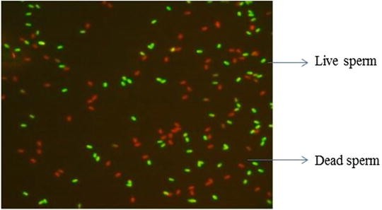
Microphotograph of spermatozoa stained with SYBR-14 and propidium iodide. Spermatozoa that fluoresced bright green with SYBR-14 were classified as live, while those stained red with PI were classified as dead

Points to Ponder
Aqueous solutions of SYBR-14 dye should not be stored or reused.
6.4 Estimating Biochemical Integrity of Plasma Membrane
6.4.1 Hypo-osmotic Swelling (HOS) Response
Principle
Hypo-osmotic swelling of spermatozoa is based on the principle of osmotic passage of solvents across the intact membrane. In an attempt to reach the osmotic equilibrium in an hypo-osmotic condition, spermatozoa will permit water molecules to cross the plasma membrane. Influx of water increases sperm volume and causes bulging of plasma membrane, giving a minimum surface-to-volume ratio. When sperm cells are subjected to hypo-osmotic solutions, because of fluid influx, the sperm tail expands and bulges characteristically, which is termed as HOS response. The osmotic pressure of seminal plasma of bull semen is 285 mOsm. The HOS assay provides a precise and repeatable measure of functional membrane integrity, defined as the ability of sperm’s outer membrane to maintain equilibrium between the sperm cell contents and surrounding environment. It measures sperm’s response to osmotic environmental stress. Intact sperm membrane, because of active biochemical function, allows passage of fluid into the intracellular space in hypo-osmotic conditions resulting into sperm tail swelling and curling. There is great variation in the pattern of curling from sperm to sperm within the same sample. When subjected to hypo-osmotic conditions, the live sperm swell (greater value in packed cell volume) in contrast to hyper-osmotic condition when they shrink to a size of 20–25 μ (lesser value in PCV). In the HOS assay, usually the total HOS response value is considered, with no emphasis given either on the response evaluation time or the value of the response subtypes. The assay can also be carried out using distilled water. This particular assay was first described by [32].
Materials
Solution A
-
0.734 g sodium citrate.
-
Dilute to 100 mL DW.
Solution B
-
1.351 g fructose.
-
Dilute to 100 mL DW.
Procedure
-
(a)
Take 0.5 mL of solution A and B (150 mOsm/L) in a clean, prewarmed (37 °C) test tube.
-
(b)
To this add 0.1 mL of semen, and mix well.
-
(c)
Incubate the sperm suspension in water bath at 37 °C for 45 min.
-
(d)
Add a drop of eosin Y solution after incubation.
-
(e)
Place a small drop of the suspension on a clean, grease-free glass slide and place a coverslip.
-
(f)
Examine under high-power magnification (400 x, PC microscope).
-
(g)
Count a minimum of 200 spermatozoa for different types of tail swelling pattern.
-
(h)
Procedure for HOS for post-thaw sample is the same as that of neat semen.
Observation
Spermatozoa are classified as per cent HOS responsiveness according to the presence of the tail swelling patterns as mentioned below. All spermatozoa stained pink are dead ones and are excluded from the count of HOS-responsive spermatozoa (Fig. 6.6, Table 6.4).

Assessing biochemical integrity of the sperm membrane using hypo-osmotic swelling assay. Microphotograph A shows HOS+ve (tail tip curl, live) and HOS-ve (dead) cells
6.4.2 Evaluation of Biochemical Membrane Integrity Using Distilled Water
Principle
The underlying principle for both assays is the same. The only difference being that instead of hypo-osmotic solution, distilled water is used in this assay. It was found that values obtained from water tests are significantly higher than those of HOS assay. This test can serve as a simple and reliable method to evaluate biochemical integrity of sperm membrane.
Procedure
-
(a)
Take 0.4 mL of DW in a clean, prewarmed (37 °C) test tube.
-
(b)
To this add 0.1 mL of semen, and mix well.
-
(c)
Incubate the sperm suspension in water bath at 37 °C for 60 min.
-
(d)
Add a drop of eosin Y solution after incubation.
-
(e)
Place a small drop of the suspension on a clean, grease-free glass slide and place a coverslip.
-
(f)
Examine under high-power magnification (400x, PC microscope).
-
(g)
Count a minimum of 200 spermatozoa for different types of tail swelling pattern.
-
(h)
For better visibility, a drop of eosin can also be added to the final sample just before microscopic evaluation.
Observation
Spermatozoa are classified as per cent positive or negative to WT (water test) similar to those of HOS values. All spermatozoa stained pink are dead ones and are excluded from the total count.
Points to Ponder
-
(a)
It is essential to maintain same temperature for HOS evaluation as that of thawing temperature for frozen-thawed samples.
-
(b)
Values obtained with the water tests are significantly higher than those obtained with the HOS test, and thus this test emerges as a simple and reliable method to assess the integrity of the membrane of spermatozoa in vitro.
6.5 Lipid Peroxidation as an Indicator of Membrane Integrity
Integrity of the plasma membrane of the spermatozoa can be determined using conventional assay like HOS and fluorescent microscopy and by detecting the lipid peroxidation. The conventional lipid peroxidation techniques usually detect the end products of lipid peroxidation, whereas on the other hand, the fluorescent probes can directly detect endogenous cholesterol and phospholipid [33]. For example, a fluorescent lipid peroxidation reporter molecule C11BODIPY (ex/em of 581/591) shifts its fluorescence from red to green when challenged with oxidizing agents and can quantify and locate the site lipid peroxidation [34, 33]. Estimation of total lipid extracts and lipid peroxidation of Percoll-washed spermatozoa can be made by following the extraction protocol [35]. The relevant assays have been described in the chapters ‘Determining Oxidative Stress to Spermatozoa’ and ‘Estimating Lipids’.
6.6 Background Information
Estimates of sperm vitality are made by assessing the integrity of the cell membrane. Since the per cent dead cells would not exceed (within sampling error) the per cent immotile sperm, the vitality assay can serve to provide a check on the motility estimates. Normally, the per cent viable cells exceed that of the per cent motile cells. One must assess sperm vitality as soon as possible after collection of the semen sample (preferably within 30 min), but never later than 1 h of ejaculation. This helps in prevention of deleterious effects of dehydration or of changes in temperature on vitality estimates.
Staining with eosin and nigrosin has long been done to assess mammalian sperm viability but has some limitations. The factor that might contribute to false high dead sperm per cent includes preservation of stained and unmounted smears under humid conditions. All sperm will be dead by the time the smears are made and dried. However, under humid environment, condensed moisture vapour could reconstitute the dye solution on the slide which then enters the cells which were unstained earlier. The dye eosin is easily incorporated into live spermatozoa, which results in underestimation of sperm viability particularly when used with frozen-thawed semen containing glycerol [5]. A probable explanation of these observations is that the higher levels of glycerol increased the permeability of the living and motile sperm cells to the stain, allowing some of them to be counted as dead cells in the differential staining procedure.
This disadvantage can be overcome by using fluorescent stains. Thus, CFDA fluorescent probe in combination with PI is used to assess the viability of sperm [28]. The drawback of CFDA- and CMFDA-based assessment of sperm viability is the time dependency. This is because fluorescent products are developed only after activity of the enzyme substrate conversion. Moreover, using CFDA as a viability probe poses several disadvantages in the accuracy in scoring of damaged/dead and live cells. For example, the presence of extracellular esterases and/or greater quantity of CFDA can lead to a high background. Many a times, the green emission peak of the fluorescent product fluorescein extends into the red region of the spectrum. In the presence of an excessive intracytoplasmic fluorescein in relation to the red dead cell stains, the red fluorescence observed may not have come from PI. Moreover, the spontaneous hydrolysis of CFDA to fluorescein at the time of storage is promoted by the traces of moisture in the solvent used earlier to resolubilize the substrate (to make stock solutions or by the gradual accumulation of moisture during storage). Often, morbid spermatozoa show residual esterase activity, which complicates the evaluation of dead cells (they exhibit staining with both dyes). Recently dead sperm may be misinterpreted as live because of exhibiting esterase activity at the time of examination.
The bisbenzimide stains, H 33342 and H 33258, are some of the more commonly used probes for assessing sperm DNA integrity and cell viability since these stains are excited with UV light to emit blue fluorescence, which in turn is detrimental to cellular function and DNA integrity. To overcome the drawbacks of enzyme-based and bisbenzimide stains, a membrane-permeant nuclear stain SYBR-14 in combination with PI has been developed as a valuable tool for sperm viability in different species. The use of SYBR-14 which has excitation spectrum with visible light (488 nm) is always advisable than the use of UV light where viability is an issue.
The advantage of using SYBR-14 over enzyme-based stains (CFDA and CMFDA) is that staining time is not as critical. The enzyme-based stains need very careful planning of time since there is a continuous increase in cellular fluorescence over time. On the other hand, SYBR-14 quickly reaches equilibrium (within 15 min) with the nucleic acid and becomes relatively stable. CFDA- and CMFDA-stained sperm samples tend to show heavy background fluorescence due to extracellular esterases. Advantageously, following staining with SYBR-14, the background fluorescence is non-existent. Thus, fluorescence staining with SYBR-14 and PI causes minimal staining artefacts [7]. Furthermore, both of the stains’ targets are the same molecules within the sperm, nucleic acids, thus making the latency of change from green to red relatively short. The moribund sperm stained with any of the carboxyfluorescein diacetate derivatives do not lose their green fluorescence as rapidly as SYBR-14 and P1 combination. SYBR-l4 is excited with visible light (488 nm), thereby avoiding exposure to UV irradiation. It is possible that sperm stained with SYBR-14 will retain their fertilizing potential.
Literature Cited
Hidalgo M, Rodríguez I, Dorado J (2006) Influence of staining and sampling procedures on goat sperm morphometry using the Sperm Class Analyzer. Theriogenology 66:996–1003
Blom E (1950) A one-minute live-dead sperm stain by means of eosin-nigrosin. Fertil Steril 1:176–177
Campbell RC, Dott HM, Glover TD (1956) Nigrosin eosin as stain for differentiating live and dead spermatozoa. J Agric Sci 48:1–8
Dott HM, Foster GC (1972) A technique for studying the morphology of mammalian spermatozoa which are eosinophilic in a differential live/dead stain. J Reprod Fertil 29:443–445
Mixner JP, Saroff J (1954) Interference of glycerol with differential staining of bull spermatozoa as used with semen thawed from the frozen state. J Dairy Sci 37:1094
Garner DL, Pinkel D, Johnson LA, Pace MM (1986) Assessment of spermatozoal function using dual fluorescent staining and flow cytometric analyses. Biol Reprod 34:127–138
Garner DL, Johnson LA (1995) Viability assessment of mammalian sperm using SYBR-14 and Propidium Iodide. Biol Reprod 53:276–284
Enciso M, Cisale H, Johnston SD, Sarasa J, Fernández JL, Gosálvez J (2011) Major morphological sperm abnormalities in the bull are related to sperm DNA damage. Theriogenology 76:23–32
Garner DL, Johnson LA, Allen CH (1988) Fluorometric evaluation of cryopreserved bovine spermatozoa extended in egg yolk and milk. Theriogenology 30:369–378
Gillian L, Evans G, Maxwell WMC (1997) Capacitation status and fertility of fresh and frozen-thawed ram spermatozoa. Reprod Fertil Dev 9:481–487
Graham JK, Kune E, Hamersledt RH (1990) Analysis of germ cell viability, acrosomal integrity and mitochondrial function using flow cytometry. Biol Reprod 43:55–64
Farrell PB, Foote RH, Zinaman MJ (1996) Motility and other characteristics of human sperm can be measured by computer-assisted sperm analysis of samples stained with Hoechst 33342. Fertil Steril 66:446–453
Maxwell WM, Johnson LA (1997) Chlortetracycline analysis of boar spermatozoa after incubation; flow cytometric sorting, cooling, or cryopreservation. Mol Reprod Dev 46:408–418
Morrell JM, Dresser DW (1989) Offspring from inseminations with mammalian sperm stained with Hoechst 33342, either with or without flow cytometry. Mutation Res 224:177
Kapuscinski J (1995) DAPI: a DNA-specific fluorescent probe. Biotech Histochem 70(5):220–233
Rathi R, Colenbrander B, Bevers MM (2001) Evaluation of in vitro capacitation of stallion spermatozoa. Biol Reprod 65:462–470
Garner DL, Thomas CA, Allen CH, Senger PL, Sasser RG (1997) Effect of cryopreservation on bovine sperm viability as determined by dual DNA staining. Reprod Domest Anim 35:279–283
Garner DL, Johnson LA (1995) Viability assessment of mammalian sperm using SYBR-14 and propidium iodide. Biol Reprod 53:276–284
Kondracki S, Banaszewska D, Wysokińska A, Iwanina M (2012) The Effect of Sperm Concentration in the Ejaculate on Morphological Traits of Bull Spermatozoa. Folia Biol (Krakow) 60:85–91
Hancock JL (1951) A staining technique for the study of temperature-shock in semen. Nature 167:323–324
Barth AD, Oko RJ (1989) Preparation of semen for morphological examination. In: Abnormal morphology of bovine spermatozoa. Iowa State University Press, Ames, IA, pp 8–18
Chemes HE, Rawe YV (2003) Sperm pathology: a step beyond descriptive morphology. Origin, characterization and fertility potential of abnormal sperm phenotypes in infertile men. Hum Reprod Update 9:405–428
Wilton LJ, Temple-Smith PD, Baker HW, de Kretser DM (1988) Hum male infertility caused by degeneration and death of sperms in the epididymis. Fertil Steril 49:1051–1058
Correa-Perez JR, Fernandez-Pelegrina R, Aslanis P (2004) Clinical management of men producing ejaculates characterized by high levels of dead sperm and altered seminal plasma factors consistent with epididymal necrospermia. Fertil Steril 81:1148–1150
Farah OI, Cuiling L, Jiaojiao W, Huiping Z (2013) Use of fluorescent dyes for readily recognizing sperm damage. J Reprod Infertil 14:120
Otto FJ, Hacker U, Zante J, Schumann J, Gohde W (1979) Flow cytometry of human spermatozoa. Histochemistry 62:249–254
Makarevich AV, Kubovicova E, Sirotkin AV, Pivko J (2010) Demonstration of the effect of epidermal growth factor on ram sperm parameters using two fluorescent assays. Vet Med 55:581–589
Watson PF, Kunze E, Cramer P, Hammerstedt RH (1992) A comparison of Critical osmolarity and hydraulic conductivity and its activation energy in fowl and bull spermatozoa. J Androl 13:131–l38
Harrison RAP, Vickers SE (1990) Use of fluorescent probes to assess membrane integrity in mammalian spermatozoa. J Reprod Fertil 88:343–352
Oko RJ, Costerton JW, Coulter GH (1976) An ultrastructural study of the head region of bovine spermatozoa. Can J Zool 54:1326–1340
Kordan W, Fraser L, Wysocki P, Strzezek R, Lecewicz M, Mogielnicka-Brzozowska DA, Soliwoda D, Koziorowska-Gilun M (2013) Semen quality assessments and their significance in reproductive technology. Pol J Vet Sci 16(4):823–833
Jeyendran RS, Vander Ven HH, Parez-Pelaez M, Crabo BG, Zaneweld LJD (1984) Development of an assay to assess the functional integrity of the human membrane and its relationship to other semen characteristics. J Reprod Fertil 70:219–228
Brouwers JF, Silva PF, Gadella BM (2005) New assays for detection and localization of endogenous lipid peroxidation products in living boar sperm after BTS dilution or after freeze-thawing. Theriogenology 63(2):458–469
Drummen GP, Gadella BM, Post JA, Brouwers JF (2004) Mass spectrometric characterization of the oxidation of the fluorescent lipid peroxidation reporter molecule C11-BODIPY(581/591). Free Radic Biol Med 36(12):1635–1644
Bligh EG, Dyer WJ (1959) A rigid method of total lipid extraction and purification. Can J Biochem Physiol 37:911–917
Key Reference
Rathi and others (2001) See above [16]. In-depth discussion and excellent background information related to merits of different staining techniques
Author information
Authors and Affiliations
Corresponding author
Editor information
Editors and Affiliations
Rights and permissions
Copyright information
© 2017 Springer Nature Singapore Pte Ltd.
About this chapter
Cite this chapter
Kumar, P., Srivastava, N., Pande, M., Prasad, J.K., Sirohi, A.S. (2017). Evaluating Sperm Cell Viability and Membrane Integrity. In: Srivastava, N., Pande, M. (eds) Protocols in Semen Biology (Comparing Assays). Springer, Singapore. https://doi.org/10.1007/978-981-10-5200-2_6
Download citation
DOI: https://doi.org/10.1007/978-981-10-5200-2_6
Published:
Publisher Name: Springer, Singapore
Print ISBN: 978-981-10-5199-9
Online ISBN: 978-981-10-5200-2
eBook Packages: Biomedical and Life SciencesBiomedical and Life Sciences (R0)