
Avoid common mistakes on your manuscript.
Erratum to: BioControl DOI 10.1007/s10526-012-9499-2
The following errors regarding the artwork in the original publication remain to be addressed:
-
Figures 1, 3 and 4 were too small in size to convey important information of this study. They are presented at their proper size below.
Fig. 1 
Light micrographs of the fungus Beauveria bassiana on the aphid host after staining with cotton blue–lactophenol. a, b By one day PI, conidia had swelled (cs) or germinated, and germ tubes had grown through a spiracle (S) (seen at different focal planes in a and b) on a living aphid. Germ tube tip (gt) is marked. Bar 10 μm. c At 36 h PI, when some aphids had already died, blue-stained conidial masses (cm) have engulfed the cauda. Bar 10 μm. d, e Conidia are present at the opening of the cornicle (Co), shown in surface plane (d) and interior plane (e), and numerous hyphae (hy) are growing within the cornicle (shown by white arrows). Bar 20 μm
Fig. 3 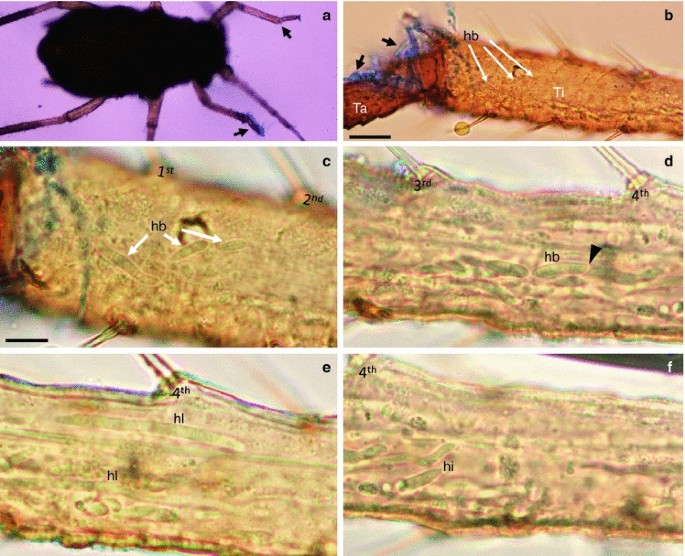
Hyphal body formation by Beauveria bassiana in the aphid leg hemocoel at 60 h PI viewed with a dissecting microscope (a) and a compound light microscope viewed with bright-field optics (b–f) with cotton blue–lactophenol staining. a The front legs of a living, sick nymph were externally colonized by the fungus at two sites (arrows). b Blue-stained, external hyphae (black arrows) in the tarsus (Ta) and numerous hyphal bodies (hb) in the tibia (Ti) near the external infection site of the leg. Bar 5 μm. c Close up of three hyphal bodies. d In the same leg, hyphae were seen fragmenting into shorter hyphal bodies/blastospores (arrowhead). e Two long hyphal bodies (hl). f Irregular-shaped hyphal body (hi, black arrow) at the predominant site of infection in this aphid. The first (1st) through fourth (4th) lateral setae of the tibia are shown. Bars are 2 μm in c–f
Fig. 4 
TEM of Beauveria bassiana cells within severely ill Myzus persicae by 72 h PI. a Hyphal bodies and blastospores (bs) of various sizes in the hemocoel. Bar 5 μm. b Hyphal bodies (hb) have penetrated the fat body (Fb) (black arrowhead). Aphid tissue around the colonized area was degraded. Muscle tissue (M). c Hyphal bodies and blastospores penetrated the gut (G) wall and grew near the embryos (E). d Fungal cells around muscle (M) of dorsal side of abdomen. e Blastospores have grown and branched in the hemolymph. f Blastospores had reproduced in SDY broth. Elongated hyphal bodies that were produced in the insect lacked a well-defined cell wall (black arrows in a–d), whereas blastospores produced in vivo (e) and in vitro in SDY broth (f) have a more defined cell wall (white arrows). Bars 2 μm in b–f
-
The wrong parts of Fig. 6 were specified in the very last paragraph of the Results section. The references should read as follows:
Hyphal bodies, mostly short, 1–2 celled, with tapered ends, had multiplied extensively, notably in the leg hemocoel in the coxa, trochanter, and femur segments (Fig. 6a, b). By seven days PI or longer, the hyphal bodies had begun to produce long, thin hyphae (Fig. 6c).


Author information
Authors and Affiliations
Corresponding author
Additional information
The online version of the original article can be found under doi:10.1007/s10526-012-9499-2.
Rights and permissions
About this article
Cite this article
Amnuaykanjanasin, A., Jirakkakul, J., Panyasiri, C. et al. Erratum to: Infection and colonization of tissues of the aphid Myzus persicae and cassava mealybug Phenacoccus manihoti by the fungus Beauveria bassiana . BioControl 58, 393–396 (2013). https://doi.org/10.1007/s10526-013-9504-4
Published:
Issue Date:
DOI: https://doi.org/10.1007/s10526-013-9504-4