
Abstract
The human skin possesses a complex structure and various functions which ensure the entity between the organism and the environment. Mechanical properties of the skin are of major importance for its protective function. They vary in accordance with age, sex and body sites, in some physiological and pathological skin conditions, and change due to different external and therapeutic influences. Considerable progress in the quantification of the skin mechanical functions had been achieved for the past 20 years through the introduction of modern non-invasive bioengineering methods and devices which provide the researchers with objective, quantitative, sensitive and reproducible measurements in vivo.
Access provided by Autonomous University of Puebla. Download chapter PDF
Keywords
These keywords were added by machine and not by the authors. This process is experimental and the keywords may be updated as the learning algorithm improves.
1 Introduction
The human skin possesses a complex structure and various functions which ensure the entity between the organism and the environment. Mechanical properties of the skin are of major importance for its protective function. They vary in accordance with age, sex and body sites, in some physiological and pathological skin conditions, and change due to different external and therapeutic influences. Considerable progress in the quantification of the skin mechanical functions had been achieved for the past 20 years through the introduction of modern non-invasive bioengineering methods and devices which provide the researchers with objective, quantitative, sensitive and reproducible measurements in vivo.
The Cutometer® (Courage + Khazaka Electronic GmbH, Cologne, Germany) is a well-recognized commercial device for measurement of the biomechanical properties of the skin. In this chapter, based on our own experience and review of the literature, we aimed to present the practical application of the Cutometer® method.
2 Measuring Equipment and Principle
2.1 Equipment
The Cutometer® is designed as separate or combined device.
-
1.
The Cutometer® SEM 575 (Fig. 29.1), which is the successor of the Cutometer® SEM 474, is an independent device that includes two basic parts [7, 9, 40, 70].
Fig. 29.1 
Cutometer® SEM 575 (With permission from Courage and Khazaka GmbH, Cologne, Germany)
-
Main unit, a metal housing containing vacuum pump with pressure sensor and microelectronics. The load of the vacuum (negative air pressure), the rate of its increase or decrease, the duration of suction (on time) and relaxation (off time) and the number of measuring cycles during one measurement can be defined through the Cutometer® software. The resolution of applied pressure is equal to 1 mbar.
-
Measuring probe, a handheld probe containing suction head, optical measuring system and microelectronics. The suction head is centred in the probe and has a standard circular aperture of 2-mm diameter (test area of about 3 mm2). Optional probes with apertures of 4, 6 and 8 mm are available on request. The measuring system inside the probe head is noncontact and consists of a light source (light-emitting diode) and a light recipient, as well as two opposing glass prisms, which project the light from emitter to recipient. The changes of the infrared light beam intensity during the measurement are converted into millimetres (from 0 to 3.0 mm) and calculated with a resolution equal to 2 μm. The measuring probe is connected to the main unit through an air tube and an electric cable. During the measurement, it is held perpendicularly to the skin surface under constant pressure ensured (provided) by an elastic spring. The calibration data are stored in the memory of each probe which allows fast and easy exchange of probes with different apertures.
-
-
2.
The Cutometer® MPA 580 (Fig. 29.2), that is currently available, combines the vacuum box for the Cutometer® with the built-in Sebumeter SM 815 and modular Multi Probe Adapter System which makes possible to connect up to 4 additional measuring probes as well as an ambient condition sensor. The Cutometer® Dual MPA 580 (Fig. 29.3) is the last generation device that allows the connection of two Cutometer® probes (with different diameters) at the same time.
Fig. 29.2 
Cutometer® MPA 580 (With permission from Courage and Khazaka GmbH, Cologne, Germany)
Fig. 29.3 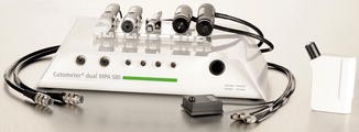
Cutometer® Dual MPA 580 (With permission from Courage and Khazaka GmbH, Cologne, Germany)


2.2 Principle
The measuring principle of the Cutometer® is based on a suction method that consists of the measurement of vertical deformation of the skin surface after application of vacuum (Fig. 29.4) [7, 9, 40, 70].

Schematic view of the measuring system within the Cutometer® probe [56] (With permission from Courage and Khazaka GmbH, Cologne, Germany)
A defined negative air pressure is applied perpendicular to the skin through the opening of the probe for a selected time period. The evaluated skin surface is sucked into the aperture of the probe, and the resulted vertical deformation is measured by the optical measuring system inside the probe. The changes of light intensity are proportionally related to the penetration depth of the skin and are displayed on the monitor as curves in a coordinate system (extension/time or pressure/extension).
3 Software
The Cutometer® is designed for operation with an IBM-compatible PC via USB port. The last version of the Windows software allows storage of various data regarding the volunteer, date and time of experiment, skin area, external temperature and relative humidity, type of probe used and mode of measuring technique. The obtained results are automatically calculated and displayed as curves and values [56].
The Cutometer® measuring cycle consists of suction phase and relaxation phase. It can be applied once or several times. The following parameters of the measurement can be exactly defined by the software:
-
Pressure. The load of the air-negative pressure (vacuum) can be chosen between 20 and 500 mbar.
-
Rate. The rate of increase or decrease of the air-negative pressure can be selected between 10 and 100 mbar/s.
-
On-Time. The time when the air-negative pressure is applied (suction interval) can be chosen between 0.1 and 60 s.
-
Off-Time. The time when the air-negative pressure is applied no longer (relaxation interval) can be chosen between 0.1 and 60 s.
-
Repetition. The number of measuring cycles (suctions) included in one measurement can be varying between 1 and 99.
-
Pre-Time. This function allows setting a short interval between pressing the start key and the beginning of the measurement.
-
Preconditioning Time. This function allows (in the strain–time mode) to pretension the skin by applying a preliminary suction during a short time (0.1 s) before the real measurement is carried out.
The most used settings include air-negative pressure between 400 and 450 mbar, on-time and off-time intervals between 2 and 5 s and repetitions between 1 and 10.
3.1 Measuring Techniques
There are two measuring techniques available [7, 9, 56]:
-
1.
In the strain–time mode, the deformation of the skin (in millimetres) is showed as a function of time (in seconds). This mode is mostly used in research studies.
-
2.
In the stress–strain mode, the deformation of the skin (in millimetres) is showed as a function of the vacuum (in millibar).
3.2 Measuring Modes
There are four measuring modes available based on different combination of the measurement parameters [7, 9, 56].
3.2.1 Mode 1: Measurement with Constant Negative Pressure (−)
The measurement cycle consists of suction and relaxation phases. During the first phase, the skin is drawn into the probe with constant negative pressure set under “Pressure” within the interval set under “On-Time”. In the second phase, the negative pressure is switched off, and the relaxation of the skin is determined within the interval set under “Off-Time”. With the command “Repetition”, the number of measuring cycles can be chosen. The skin deformation is displayed as a function of time (Fig. 29.5). The measurement mode 1 is most important and is predominantly used for research studies in the field of dermatology and cosmetics.

An example of a curve obtained in mode 1 (E/T mode; extension/time)
3.2.2 Mode 2: Measurement with Linear Increase and Linear Decrease in Negative Pressure (/\)
The measurement cycle consists of three phases. At the start, the negative pressure is zero. During the first phase, the skin is drawn into the probe with linearly increasing negative pressure set under commands “Pressure” and “Rate” in the menu “Parameter”. This phase is succeeded by linearly decreasing negative pressure. In the last phase, the skin properties are evaluated when no negative pressure is applied for an interval set under “Off-Time”. With the command “Repetition” the number of measuring cycles can be chosen. The skin deformation can be displayed both as a function of time (Fig. 29.6a) and as a function of negative pressure (Fig. 29.6b).

(a) An example of a curve obtained in mode 2 (E/T mode; extension/time). (b) An example of a curve obtained in mode 2 (P/E mode; pressure/extension)
3.2.3 Mode 3: Measurement with First Constant and Then Linear Decrease in Negative Pressure (−\)
The measurement consists of one measuring cycle which combines the first phase of mode 1 and the second phase of mode 2. The “On-Time” can be selected, whereas the “Off-Time” results from the selected “Pressure” and “Rate”. A relaxation time (here “Off-Time”) cannot be set as repetitions due to the combination of full pressure and slight release are impossible. The skin deformation is displayed as a function of time (Fig. 29.7).

An example of a curve obtained in mode 3 (E/T mode; extension/time)
3.2.4 Mode 4: Measurement with a Linear Increase in Negative Pressure and Then a Sudden Cessation of the Negative Pressure (/)
The measurement consists of one measuring cycle which combines the first phase of mode 2 and the second phase of mode 1. The “On-Time” results from the selected “Pressure” and “Rate”, whereas the “Off-Time” can be set. A repetition is impossible due to the combination of slightly increasing pressure and full release. The skin deformation is displayed as a function of time (Fig. 29.8).

An example of a curve obtained in mode 4 (E/T mode; extension/time)
In the literature measuring modes 2, 3 and 4 have no significance. There are few studies using measuring mode 2. According to Dobrev [18], the application of measuring mode 2 does not provide any advantage and only burdens the vacuum pump of the device.
3.3 Skin Mechanical Parameters
The new version of Cutometer® MPA 580Q software (v.1.3.6.16) allows calculating three groups of skin mechanical parameters designated as “R-parameters”, “F-parameters” and “Q-parameters”. In addition, there is a possibility for advanced users to edit the calculation formula for “R-parameters” according to their needs [56].
In this section, we describe in details the mechanical parameters derived from skin deformation curves obtained using measurement mode 1. Most detailed information concerning the mechanical parameters determined by measurement modes 2, 3 and 4 can be obtained from the manufacturer’s Information and Operating Instructions for the Cutometer® [56].
3.3.1 Mode 1 (Single Strain–Time Curve)
3.3.1.1 R-Parameters
The skin deformation curve obtained with Cutometer® includes two main parts generated during the suction phase and relaxation phase, respectively (Fig. 29.9). Each of them is composed of rapid deformation representing an elastic section, followed by a viscoelastic and finally a viscous section. These parts are designated as follows: immediate deformation (Ue), delayed deformation (Uv), immediate retraction (Ur) and delayed retraction (Ua–Ur). Values of Ue and Ur are taken at fixed intervals of time, respectively, 0.1 s after application of a suction and 0.1 s after removal of negative pressure [5, 7, 9, 56].

Skin deformation curve obtained with CutoMPA 580. Aperture, 2 mm; suction time, 5 s, relaxation time, 5 s, repetition, 1
Based on these parts, the Cutometer® software calculates automatically the following parameters:
-
R0 = Uf, the final deformation (skin distensibility or skin extensibility).
-
R1 = Uf – Ua, the residual deformation at the end of 1st measuring cycle (resilient distension).
-
R2 = Ua/Uf, the ratio of total retraction to total deformation, which is called gross elasticity of the skin, including viscous deformation = gross elasticity, including viscous deformation) (overall elasticity).
-
R5 = Ur/Ue, the net elasticity without viscous deformation.
-
R6 = Uv/Ue, the ratio between delayed and immediate deformation, which indicates the relative contributions of the viscoelastic plus viscous and the elastic distension to the total deformation (viscoelastic ratio, the ratio of viscoelastic to elastic distension).
-
R7 = Ur/Uf, the ratio of immediate retraction to the total deformation, which is called biological elasticity (the ratio of immediate retraction to total distension).
-
R8 = Ua, the final retraction after removal of the vacuum (total recovery of the skin).
3.3.1.2 F-Parameters
The software calculates two surfaces (“areas”) designated as F-parameters: (Fig. 29.8)
-
F0 = the surface between the real curve and the value corresponding to the maximal deformation Uf when going from start of suction to cease of suction.
-
F1 = the surface between the real recovery curve and the value corresponding to the maximal recovery going from cease of suction to cease of measurement.
3.3.1.3 Q-Parameters
The Q-parameters are developed by the scientist Di Qu et al. [56, 83] and have recently been added in the calculation formula. They could only be obtained in mode 1 for an equal suction and relaxation time. To receive these parameters, two horizontal lines have to be spread at the graph – the first one spreads through the highest point (R0), and the second one spreads through the inflexion point that is the point in time at which the recovery curve deviates from its initial linearity. At this point, the as QE and QR are divided (Fig. 29.10).

Skin deformation curve obtained with CutoMPA 580. Aperture, 2 mm; suction time, 5 s, relaxation time, 5 s, repetition, 1
The Q-parameters include:
-
Q0 = the maximum recovery area, i.e. the area under the highest point (R0).
-
QE = the elastic recovery area of the skin.
-
QV = the viscous recovery area of the skin.
The parameters calculated by the Cutometer® software include:
-
Q0 = Q0; the maximum recovery area, i.e. the area under the highest point (R0).
-
Q1 = QE/Q0; the elastic recovery of the skin.
-
Q2 = Q2 = QV/Q0; the viscous recovery of the skin.
-
Q3 = (QE + QV)/Q0; the total viscoelastic recovery of the skin, i.e. overall skin elasticity.
3.3.2 Mode 1 (Repetitive Strain–Time Curve)
When repetitive suctions are applied, the subsequent curves are similar to the first one. However, they are progressively shifted vertically upward as a consequence of the slow return of the skin to the original state (Fig. 29.11) [56].

Skin deformation curve obtained with CutoMPA 580. Aperture, 2 mm; suction time, 4 s, relaxation time, 2 s, repetition, 5
3.3.2.1 R-Parameters
The following additional parameters are calculated by the software:
-
R3 = last maximum amplitude (last maximal deformation).
-
R4 = last minimum amplitude (last residual deformation).
-
R9 = R3 – R0, the difference in maximal skin deformation between the last and the first suction called hysteresis (H).
3.3.2.2 F-Parameters
For curves taken in mode 1 with a minimum of 10 repetitions, the software calculates three additional surface parameters (“areas”). For this purpose, the curves are wrapped with an “envelope” function. Above and below the curves, the envelope curves appear as logarithmical average of maximum and minimum amplitudes. The F-parameters are designated as follows (Fig. 29.12):

Skin deformation curve obtained with CutoMPA 580. Aperture, 2 mm; suction time, 1 s, relaxation time, 1 s, repetition, 30 (F2 = area A; F3 = area B; F4 = area B + area C)
-
F2 = area above the upper envelope curve (the surface between the real curve and the value corresponding to the maximal deformation R3 after 10 cycles when going from start of suction to cease of the 10 cycles).
-
F3 = area within the envelope curves (the surface between the repetitive curves).
-
F4 = area below the upper envelope curve (the complete area, limited by the upper envelope curve).
4 Use of Cutometer®
4.1 Factors Influencing Measurements
4.1.1 Probe Aperture
At a constant vacuum, the degree of skin deformation (i.e. absolute parameters) directly correlates with the aperture diameter of the measuring probe and inversely correlates with the skin thickness. When using identical vacuum, the values of the absolute parameters Ue, Uv, Uf, Ur and R measured with 2-mm diameter probe are lower compared to values measured with 8-mm diameter probe. The differences in the values of relative parameters Ua/Uf, Ur/Ue, Ur/Uf and Uv/Ue measured with both probes are minimal [5, 26, 74].
The small aperture (2-mm diameter) measuring probe determines the mechanical properties of the epidermis and partly of the papillary dermis. It is applicable at any anatomical region and is most appropriate for measurement of healthy skin and studying the changes after the application of topical products.
The medium aperture (4- and 6-mm diameter) measuring probes determine the mechanical properties of the outer skin layers.
The large aperture (8-mm diameter) measuring probe determines the mechanical properties of the whole skin (derma and hypoderma). It is appropriate for evaluation of skin diseases with predominantly changes in the dermis (systemic sclerosis, scleredema of Buschke, psoriasis, keloids and erysipelas). The use of 8-mm diameter probe may be difficult for measurements at anatomic regions with thin and flabby skin (i.e. medial surfaces of the arm in elderly individuals), skin over bones or convex areas (i.e., forehead, temporal region, chest, dorsum of the hand and phalanx) as well as at the presence of residual lipid film on the skin surface shortly after the application of topical products [26, 30].
We consider that the simultaneous use of at least two probes with different apertures, i.e. both 2-mm and 8-mm probes, gives more complex information about the mechanical properties of the skin [26, 30].
4.1.2 Pressure (Vacuum)
For a constant opening of the probe, the absolute mechanical parameters are directly correlated with the intensity of vacuum applied, whereas the relative parameters are less independent of load for most anatomical regions [5]. Cua et al. [12] have found that Ur/Uf and Uv/Ue tended to increase with increasing loads. According to Wickett [99], the application of 200 mbar of vacuum leads to more sensitivity to moisturizing effects compared to 500 mbar of negative pressure.
4.1.3 Test Site
Volar forearms are considered the most appropriate test site generating reproducible measurement results using probes with different apertures. Significant differences in the skin mechanical parameters between both forearms have not been established [26, 30].
4.1.4 Measurement Scheme
The application of one and the same measurement scheme for study of one and the same skin condition makes the obtained results comparable. According to us, the most appropriate measurement scheme comprises the single application of constant vacuum of 400 mbar for 5 s followed by a 5-s relaxation time. This scheme produces enough meaningful results, does not overburden the device and shortens the duration of measurements [26, 30].
4.1.5 Preconditioning of the Skin
Barel et al. [5] have measured higher values of relative elastic parameters Ur/Ue and Ur/Uf using the pretension mode of the Cutometer®, indicating that preconditioned skin recovers more of its elastic deformation. They also consider that under pretension, the values of the skin deformation parameters are more reproducible and accurate. Dobrev [26] did not find any significant changes in the skin mechanical parameters using pretension of the skin, except for a nonsignificant tendency toward higher values.
4.1.6 Environmental Factors
It is recommended to perform the measurements under the same controlled room conditions. Temperature of 20–24 °C and relative humidity of 40–50 % are preferable. The tested person needs at least 15–20 min to acclimatize [56, 99].
4.2 Results Interpretation
The mechanical parameters determined by Cutometer® reflect the condition and the changes in skin structure and composition. They provide meaningful information about its major properties such as [5, 7, 30, 32, 70, 90]:
-
Skin distension (stiffness), i.e. the ability of the skin to undergo distension or the skin resistance to change of shape under the influence of stress.
-
Skin elasticity, i.e. the ability of the skin to recover the original shape after deformation.
-
Skin viscoelasticity, i.e. the time-dependent deformation with a “creep” phenomenon and nonlinear stress–strain properties with “hysteresis”. The creep is characterized as a slowly increasing deformation of the skin in function of the time when a constant stress is applied. The hysteresis is related to the observation that after interrupting the stress the skin does not immediately return to its initial position and remains slightly deformed. In this way, the stress–strain curve obtained during suction time will not be superposed by the curve obtained during relaxation time.
The measurement mode 1 is most used in research studies. That is why the results obtained using mode 1 are explained in details in this section.
4.2.1 R-Parameters
According to their calculation, the R-parameters are divided into two groups [7, 56]:
-
Absolute parameters: Ue, Uv, Uf, Ur, Ua and R1.
-
Relative parameters: Ua/Uf, Ur/Uf, Ur/Ue and Uv/Ue.
The absolute parameters are measured in millimetres, while the relative parameters are presented with a number, which represents a ratio between the values of two absolute parameters – the maximal value is 1 (100 %).
It is considered that the absolute parameters are dependent on the skin thickness, which varies with age, sex and body region. That is why for comparison studies, they should firstly be standardized for skin thickness determined by ultrasound. Because this is not always possible, the ratios of absolute parameters, i.e. the relative parameters, should be compared. It is accepted that they do not depend on skin thickness and can be compared between subjects, anatomical regions and time points [5, 9].
Nevertheless, we suggest all measured skin mechanical parameters to be considered simultaneously. The reason is that relative parameters are composed of two parts – numerator and denominator, and one and the same value could be found as a result of an increase of a numerator or a decrease of a denominator and vice versa [26, 30].
According to their biological informativeness, R-parameters are divided into three groups [7, 26, 30]:
-
1.
Elastic parameters:
-
Absolute parameters – Ue, Uf and Ur.
-
Relative parameters – Ua/Uf, Ur/Ue and Ur/Uf.
-
-
2.
Viscoelastic parameters:
-
Absolute parameters – Uv and H.
-
Relative parameters – Uv/Ue.
-
-
3.
Mixed parameters:
-
R1 and R4
-
The final skin distension Uf consists of two components – immediate distension Ue (elastic part) and delayed distension Uv (viscoelastic part).
Ue is related to the stretching of collagen and elastic fibres and reflects the skin thickness and rigidity.
Ue decreases during the skin ageing. At sun-protected areas, this is due to the decrease in elastic properties of collagen bundles as a result of fragmenting and increased number of intermolecular binds, whereas at sun-exposed areas this is due to the thickening of the skin as a result of advanced elastosis. Ue is also decreased in disorders characterized by skin thickening and induration. The enlarged volume of the skin as a result of dermal oedema (oedematous phase of scleroderma, psoriasis, erysipelas) or deposition of collagen bundles and glycosaminoglycans in the dermis (indurative phase of scleroderma, scleredema of Buschke, keloids) restricts the skin possibilities for deformation after application of vacuum [17–19, 45].
Ue is increased in some inherited diseases of connective tissue such as Ehlers–Danlos syndrome, which is due to alterations in collagen tissue and thinning of the skin [27, 32, 50]. The application of moisturizers and emollients induces an increase in Ue which is due to the softening of corneal layer and improvement of the plasticity of epidermal layer [25, 65].
Delayed distension (Uv) is attributed to the movement of the interstitial fluid throughout the fibrous network in the dermis.
Uv increases after application of moisturizing agents [6, 25], which is related to the epidermal hydration and improved plasticity of corneal layer. Uv increases in the presence of inflammatory dermal oedema (erysipelas, lymphoedema) [4, 18]. It is also increased in elderly skin because of the decrease of the viscosity due to the decrease in proteoglycans content during skin ageing [22, 26].
Uv could be decreased in scleredema of Buschke and keloids because of the increase of interstitial fluid viscosity as a result of accumulation of proteoglycans [17, 20, 45]. Lower Uv is also observed in the indurative phase of scleroderma and Ehlers–Danlos syndrome. In the first case, this is due to the increased stiffness of the skin, whereas in the second case, this is due to the thinning of the skin [19, 27, 50].
The alterations in Ue and Uv could be unidirectional (epidermal hydration, keloids) [20, 25] or heterogeneous (chronological and photoaging, UV-light irradiation, systemic sclerosis, scleredema of Buschke, erysipelas and lymphoedema, psoriasis, Ehlers–Danlos) [4, 18, 19, 24, 25, 27, 30, 50]. The changes in both elastic and viscoelastic part could influence the value of final skin distension (Fig. 29.13).

Changes in immediate distension Ue and delayed distension Uv of the skin
Uf could be increased at the expense of the increase in:
-
Both parts Ue and Uv (epidermal hydration) [25].
-
The elastic part Ue (Ehlers–Danlos syndrome) [27].
-
The viscoelastic part Uv (dermal oedema in erysipelas) [18].
Uf could be decreased at the expense of the decrease in:
-
Both parts Ue and Uv (keloids) [20].
-
The elastic part Ue (photoaging, scleroderma, scleredema of Buschke, psoriasis) [17, 19, 22, 24, 26].
Uf could be preserved despite the changes in its parts:
-
Decrease in Ue is accompanied with increase in Uv (chronological ageing, UV-light irradiation) [22, 26, 29].
Viscoelastic to elastic ratio (Uv/Ue) represents the distribution between elastic and viscoelastic parts of skin deformation. The increase of Uv/Ue indicates the prevalence of viscoelastic over elastic part of skin deformation. This can mainly be due to the increase of Uv, for example, in erysipelas [18], or decrease of Ue, in oedematous phase of scleroderma [19], scleredema of Buschke [17, 45] and lymphoedema of the lover limbs [4], for example. During skin ageing, Uv/Ue progressively increases because of the simultaneously increase of Uv and decrease of Ue [21, 22]. Identical changes are observed after UV-light irradiation [29].
A direct correlation has been established between Uv and Uv/Ue [26, 30].
Immediate retraction (Ur), gross elasticity (Ua/Uf), net elasticity (Ur/Ue) and biological elasticity (Ur/Uf) are related to the function of elastic fibres and represent the skin ability to restore its initial position after deformation. A direct correlation has been established between them [26, 30].
The elastic parameters are decreased in elderly individuals due to chronological ageing and photoaging of the skin [21, 22], after UV-light irradiation [29] and in diseases, which are characterized by increased thickness and indurations of the epidermis, dermis or whole skin such as psoriasis [24], erysipelas [18] and colloids [20].
The elastic parameters increase in varying degree after the application of moisturizers and emollients, which is due to the effects on the mechanical properties of the corneal layer rather than the effects on the elastic fibres and in inherited diseases of the connective tissue such as Ehlers–Danlos syndrome [27, 50].
In oedematous phase of scleroderma and in scleredema of Buschke, the measurements with 8-mm diameter probe explore a relative increase in the elastic parameters regardless of skin thickening. This phenomenon can be explained by the “lubricating” action of the dermal oedema and decreased friction between the fibres [17, 19].
Mechanical parameters Ua/Uf and Ur/Uf better characterize the elastic properties of the skin than Ur/Ue, because they include the viscous part of skin deformation, too [30].
Hysteresis (H) is a viscoelastic parameter. When a few consecutive suctions are applied (i.e. 3–10 suctions, 3 s/3 s), H reflects the water content of the skin. Using 8-mm diameter probe, higher values of H have been found on psoriasis and erysipelas plaques which is due to the inflammatory dermal oedema [18, 24]. H and Uv were decreased on the irradiated skin in patients undergoing telegamma therapy for breast cancer [23].
When a lot of repeated suctions are applied (i.e. 30 suctions, 1 s/1 s) at one and the same anatomic region, H characterizes skin fatigue. The age-related decline in skin elasticity results in marked fatigue of adult skin rather than of young skin [33].
Residual deformation R is a mixed parameter because it reflects both elastic and viscous properties of the skin. Its interpretation is somewhat difficult and it has been little reported in the literature. In healthy individuals R increases with age as well as directly correlates with the parameters Uv, Uv/Ue, R8 and H [26]. In some diseases, which are characterized by thickening of the skin such as psoriasis, scleredema of Buschke and keloids, using 8-mm diameter probe R is decreased, while in others such as erysipelas R is increased [17, 20, 24].
In general, an inverse correlation exists between the changes in elastic and viscoelastic parameters, while between the single parameters in each group, the relationship is direct [30].
We suggest the following Cutometer® R-parameters, which characterize the main mechanical properties of the skin, to be always analysed: Ue and Uf (distensibility), Ua/Uf and Ur/Uf (elasticity) and Uv and Uv/Ue (viscoelasticity) [26, 30, 32].
4.2.2 F-Parameters (Area-Parameters)
The surface parameters F0 and F1 reflect the viscous part of skin deformation. A completely elastic material will show the complete area (total area and F1 are the same). However, these parameters are not well-known in the scientific literature [56].
The surface parameters F2, F3 and F4 are also not familiar to scientists. We studied the age-related changes in skin fatigue applying multiple suctions at one and the same anatomic region and found that adult skin is characterized by significantly higher values of F2 and lower F3 compared to young skin [33]. F4 is considered a firmness parameter [56].
4.2.3 Q-Parameters
The surface Q-parameters reflect the elastic and viscous recovery of the skin. It is considered that Q0 (maximum recovery area) will go down with more firmness of the skin. Qu et al. [83] found that overall skin elasticity (Q3) and elastic recovery (Q1) decreased significantly, whereas the viscous recovery (Q2) did not show significant change with age. There was a marked decrease in Q3 and Q1, whereas Q2 was higher in the sun-exposed skin.
5 Practical Applications
The Cutometer® is widely used for study in the mechanical properties of healthy skin, their changes under the influence of various internal and external factors, for clinical diagnosis and monitoring, efficacy testing and claim support for medical and cosmetic topical products.
5.1 Study of Healthy Skin
5.1.1 Influence of Sex
Generally, no significant sex-dependent differences in skin mechanical parameters have been reported [5, 12, 21]. However, the menopause is associated with more expressed increase in distensibility and viscosity and decrease in elasticity of the female skin. The application of hormone replacement therapy is able to significantly reduce the climacteric-associated loss of skin elasticity [51, 75, 78, 82, 93]. Using the new introduced Q-parameters, Qu et al. [83] found that female subjects exhibited greater elastic recovery and lower viscous recovery than male subjects.
5.1.2 Influence of Age
Aged skin is characterized by significantly lower elastic and higher viscoelastic parameters. At all anatomic regions, the decrease in skin elasticity and the increase in skin viscoelasticity significantly correlate with the age [5, 9, 12, 21, 51, 58, 87, 94].
5.1.3 Influence of Body Region
The regional differences in skin mechanical properties determined by Cutometer® are mainly due to the differences in skin thickness and sun exposure. The absolute parameters are more influenced than the relative mechanical parameters [5, 12, 26, 41, 48, 53, 94, 100].
5.1.4 Influence of External Factors
Chronic sun exposure and UV-light irradiation produce a decrease in skin extensibility and elasticity and an increase in skin viscoelasticity. These alterations have been reported at facial and dorsal vs. volar forearm skin and are accompanied with increased skin thickness [22, 26, 94]. Similar changes in skin mechanical properties have been observed on irradiated skin in patients undergoing telegamma therapy for breast cancer [23].
Cutometer® has been used for investigation of the skin mechanical properties in astronauts before and after a long-term mission in the International Space Station [95].
5.2 Study of Diseased Skin
Mechanical properties of the skin are altered in many dermatological diseases. The Cutometer® allows quantifying these alterations in details, and the changes of mechanical parameters determined are valuable for diagnosis, assessment of severity, monitoring of progression and evaluation of treatment in skin diseases characterized by thickening or thinning and induration or softness of the skin (Fig. 29.14).


Examples of skin deformation curves obtained with Cutometer® in some skin diseases: (a) Systemic sclerosis (measuring mode 1; skin deformation mode extension/time). (b) Scleredema of Buschke (measuring mode 1; skin deformation mode extension/time). (c) Keloids (measuring mode 1; skin deformation mode extension/time). (d) Erysipelas of the lower leg (measuring mode 1; skin deformation mode extension/time). (e) Erysipelas of the lower leg (measuring mode 2; skin deformation mode extension/time). (f) Erysipelas of the lower leg (measuring mode 2; skin deformation mode pressure/extension). (g) Psoriasis (measuring mode 1; skin deformation mode extension/time). (h) Skin fatigue (measuring mode 1; skin deformation mode extension/time)
The Cutometer® has been used to study mechanical properties of the skin affected by systemic sclerosis [19, 42, 54, 69], Raynaud’s phenomenon [36, 80], localized scleroderma [2, 14], scleredema of Buschke [17, 45], eosinophilic fasciitis [28, 86], psoriasis vulgaris [24], erysipelas [18] and lymphoedema of the lower legs [4], keloids [20] and hypertrophic scars [39, 44, 66, 67], eczema [37], striae distensae [52, 77], Ehlers–Danlos syndrome [27, 43, 50], diabetes mellitus [47, 68, 80, 96], acromegaly [10], gravitational syndrome [81], type 1 neurofibromatosis [64], spinal cord injury [73] and adult groin hernias [72].
Results observed suggest that:
-
The Cutometer® is more sensitive than the human perceptions and could detect minimal and initial changes in skin mechanical properties. The measurements could identify patients with secondary Raynaud’s phenomenon at risk of developing subsequently systemic sclerosis [19, 36].
-
The measurement values correlate well with clinical scoring systems and could be used for evaluation of the degree of skin involvement [17, 19, 42].
-
Since the improvement in skin condition is accompanied by changes in skin mechanical parameters toward values of normal healthy skin, their measurements could be used for monitoring disease progress and treatment response, also [17, 18, 20, 28, 31, 45].
-
Moreover, the Cutometer® measurements are able to differentiate the oedematous from the indurative phase of scleroderma as well as indurative phase of scleroderma from SB as well as the firm no pitting oedema in SB from the soft pitting oedema in erysipelas [17–19].
5.3 Study of Product Efficacy
The Cutometer® measurements could be useful for product efficacy and claim support studies. They have been used for evaluation of the effects of cosmetic products such as moisturizers and emollients [3, 6, 25, 65], anti-ageing creams containing different active ingredients [8, 34, 46, 62, 89], photoprotective creams [29], plant extracts [1, 35, 55, 88], chemical peelings [76], nutritional supplementation [60, 63], dietary bee pollen supplementation [92], intradermal [71] and oral [79] administration of growth factor and mucopolysaccharide polysulphate [98].
Measurements with Cutometer® were useful for exploring the mechanisms for improving skin mechanical properties after short-term and long-term application of cosmetic products. The single application of emulsions improves the plasticity of epidermal corneal layer by increasing its hydration (urea and other humectants; predominantly raised viscoelastic parameters) or by decreasing the intracorneal cohesion (alpha hydroxy acids; predominantly raised elastic parameters). Multiple applications of moisturizing cream containing plant extracts and oils improve the plasticity of the skin by increasing its water content (both raised elastic and viscoelastic parameters), while the cream containing pentapeptides increases skin firmness by inducing the accumulation of newly synthesized collagen (raised elastic and reduced viscoelastic parameters) [25, 34, 35, 38].
Cutometer® measurements have been used for assessment of the activity of different topical corticosteroids as ointments and creams. However, the alteration in skin mechanical properties observed was related to the effects of vehicles rather than to the effects of active substances [16].
Distante et al. [15] have conducted an interesting study using Cutometer® and other objective measurements. They aimed to evaluate if cosmetic product’s packaging and strongly claimed efficacy attributes can influence the objectively measured efficacy. The results obtained suggest that the packaging characteristics cannot be a key factor for improving the biophysical skin properties related to anti-ageing and restoring effects.
5.4 Study of Treatment Efficacy
The Cutometer® measurements have been used for objective and quantitative evaluation of disease progress and treatment efficacy in many skin diseases such as localized scleroderma treated with phototherapy [14], scleredema of Buschke [17, 45], eosinophilic fasciitis treated with corticosteroids [28], psoriasis treated with dithranol ointment [24] and with topical corticosteroids and hydrocolloid occlusive dressings [31], erysipelas treated with regular treatment [18], eczema treated with corticosteroids [37], keloids treated with intralesional triamcinolone acetonide [59] and cryosurgery [20], anti-keloidal products [101], haemodialysis [13], burn wounds treated with composite and split-thickness skin grafts [44, 91, 97], topical dressings for wounds [84], CO2 therapy and liposuction for adipose tissue accumulation [11], skin resurfacing with pulsed carbon dioxide laser [57], skin rejuvenation treatment using hyaluronic acid-based gel of non-animal origin [85], liposuction [49] and cellulite treatment with a TriPollar radiofrequency device [61].
6 Conclusions
The Cutometer® is an easy to handle non-invasive suction device for evaluating the mechanical properties of the skin. It provides the users with objective, quantitative, reproducible and meaningful data on the elastic and viscoelastic properties of healthy and diseased human skin as well as their changes under the influence of various external factors, therapeutical and cosmetic products. The Cutometer® is now recognized as a standard tool in dermatological and cosmetic research.
References
Akhtar N, Zaman SU, Khan BA, Amir MN, Ebrahimzadeh MA (2011) Calendula extract: effects on mechanical parameters of human skin. Acta Pol Pharm 68(5):693–701
Andres C, Kollmar A, Mempel M, Hein R, Ring J, Eberlein B (2010) Successful ultraviolet A1 phototherapy in the treatment of localized scleroderma: a retrospective and prospective. Br J Dermatol 162(2):445–447, Epub 2009 Aug 8
Auriol F, Vaillant L, Machet L, Diridollou S, Lorette G (1993) Effects of short-time hydration on skin extensibility. Acta Derm Venereol 73(5):344–347
Auriol F, Vaillant L, Pelucio-Lopes C, Machet L, Diridollou S, Berson M, Lorette G (1994) Study of cutaneous extensibility in lymphoedema of the lower limbs. Br J Dermatol 131(2):265–269
Barel AO, Lambrecht R, Clarys P (1998) Mechanical function of the skin: state of the art. In: Elsner P, Barel AO, Berardesca E, Gabard B, Serup J (eds) Skin bioengineering. Techniques and applications in dermatology and cosmetology, vol 26. Karger, Basel, pp 69–83, Curr Probl Dermatol
Barel AO (2002) Product testing: moisturizers. In: Elsner P, Berardesca E, Wilhelm K-P, Maibach HI (eds) Skin biomechanics. CRC Press, Boca Raton, pp 241–256
Barel AO, Courage W, Clarys P (2006) Suction chamber method for measurement of skin mechanics: the new digital version of the cutometer. In: Serup J, Jemec GBE, Grove G (eds) Handbook of non-invasive methods and the skin. CRC Taylor & Francis, Boca Raton, pp 583–591
Berardesca E, Gabba P, Farinelli N, Borroni G, Rabbiosi G (1990) In vivo tretinoin-induced changes in skin mechanical properties. Br J Dermatol 122(4):525–529
Berndt U, Elsner P (2002) Hardware and measuring principle: the cutometer. In: Elsner P, Berardesca E, Wilhelm K-P, Maibach HI (eds) Bioengineering and the skin. Skin biomechanics. CRC Press, Boca Raton, pp 91–97
Braham C, Betea D, Piйrard-Franchimont C, Beckers A, Piйrard GE (2002) Skin tensile properties in patients treated for acromegaly. Dermatology 204(4):325–329
Brandi C, D’Aniello C, Grimaldi L, Caiazzo E, Stanghellini E (2004) Carbon dioxide therapy: effects on skin irregularity and its use as a complement to liposuction. Aesthetic Plast Surg 28(4):222–225
Cua AB, Wilhelm KP, Maibach HI (1990) Elastic properties of human skin: relation to age, sex, and anatomical region. Arch Dermatol Res 282(5):283–288
Deleixhe-Mauhin F, Piйrard-Franchimont C, Rorive G, Piйrard GE (1994) Influence of chronic haemodialysis on the mechanical properties of skin. Clin Exp Dermatol 19(2):130–133
de Rie MA, Enomoto DN, de Vries HJ, Bos JD (2003) Evaluation of medium-dose UVA1 phototherapy in localized scleroderma with the cutometer and fast Fourier transform method. Dermatology 207(3):298–301
Distante F, Pagani V, Bonfigli A, Rigano L, Fluhr J (2007) Objective evaluation of the placebo effect in cosmetic treatments. A randomized controlled study. Int J Cosmet Sci 29:64
Dobrev H (1996) In vivo noninvasive study of the mechanical properties of the human skin after single application of topical corticosteroids. Folia Med (Plovdiv) 38(2):11–17
Dobrev H (1998) In vivo study of skin mechanical properties in scleredema of Buschke. Acta Derm Venereol 78(2):103–106
Dobrev H (1998) Use of cutometer to assess dermal oedema in erysipelas of the lower legs. Skin Res Technol 4(3):155–159
Dobrev HP (1999) In vivo study of skin mechanical properties in patients with systemic sclerosis. J Am Acad Dermatol 40(3):436–442
Dobrev H (1999) Non-invasive monitoring of the mechanical properties of keloids during cryosurgery. Acta Derm Venereol 79(6):487–488
Dobrev H (1999) Age related changes in the mechanical properties of human skin. Dermatol Venereol (Bulgaria) 38(2):21–25
Dobrev H (2000) Photoaging and skin elasticity. Research Reports of the Union of Scientists in Bulgaria – Plovdiv. Annual, Series B. Nat Sci Humanit 1:117–120
Dobrev H (2000) Influence of telegammatherapy on the skin physiology. In: 7the national congress of dermatology and venereology, Sofia, 11–13 May 2000. pp 28 (Abstract)
Dobrev H (2000) In vivo study of skin mechanical properties in psoriasis vulgaris. Acta Derm Venereol 80(4):263–266
Dobrev H (2000) Use of cutometer to assess epidermal hydration. Skin Res Technol 6(4):239–244
Dobrev H (2000) Value of non-invasive bioengineering investigation of the skin mechanical properties in vivo. Doctoral thesis, Plovdiv
Dobrev H (2001) Syndrome Ehlers-Danlos – mechanical properties of the skin. In: 10th annual Sofia dermatological days, vol 40(2). Sofia, 2–3 Nov 2001, p 27. Dermatol Venereol (Bulgaria) (Summary)
Dobrev H. (2001) Fasciitis eosinophilica – mechanical properties of the skin. In: 10th annual Sofia dermatological days, vol 40(2), Sofia, 2–3 Nov 2001, p 28. Dermatol Venereol (Bulgaria) (Summary)
Dobrev H (2001) Evaluation of the inhibitory activity of topical indomethacin, betamethasone valerate and emollients on UVL-induced inflammation by means of non-invasive measurements of the skin elasticity. Photodermatol Photoimmunol Photomed 17(4):184–188
Dobrev HP (2002) A study of human skin mechanical properties by means of cutometer. Folia Med (Plovdiv) 44(3):5–10
Dobrev H (2002) Treatment of psoriasis vulgaris with hydrocolloid occlusive dressings in combination with betamethasone dipropionate 0.05% cream. Scientific Researches of the Union of Scientists – Plovdiv. Series D Medicine. Pharm Stomatol 1:103–106
Dobrev H (2002) Mechanical properties in other dermatological diseases. In: Elsner P, Berardesca E, Wilhelm K-P, Maibach HI (eds) Skin biomechanics. CRC Press, Boca Raton, pp 215–228
Dobrev H (2005) Application of cutometer area parameters for the study of human skin fatigue. Skin Res Technol 11(2):120–122
Dobrev H (2005) The effects of topically applied Matrixyl, natural grape seed and avocado oils on skin surface, hydration and elasticity. III. Spring symposium of the European Academy of Dermatology & Venereology (EADV), Sofia, 19–22 May 2005. Book of Abstracts: p 73
Dobrev H (2005) Evaluation of the efficacy of a Rooibos extract containing anti-wrinkle cream. III. Spring symposium of the European Academy of Dermatology & Venereology (EADV), Sofia, 19–22 May 2005. Book of Abstracts: p 84
Dobrev H (2007) In vivo study of skin mechanical properties in Raynaud’s phenomenon. Skin Res Technol 13(1):91–94
Dobrev H. (2008) Evaluation of therapy effect in patients with eczema by measuring of water content and mechanical properties of lesional skin. In: VIII national congress of the Bulgarian Dermatological Society with international participation, Albena, 2–5 Oct 2008 (Poster 25)
Dobrev H (2009) How do cosmetics improve the skin mechanical properties? In: 18th congress of the European Academy of Dermatology & Venereology (EADV), Berlin, 7–11 Oct 2009.. J Eur Acad Dermatol Venerol (Suppl.)
Draaijers LJ, Botman YA, Tempelman FR, Kreis RW, Middelkoop E, van Zuijlen PP (2004) Skin elasticity meter or subjective evaluation in scars: a reliability assessment. Burns 30(2):109–114
Elsner P (1995) Skin elasticity. In: Berardesca E, Elsner P, Wilhelm K-P, Maibach HI (eds) Bioengineering of the skin: methods and instrumentation. CRC Press, Boca Raton, pp 53–64
Elsner P, Wilhelm D, Maibach HI (1990) Mechanical properties of human forearm and vulvar skin. Br J Dermatol 122(5):607–614
Enomoto DNH, Mekkes JR, Bossuyt PMM, Hoekzema R, Bos JD (1996) Quantification of cutaneous sclerosis with a skin elasticity meter in patients with generalized scleroderma. J Am Acad Dermatol 35:381–387
Flagothier C, Goffin V, Hermanns-Lк T, Piйrard GE, Quatresooz P (2007) A four-generation Ehlers-Danlos syndrome with vascular dissections. Skin ultrastructure and biomechanical properties. J Med Eng Technol 31(3):175–180
Fong SS, Hung LK, Cheng JC (1997) The cutometer and ultrasonography in the assessment of postburn hypertrophic scar–a preliminary study. Burns 23(Suppl 1):S12–S18
Grudeva-Popova J, Dobrev H (2000) Biomechanical measurement of skin distensibility in scleredema of Buschke associated with multiple myeloma. Clin Exp Dermatol 25(3):247–249
Heinrich U, Garbe B, Tronnier H (2007) In vivo assessment of Ectoin: a randomized, vehicle-controlled clinical trial. Skin Pharmacol Physiol 20(4):211–218, Epub 2007 May 23
Hashmi F, Malone-Lee J, Hounsell E (2006) Plantar skin in type II diabetes: an investigation of protein glycation and biomechanical properties of plantar epidermis. Eur J Dermatol 16(1):23–32
Hashmi F, Malone-Lee J (2007) Measurement of skin elasticity on the foot. Skin Res Technol 13(3):252–258
Henry F, Van Look R, Goffin V, Fissette J, Pierard GE (1996) Mechanical properties of skin and liposuction. Dermatol Surg 22(6):566–568
Henry F, Goffin V, Piйrard-Franchimont C, Piйrard GE (1996) Mechanical properties of skin in Ehlers-Danlos syndrome, types I, II, and III. Pediatr Dermatol 13(6):464–467
Henry F, Piйrard-Franchimont C, Cauwenbergh G, Piйrard GE (1997) Age-related changes in facial skin contours and rheology. J Am Geriatr Soc 45(2):220–222
Henry F, Piйrard-Franchimont C, Pans A, Piйrard GE (1997) Striae distensae of pregnancy. An in vivo biomechanical evaluation. Int J Dermatol 36(7):506–508
Ishikawa T, Ishikawa O, Miyachi Y (1995) Measurement of skin elastic properties with a new suction device (I): relationship to age, sex and the degree of obesity in normal individuals. J Dermatol 22(10):713–717
Ishikawa T, Tamura T (1996) Measurement of skin elastic properties with a new suction device (II): systemic sclerosis. J Dermatol 23(3):165–168
Kapoor S, Saraf S (2010) Assessment of viscoelasticity and hydration effect of herbal moisturizers using bioengineering techniques. Pharmacogn Mag 6(24):298–304
Khazaka D (2010) Information and operating instructions for the cutometer® MPA 580 and the software cutometer® MPA Q. Courage + Khazaka Electronic GmbH, Cologne
Koch RJ, Cheng ET (1999) Quantification of skin elasticity changes associated with pulsed carbon dioxide laser skin resurfacing. Arch Facial Plast Surg 1(4):272–275
Krueger N, Luebberding S, Oltmer M, Streker M, Kerscher M (2011) Age-related changes in skin mechanical properties: a quantitative evaluation of 120 female subjects. Skin Res Technol 17(2):141–148
Krusche T, Worret WI (1995) Mechanical properties of keloids in vivo during treatment with intralesional triamcinolone acetonide. Arch Dermatol Res 287(3–4):289–293
Manosroi A, Chutoprapat R, Abe M, Manosroi W, Manosroi J (2012) Anti-aging efficacy of topical formulations containing niosomes entrapped with rice bran bioactive compounds. Pharm Biol 50(2):208–224
Manuskiatti W, Wachirakaphan C, Lektrakul N, Varothai S (2009) Circumference reduction and cellulite treatment with a TriPollar radiofrequency device: a pilot study. J Eur Acad Dermatol Venereol 23(7):820–827, Epub 2009 Apr 8
Marini A, Grether-Beck S, Jaenicke T, Weber M, Burki C, Formann P, Brenden H, Schцnlau F, Krutmann J (2012) Pycnogenol® effects on skin elasticity and hydration coincide with increased gene expressions of collagen type I and hyaluronic acid synthase in women. Skin Pharmacol Physiol 25(2):86–92, Epub 2012 Jan 21
McCall-Perez F, Stephens TJ, Herndon JH Jr (2011) Efficacy and tolerability of a facial serum for fine lines, wrinkles, and photodamaged skin. J Clin Aesthet Dermatol 4(7):51–54
Mimoun N, Razzouq N, Wolkenstein P, Moreno JC, Marty JP, Lantieri L, Astier A, Paul M (2006) Evaluation of skin viscoelasticity in type 1 neurofibromatosis patients. Skin Pharmacol Physiol 19(1):22–27, Epub 2005 Oct 20
Murray BC, Wickett RR (1996) Sensitivity of cutometer data to stratum corneum hydration level. A preliminary study. Skin Res Technol 2:167–172
Nedelec B, Correa JA, Rachelska G, Armour A, LaSalle L (2008) Quantitative measurement of hypertrophic scar: intrarater reliability, sensitivity, and specificity. J Burn Care Res 29(3):489–500
Nedelec B, Correa JA, Rachelska G, Armour A, LaSalle L (2008) Quantitative measurement of hypertrophic scar: interrater reliability and concurrent validity. J Burn Care Res 29(3):501–511
Nikkels-Tassoudji N, Henry F, Letawe C, Pierard-Franchimont C, Lefebvre P, Pierard GE (1996) Mechanical properties of the diabetic waxy skin. Dermatology 192(1):19–22
Nikkels-Tassoudji N, Henry F, Piйrard-Franchimont C, Piйrard GE (1996) Computerized evaluation of skin stiffening in scleroderma. Eur J Clin Invest 26(6):457–460
O’Goshi K (2006) Suction chamber method for measurement of skin mechanics: the cutometer. In: Serup J, Jemec GBE, Grove G (eds) Handbook of non-invasive methods and the skin. CRC Taylor & Francis, Boca Raton, pp 579–582
Ono I (2011) A study on the alterations in skin viscoelasticity before and after an intradermal administration of growth factor. J Cutan Aesthet Surg 4(2):98–104
Pans A, Pierard GE, Albert A, Desaive C (1997) Adult groin hernias: new insight into their biomechanical characteristics. Eur J Clin Invest 27(10):863–868
Park JW, Seo CH, Han SH, Lee YG (2011) Sympathetic influence on biomechanical skin properties after spinal cord injury. Spinal Cord 49(2):236–243, Epub 2010 Sep 21
Piйrard GE, Nikkels-Tassoudji N, Piйrard-Franchimont C (1995) Influence of the test area on the mechanical properties of skin. Dermatology 191(1):9–15
Piйrard GE, Letawe C, Dowlati A, Piйrard-Franchimont C (1995) Effect of hormone replacement therapy for menopause on the mechanical properties of skin. J Am Geriatr Soc 43(6):662–665
Piйrard GE, Henry F, Piйrard-Franchimont C (1996) Comparative effect of short-term topical tretinoin and glycolic acid on mechanical properties of photodamaged facial skin in HRT-treated menopausal women. Maturitas 23(3):273–277
Piйrard GE, Nizet JL, Adant JP, Camacho MA, Pans A, Fissette J (1999) Tensile properties of relaxed excised skin exhibiting striae distensae. J Med Eng Technol 23(2):69–72
Piйrard GE, Vanderplaetsen S, Piйrard-Franchimont C (2001) Comparative effect of hormone replacement therapy on bone mass density and skin tensile properties. Maturitas 40(3):221–227
Piйrard-Franchimont C, Henry F, Crielaard JM, Piйrard GE (1996) Mechanical properties of skin in recombinant human growth factor abusers among adult bodybuilders. Dermatology 192(4):389–392
Piйrard-Franchimont C, Nikkels-Tassoudji N, Lefиbvre P, Piйrard GE (1998) Subclinical skin stiffening in adults suffering from type 1 diabetes mellitus. A comparison with Raynaud’s syndrome. J Med Eng Technol 22(5):206–210
Piйrard-Franchimont C, Letawe C, Fumal I, Van Cromphaut I, Piйrard GE (1998) Gravitational syndrome and tensile properties of skin in the elderly. Dermatology 197(4):317–320
Piйrard-Franchimont C, Cornil F, Dehavay J, Deleixhe-Mauhin F, Letot B, Piйrard GE (1999) Climacteric skin ageing of the face–a prospective longitudinal comparative trial on the effect of oral hormone replacement therapy. Maturitas 32(2):87–93
Qu D, Masotti CJ, Seehra GP (2007) Effect of age and gender on the viscoelastic properties of skin. J Soc Cosmet Chem 58(2):197–198
Rennekampff HO, Rabbels J, Reinhard V, Becker ST, Schaller HE (2006) Comparing the Vancouver scar scale with the cutometer in the assessment of donor site wounds treated with various dressings in a randomized trial. J Burn Care Res 27(3):345–351
Reuther T, Bayrhammer J, Kerscher M (2010) Effects of a three-session skin rejuvenation treatment using stabilized hyaluronic acid-based gel of non-animal origin on skin elasticity: a pilot study. Arch Dermatol Res 302(1):37–45
Romano C, Rubegni P, De Aloe G, Stanghellini E, D’Ascenzo G, Andreassi L, Fimiani M (2003) Extracorporeal photochemotherapy in the treatment of eosinophilic fasciitis. J Eur Acad Dermatol Venereol 17(1):10–13
Ryu HS, Joo YH, Kim SO, Park KC, Youn SW (2008) Influence of age and regional differences on skin elasticity as measured by the cutometer. Skin Res Technol 14(3):354–358
Saraf S, Jeswani G, Deep Kaur C, Saraf S (2011) Development of novel herbal cosmetic cream with curcuma longa extract loaded transfersomes for antiwrinkle effect. Afr J Pharm Pharmacol 5(8):1054–1062
Segger D, Schцnlau F (2004) Supplementation with Evelle improves skin smoothness and elasticity in a double-blind, placebo-controlled study with 62 women. J Dermatolog Treat 15(4):222–226
Serup J (2002) Mechanical properties of human skin: elasticity parameters and their relevance. In: Elsner P, Berardesca E, Wilhelm K-P, Maibach HI (eds) Bioengineering and the skin. Skin biomechanics. CRC Press, Boca Raton, pp 41–47
Sнn P, Stupka I, Brychta P (2010) Evaluation and comparison of composite and split-thickness skin grafts using cutometer mpa 580. Ann Burns Fire Disaster 23(4):208–213
Soyun C, Chong-Won C, Chong-Hyun W, Dong HL, Kwang HC, Chung JH (2006) Dietary bee pollen supplementation does not improve signs of photoaging in human skin in vivo. Korean J Invest Dermatol 13(4):120–124
Sumino H, Ichikawa S, Abe M, Endo Y, Ishikawa O, Kurabayashi M (2004) Effects of aging, menopause, and hormone replacement therapy on forearm skin elasticity in women. J Am Geriatr Soc 52(6):945–949
Takema Y, Yorimoto Y, Kawai M, Imokawa G (1994) Age-related changes in the elastic properties and thickness of human facial skin. Br J Dermatol 131(5):641–648
Tronnier H, Wiebusch M, Heinrich U (2008) Change in skin physiological parameters in space–report on and results of the first study on man. Skin Pharmacol Physiol 21(5):283–292, Epub 2008 Jul 28
Yoon HS, Baik SH, Oh CH (2002) Quantitative measurement of desquamation and skin elasticity in diabetic patients. Skin Res Technol 8(4):250–254
van Zuijlen PP, van Trier AJ, Vloemans JF, Groenevelt F, Kreis RW, Middelkoop E (2000) Graft survival and effectiveness of dermal substitution in burns and reconstructive surgery in a one-stage grafting model. Plast Reconstr Surg 106(3):615–623
Wanitphakdeedecha R, Eimpunth S, Manuskiatti W (2011) The effects of mucopolysaccharide polysulphate on hydration and elasticity of human skin. Dermatol Res Pract 2011:807906, Epub 2011 Jun 30
Wickett RR (2002) Standardization of skin biomechanical measurements. In: Elsner P, Berardesca E, Wilhelm K-P, Maibach HI (eds) Bioengineering and the skin. Skin biomechanics. CRC Press, Boca Raton, pp 179–185
Wilhelm K-P, Maibach HI (2002) Mapping mechanical properties of human skin. In: Elsner P, Berardesca E, Wilhelm K-P, Maibach HI (eds) Skin biomechanics. CRC Press, Boca Raton, pp 187–197
Worret W-I (2002) Antikeloidal products. In: Elsner P, Berardesca E, Wilhelm K-P, Maibach HI (eds) Bioengineering and the skin. Skin biomechanics. CRC Press, Boca Raton, pp 257–268
Author information
Authors and Affiliations
Corresponding author
Editor information
Editors and Affiliations
Rights and permissions
Copyright information
© 2014 Springer Berlin Heidelberg
About this chapter
Cite this chapter
Dobrev, H. (2014). Cutometer®. In: Berardesca, E., Maibach, H., Wilhelm, KP. (eds) Non Invasive Diagnostic Techniques in Clinical Dermatology. Springer, Berlin, Heidelberg. https://doi.org/10.1007/978-3-642-32109-2_29
Download citation
DOI: https://doi.org/10.1007/978-3-642-32109-2_29
Published:
Publisher Name: Springer, Berlin, Heidelberg
Print ISBN: 978-3-642-32108-5
Online ISBN: 978-3-642-32109-2
eBook Packages: MedicineMedicine (R0)