
Abstract
The word cryotherapy comes from Greek word cryo—cold and therapy—to cure. With the advent of liquefied gases in 1899, it was used for skin and inoperable brain tumors. Introduction to ophthalmology was pioneered by Bietti in 1933 to seal a retinal hole. Since then, cryotherapy was used for intracapsular cataract extraction, retinal detachment and benign and malignant tumors of the eye. Based on Joule-Thompson effect, it causes ischemia, ice crystal formation, lipid protein complexes denaturation, osmotic stress leading to tissue necrosis and cellular apoptosis. The ophthalmic use of cryotherapy is indicated both for the ocular surface diseases and for intraocular use with a few contraindications and complications. This chapter describes in detail the medical background of the cryotherapy with the principle, mechanism of action, indications, contraindications, equipment, technique with complications and the important diseases for which cryotherapy is done with result outcomes.
Access provided by Autonomous University of Puebla. Download chapter PDF
Keywords
- Cryotherapy
- Joule-Thompson effect
- Retinopathy of prematurity
- Coats disease
- Retinoblastoma
- Toxoplasmosis
- Rhegmatogenous retinal detachment
Introduction
Background
Medical cryotherapy’s fascinating history (also known as cryocautery, cryogenic surgery, and cryosurgery) has been thoroughly recorded by Lubritz [1], Cooper and Dawber [2], and Freiman and Bouganim [3], among others. The advantages of cryotherapy have been documented dating all the way back to 2500 B.C., when the ancient Egyptians utilised cold to ease injuries [1, 4]. Hippocrates advocated for the use of cold to alleviate swelling, bleeding, and discomfort [5]. Arnott made the earliest report on freezing biologic tissue, in which he described freezing tissue locally in the presence of malignancies [6]. Arnott employed cryotherapy to relieve pain in patients with breast cancer, uterine malignancies, and certain skin cancers by using a salt and crushed ice combination. He also utilised his cold therapy to cure acne, neuralgia, and migraines, achieving temperatures of −24°C.
With the introduction of liquefied gases, it was discovered that rapidly freezing tumours with a colder cryogen was an efficient treatment method. White [7, 8] pioneered the use of commercially accessible refrigerants (liquefied gases) in medical treatment in 1899. He produced articles describing the use of liquid air to treat lupus, herpes zoster, nevi, warts, chancroid, varicose leg ulcers, carbuncles, and epitheliomas. Irvine and Turnacliffe extended the indications for liquid air therapy to include seborrheic keratosis, senile keratosis, lichen simplex, and poison ivy dermatitis [9].
Allington pioneered the use of liquid nitrogen to treat skin lesions [10]. Cooper developed a liquid nitrogen probe capable of reaching a temperature of −195.6°C [11]. Torre and Zacarian took the field further by constructing one-handed cryospray and cryoprobe devices [12].
In the early and mid-1930s, Bietti [13] and Deutschmann [14] pioneered the use of cryotherapy to treat retinal detachments. They employed cryotherapy in place of diathermy in a retinopexy operation that focused on treating the choroid and retina and draining subretinal fluid.
Cryotherapy was first employed in ophthalmic surgery in 1961, when Krwawicz [15] developed an intracapsular cataract extraction probe made of metal. The probe was chilled using an alcohol/solid carbon dioxide combination, but lacked a means for quick defrosting. Following that, other cryotherapy devices were created, the most significant of which being the Cooper-Linde unit, which was initially intended to treat parkinsonism. The instrument’s design comprised an insulated probe through which liquid nitrogen flowed, resulting in a temperature control of up to −196 °C. The temperature at the probe’s tip may be adjusted manually. Kelman and Coopers first described the use of this instrument to remove cataracts in 1963. Additionally, they documented the formation of a chorioretinal scar in a cat after transscleral freezing.
The original Cooper-Linde equipment was large, and Lincoff and McLean designed a smaller one for treating retinal detachments. Lincoff et al. had previously conducted cryotherapy experiments with solid carbon dioxide applicators, but preferred the Cooper-Linde device due to its ability to generate lower temperatures. Their original study reported the clinical and histopathologic examination of rabbit, dog, and cat transscleral cryotherapy lesions. They decided that a temperature range of −35 to −40° C should be utilised therapeutically after studying the effects of different temperatures.
Lincoff performed the first cryotherapy treatment on patients in 1963. The early patients had acute retinal tears coupled with regions of vitreoretinal degeneration, with or without accompanying minor detachments; a silicone sponge was sometimes sutured to the episcleral surface.
Amoils invented a cryoprobe in 1965 that used carbon dioxide gas and was first used for cataract surgery. Amoils described a retinal cryoprobe in 1968 that could be adjusted to a temperature range of −40 to −70° C by altering the pressure valve. Due to its simplicity, this kind of carbon dioxide-based cryosurgical tool became the instrument of choice for retinal surgery.
Principle
The fundamental physical principle is the Joule–Thomson effect, which states that when a gas such as compressed carbon dioxide or nitrous oxide gas is quickly expanded to atmospheric pressure, a rapid reduction in temperature occurs.
The tip is often made up of two tubes: an inner delivery tube and an outer exhaust tube. Compressed nitrous oxide (N2O) gas with a pressure range of 400–625 psi flows via the inner delivery tube and out through a tiny hole at the probe tip. The gas then expands as the pressure in the huge exhaust tube decreases. The expanding gas cools the tip as it expands. The same gas is also used to thaw the cryoprobe tip.
Defrosting is accomplished in certain probes by passing a warm, lower-pressure gas at 85–90 psi via a wider aperture that is not affected by the Joule-Thompson effect. In some probes, high-pressure gas travels through a tiny aperture to cool the tip, but the exhaust tube is occluded, compressing the gas again and heating the probe.
The Joule-Thompson principle, which regulates the cooling of a cryoprobe, may be described using simple physical and thermodynamic ideas, and its mathematical derivation yields a complicated formula. The Joule-Thompson effect may be reduced to ΔT = µ兀Δp, however the simplified formula is neither accurate or rigors mathematically or thermodynamically. The change in temperature ΔT is equal to the product of the Joule-Thompson coefficient µ兀 and the change in pressure Δp, according to the simplified formula. The difference between the effort needed to force gas through the cryoprobe’s aperture and the work created by the expanding gas controls the temperature change’s direction and amplitude. For the majority of gases, µ兀 has a positive value, which means that the temperature of the gas falls as the pressure of the gas reduces as it passes through the cryoprobe’s narrow aperture. For an ideal gas, µ兀 equals zero, and the temperature does not vary in response to pressure reduction. As a result, if a perfect gas were available, the cryoprobe’s temperature would remain constant. At ambient temperature, a few gases, such as helium, have a negative µ兀 value. These gases would, ironically, cause the probe’s tip to heat up.
Gases having a big, positive Joule-Thompson coefficient experience a significant temperature reduction when the pressure is decreased. Nitrous oxide and carbon dioxide both have a positive µ兀 value, which makes them good refrigerants. Although both nitrous oxide and carbon dioxide have been employed in cryoprobe units, nitrous oxide is the more efficient refrigerant. Nitrous oxide has a boiling point of −88.5 °C, which is lower than carbon dioxide’s triple point of −56.6 °C. Thus, nitrous oxide has a stronger cooling capacity than carbon dioxide. Allowing pressurised carbon dioxide to expand to a pressure of one atmosphere results in the formation of a solid, dry ice, at −56.6 °C without passing through a liquid phase. Carbon dioxide’s transformation from a gas to a solid restricts its usefulness to temperatures below −56.6 °C, but nitrous oxide operates at all pressures down to its freezing point.
Mechanism of Action
Cryotherapy’s medical use is based on the changes in tissue caused by subfreezing temperatures. When exposed to very low temperatures, living tissue reacts by forming ice, both inside cells and in the extracellular fluid that surrounds them. Additionally, subfreezing temperatures result in the production of ice inside tiny blood vessels, cutting off blood flow to neighbouring cells. Through ischemia and necrosis, a combination of these factors damages live tissue and induces inflammation in reaction to cell death [16].
When the choroid and retinal pigment epithelium are exposed to cold, cell death and scarring occur, resulting in the closing of the borders of retinal breaks.
Cryotherapy has the following effects:
-
Ischemia caused by vascular stasis and the destruction of small calibre blood vessels
-
Ice crystal formation within cells, resulting in cell wall rupture
-
Denaturation of lipid-protein complexes
-
Osmotic stress
-
Tissue necrosis
-
Cellular apoptosis following freezing injury caused by the accumulation of toxic solute concentrations inside cells.
Indications
Cryotherapy for Surface Eye Pathology
Benign Pathology
-
Advancing wavelike epitheliopathy
-
Conjunctival amyloidosis
-
Conjunctival lymphangiectasia
-
Conjunctival sarcoidosis
-
Conjunctival vascular tumors
-
Pterygia
-
Superior limbic keratoconjunctivitis
-
Trichiasis: Freezing of lash roots for recurrent trichiasis
-
Vernal keratoconjunctivitis.
Pre-Malignancy and Malignancy
-
Primary acquired melanosis and melanoma of the conjunctiva
-
Conjunctival intraepithelial neoplasia
-
Squamous cell carcinoma
-
Basal cell carcinomas
-
Reactive lymphoid hyperplasia and conjunctival lymphoma
-
Cryoablation of malignant peripheral melanomas of the choroid or ciliary body—This allows salvage of vision and the eye in selected cases
-
Cryoablation of retinoblastomas—Peripheral retinoblastomas can be successfully treated with transconjunctival or transscleral cryopexy
-
Cryoablation of metastatic lesions to the choroid. These secondary malignancies (most commonly from the breast or lung) can be destroyed with cryosurgery if their location is peripheral enough.
Cryotherapy for Intraocular Pathology
-
Used to repair retinal breaks (holes or tears)
-
Used during scleral buckling procedures and pneumatic retinopexies for retinal detachment [17].
-
Utilised during pars plana vitrectomy to create chorioretinal adhesions, albeit it has been mostly supplanted in this situation by endolaser methods.
-
Cyclocryopexy for advanced glaucoma—In patients with severe intractable glaucoma who are not candidates for standard glaucoma medication or surgery, ocular cryopexy given transscleral to the ciliary body may inhibit aqueous production, therefore decreasing intraocular pressure.
-
Cryoablation of the peripheral retina for neovascular glaucoma—Cryotherapy may induce the iris neovascularization to shrink in neovascular glaucoma.
-
Cryoablation of the peripheral retina in premature infants with retinopathy of prematurity (ROP)—In multicenter prospective clinical trials, destruction of the peripheral retina slows disease progression and increases the likelihood of maintaining vision in premature infants with ROP; ocular cryotherapy has significantly improved the prognosis of ROP.
-
Retinal cryoablation for peripheral uveitis (intermediate uveitis or pars planitis)—Destruction of the far peripheral retina can alleviate peripheral uveitis and improve macular edoema secondary to peripheral uveitis.
-
Transconjunctival cryotherapy for retinal toxoplasmosis—Active Toxoplasma gondii lesions in the peripheral retina can be treated with transconjunctival cryotheraphy.
-
Cryoablation of the retina for Coats disease—The most probable reason for its application is a reduction in vascular endothelial growth factor (VEGF) synthesis by the peripheral retina, which results in a decrease in vascular proliferation [18].
-
Cryoablation of the peripheral retina to promote regression of proliferative diabetic retinopathy—Although this technique has been effectively employed in the past, it has been completely superseded by panretinal photocoagulation with an argon laser, which is more effective [19,20,21].
-
Transconjunctival cryopexy for ocular larva migrans—If the intraocular nematode (Toxocara canis or Toxocara cati) is located away from the posterior retina, transconjunctival cryopexy can be used to destroy it.
-
Peripheral cryoablation of the retina and choroid for retinal vasculitis of various etiologies
-
Retinal capillary hemangiomas
Contraindications
Contraindications are:
-
Active infection of the ocular surface or eyelids (bacterial, viral, or fungal)
-
The patient’s reluctance to cooperate.
Technical Consideration
Cryotherapy needs a very cold material (i.e., a cryogen). Cryogens that are often utilised include the following:
-
Liquid nitrogen, which has a boiling point of –196°C.
-
Carbon dioxide snow, with a melting point of –79°C.
-
Argon or Freon liquid, which has a boiling point of –35°C.
These compounds may be sprayed or probed into tissue using an aerosol spray or a cryoprobe.
Today, liquid nitrogen is the most often used cryogen in medicine. Carbon dioxide (melting point = −79°C) is still widely utilised around the globe due to its relative ease of storage. Carbon dioxide, on the other hand, is often only useful for treating benign illnesses because to its higher boiling point.
A cryoprobe is a closed system that circulates cryogen inside a metal probe and applies the cold probe to the tissue. Specifically, a cryogen (e.g., liquid nitrogen) is delivered to the probe through a pressurised source. Within the probe, liquid nitrogen turns to gaseous nitrogen, chilling it to very low temperatures.
The probe is constructed from three long concentric tubes. The inner tube acts as a conduit for the liquid nitrogen to reach the probe’s tip. The gap between the inner and middle tubes provides a conduit for gaseous nitrogen to be returned from the tip. The probe’s tip is a chamber into which liquid nitrogen enters from the inner tube and out of which gaseous nitrogen returns through the gap between the inner and middle tubes. Freezing occurs in the tissue around the chamber on the probe’s tip.
The quantity and pace of tissue death are dependent on the cryogen’s temperature, the cryoprobe’s size, the circulation to the treated tissue, the kind of tissue treated, and the length of the cryogen application. On tissue, cryotherapy has both immediate and delayed effects.
Equipment
Cryotherapy equipment comprises the following:
-
Cryoconsole
-
Appropriately sized cryoprobe for the procedure—The cryoprobe is connected to the cryoconsole via insulated tubing that is integrated into the probe; probes with varying tip sizes and angulations have been developed for various applications; generally, the larger the probe tip is, the colder it will become.
-
Power source for the cryoconsole.
-
Tank of gas, typically carbon dioxide, that is connected to the cryoconsole via valves and tubing to the cryoconsole.
-
In all situations involving the retina, an indirect ophthalmoscope and condensing lens are used (Figs. 12.1 and 12.2).
Fig. 12.1 
Cryoprobe with insulated tubing
Fig. 12.2 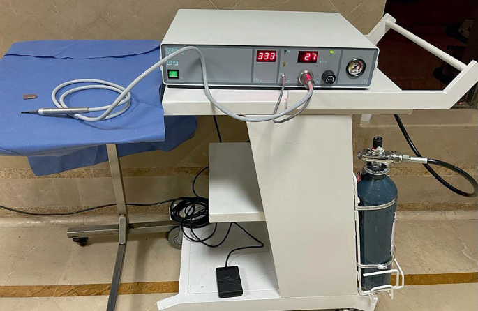
Cryoconsole with cryoprobe and gas cylinder


Cryotherapy Probes
-
Freezing Temperature (°C)-89
-
Probe Type: CE-2000™ Straight Hammerhead Retinal Probe
-
Probe Specs: Dimensions: shaft 40 mm long × 2.5 mm dia.; tip 6.5 mm long × 4.5 mm dia.
-
Probe # 145
-
Probe Type: CE-2000™ Super Cool Curved Retinal
-
Probe Specs: Completely non-electric and autoclavable for safety and ease of use the super cool probes achieve their operating temperature 30% faster than standard probes. Dimensions: shaft 38 mm long × 2.5 mm dia.; tip 2.5 mm dia.
-
Probe # 161
-
Probe Type: CE-2000™ Super Cool 3.2 mm Straight Retinal
-
Probe Specs: Completely non-electric and autoclavable for safety and ease of use the super cool probes achieve their operating temperature 30% faster than standard probes. Dimensions: shaft 38 mm long × 3.2 mm dia.; tip 3.2 mm dia.
-
Probe # 166
-
Probe Type: CE-2000™ Straight Retinal Probe
-
Probe Specs: Dimensions: shaft 40 mm long × 4.0 mm dia.; tip 3.2 mm dia. Precise freeze location with fibre optic illumination.
-
Probe # 708–4
-
Probe Type: CE-2000™ Vitreous Intraocular Probe 20 Gauge
-
Probe Specs: Dimensions: shaft 35 mm long × 0.89 mm dia.; tip 0.89 mm dia. Thermocouple absent.
-
Probe # 134
-
Probe Type: CE-2000™ Straight Retinal Probe
-
Probe Specs: Dimensions: shaft 38 mm long × 2.5 mm dia.; tip 2.5 mm dia.
-
Probe # 124
-
Probe Type: CE-2000™ 3 mm Disc Pediatric Retinal Probe
-
Probe Specs: Dimensions: shaft 35 mm long × 1.5 mm dia.; tip 3 mm long × 15 mm dia. Thermocouple absent.
-
Probe # 123
-
Probe Type: CE-2000™ 6 mm Pediatric Hammerhead Retinal Probe without thermocouple
-
Probe Specs: Dimensions: shaft 34 mm long × 1.5 mm dia.; tip 6 mm long × 1.5 mm dia. Thermocouple absent.
-
Probe # 149
-
Probe Type: CE-2000™ 1.88 mm Curved Pediatric Retinal Probe
-
Probe Specs: Dimensions: shaft 26 mm long × 2.5 mm dia.; tip 1 mm long × 1.88 mm dia.
-
Probe # 120
Patient Preparation
Anaesthesia
-
For the majority of instances of retinal cryopexy, topical anaesthetic with proparacaine drops is sufficient.
-
Lidocaine is used locally in cryotherapy to treat conjunctival neoplasms, lid neoplasms, and trichiasis.
-
Retrobulbar or peribulbar anaesthesia is used to perform cryoablation of choroidal cancers (melanoma or metastatic tumours), peripheral cryoablation of the retina or choroid, cryotherapy for ocular toxoplasmosis or Coats disease, and cyclocryotherapy for neovascular glaucoma.
-
Cryotherapy for retinopathy of prematurity and retinoblastoma is performed under general anaesthesia.
Positioning
The majority of ocular cryotherapy treatments are conducted with the patient recumbent on a narrow table, allowing the surgeon to approach the patient from either side or from the head of the table.
Application of Cryoprobe
Ascertain that there is sufficient gas in the tank and that all connections have been made and fastened properly. The appropriate cryoprobe must be used for the job. Before beginning, check that the cryotherapy equipment is operating properly by depressing the foot switch and noting that the tip is properly cooled.
After administering the required anaesthetic, the cryoprobe is introduced to the tissue receiving treatment while it is still warm. After that, the footswitch is depressed to initiate coolant flow to the tip. At the tip, an ice ball should develop. After tissue begins to adhere to the tip, the probe should not be moved to avoid ripping or breaking the tissue.
The probe is put to the conjunctiva for retinal cryopexy while the surgeon examines the eye with an indirect ophthalmoscope and gently pushes on the probe. The probe’s pressure may be seen as an indentation in the retina. When the probe is positioned properly in relation to the retinal break, cryogen is released into the probe, and the retina whitens as it freezes. After the probe is withdrawn, swelling of the retina may be noticed, and scarring occurs within approximately a week.
Complications
Cryotherapy complications include the following:
-
Over freezing—Because cryotherapy destroys tissue, extremely low temperatures or prolonged cryotherapy may cause harm to normal tissue; the quantity and duration of cryotherapy must be acceptable for the intended goal.
-
Under-freezing—Cryotherapy will not provide the intended outcomes if the tissue temperature is not sufficiently low.
-
Freezing nearby tissue that was not intended to be treated—This may occur if the cryoprobe is applied accidently to adjacent tissue that was not intended to be treated.
-
Tissue breaking—This may occur if the probe is moved after adhering to the tissue; the probe should be allowed to fully thaw before attempting to remove it.
-
Additional complications include visual loss, lid notching, corneal ulceration, accelerated symblepharon formation, xerosis, trichiasis, cellulitis, herpes zoster activation, skin depigmentation, severe soft-tissue reaction, intravitreal dispersion of viable retinal pigment epithelial cells, and retinal tear extension through the cryosurgical scar.
-
New retinal tears have been documented as a late consequence of cryotherapy for retinopathy of prematurity and more recently as an early complication of retinal detachment surgery and retinal necrosis in the Japanese literature
Indications of Cryotherapy in Retinopathy of Prematurity
-
Ablative surgery should be done within 72 h if threshold disease is present, defined as five contiguous clock hours of stage III illness with plus disease or eight non-contiguous clock hours of stage III disease with plus disease.
Outcome
-
Cryotherapy for Retinopathy of Prematurity (CRYO-ROP), a prospective randomised study of cryotherapy for threshold ROP, demonstrated a 50% decrease in unfavourable outcome for zone 2 and 3 disease [22].
-
Early treatment of high-risk pre-threshold ROP substantially decreased adverse ROP outcomes at 9 months and 2 years, according to the Early Treatment for Retinopathy of Prematurity (ET-ROP) Trial [23, 24].
-
Although therapy was beneficial in newborns with threshold disease, infants with ROP in zone 1 had a poor result despite treatment.
Indications of Cryotherapy in Coats Disease
-
For exudation with subretinal fluid of such thickness that cryo reaction can reach the retina
-
Exudative detachment
-
Peripheral lesions, stage 3A and 3B.
Indications of Cryotherapy in Retinoblastoma
-
Pre-equatorial lesions without either deep invasion or vitreous seedings
-
Lesions up to 3.5 mm in diameter and 2.0 mm in thickness.
Outcome
-
Abramson concluded that overall, 70% of tumors were cured with cryotherapy and 93% of patients treated with cryotherapy survived [25].
Indications of Cryotherapy in Toxoplasmosis
-
Transconjunctival cryotherapy for active lesions in the peripheral retina.
Outcome
-
It decreases the number of recurrences.
Indications of Cryotherapy in Rhegmatogenous Retinal Detachment
-
Far anterior retinal breaks
-
If retinal visibility is difficult due to hazy media, not amenable to laser application
-
Pneumatic retinopexy
-
Scleral buckling
-
Pars plana vitrectomy.
Review Questions
1. Cryotherapy is indicated in which stage of Coats disease?
-
(a)
Stage 2
-
(b)
Stage 3A
-
(c)
Stage 3A and 3B
-
(d)
Stage 3B
2. Which cryogen(s) is used in cryotherapy?
-
(a)
Liquid nitrogen
-
(b)
Carbon dioxide snow
-
(c)
Argon or Freon liquid
-
(d)
All of the above
3. Which of the following is NOT an indication for cryotherapy.
-
(a)
Pneumatic retinopexy
-
(b)
Scleral buckling
-
(c)
A large posterior break
-
(d)
Pars plana vitrectomy
4. All of the followings are the effects of cryotherapy EXCEPT:
-
(a)
Ischemia caused by vascular stasis
-
(b)
Decrease retinal demand of oxygen
-
(c)
Cell wall rupture
-
(d)
Osmotic stress
5. Nitrous oxide is more efficient refrigerant than carbon oxide because:
-
(a)
It operates at all pressures down its freezing point
-
(b)
It has a positive Joule-Thompson coefficient value
-
(c)
Its boiling point is higher than that of carbon dioxide
-
(d)
Its transformation from a gas to solid state.
Answers
1. C
2. D
3. C
4. B
5. A
References
Lubritz RR. Cryosurgery. Clin Dermatol. 1987;5(4):120–7. https://doi.org/10.1016/0738-081X(87)90033-2.
Cooper SM, Dawber RP. The history of cryosurgery. J R Soc Med. 2001;94(4):196–201.
Freiman A, Bouganim N. History of cryotherapy. Dermatol Online J. 2005;11(2):9.
Squazzi A, Bracco D. A historical account of the technical means used in cryotherapy. Minerva Med. 1974;65(70):3718–22.
Brock A. Greek medicine, being extracts illustrative of medical writers from Hippocrates to Galen. 2nd ed. New York: Dutton; 1972.
Arnott J. On the treatment of cancer by the regulated application of an anaesthetic temperature. London: Churchill; 1851.
White A. Liquid air: its application in medicine and surgery. Med Rec. 1899;56:109–12.
White A. Possibilities of liquid air to the physcian. JAMA. 1901;36(7):426–9. https://doi.org/10.1001/jama.1901.52470070012001d.
Irvine HG, Turnacliff DD. Liquid oxygen in dermatology. Arch Dermatol Syphilol. 1929;19(2):270–80. https://doi.org/10.1001/archderm.1929.02380200098007.
Allington H. Liquid nitrogen in the treatment of skin disease. Calif Med. 1950;72(3):153.
Cooper IS, Hirose T. Application of cryogenic surgery to resection of parenchymal organs. N Engl J Med. 1966;274(1):15–8. https://doi.org/10.1056/NEJM196601062740103.
Zacarian S. Cryogenics: the cryolesion and the pathogenesis of cryonecrosis, in cryosurgery for skin cancer and cutaneous disorders. St. Louis: Mosby; 1985. p. 1–30.
Bietti GB. Research on the variation of temperatures of some areas of the eyeball for episcleral diathermocoagulation, thermocauterization and cryocoustication. Boll Ocul. 1933;12:1427–57.
Deutschmann R. The treatment of detachment of the retina with tincture of iodine and carbonic acid snow. Klin Montasbl Augenh. 1935;94:349.
Krwawicz T. Intracapsular extraction of intumescent cataract by application of low temperature. Br J Ophthalmol. 1961;45(4):279–83. https://doi.org/10.1136/bjo.45.4.279.
Prohaska J, Bermudez R. Cryotherapy. Treasure Island;2018
Stewart S, Chan W. Pneumatic retinopexy: patient selection and specific factors. Clin Ophthalmol. 2018;12:493–502.
Bhat V, D'Souza P, Shah PK, Narendran V. Risk of tractional retinal detachment following intravitreal bevacizumab along with subretinal fluid drainage and cryotherapy for stage 3b coats’ disease. Middle East Afr J Ophthalmol. 2016;23(2):208–11.
Veckeneer M, Van Overdam K, Bouwens D, Feron E, Mertens D, Peperkamp E. Randomized clinical trial of cryotherapy versus laser photocoagulation for retinopexy in conventional retinal detachment surgery. Am J Ophthalmol. 2001;132(3):343–7.
Shalev B, Farr AK, Repka MX. Randomized comparison of diode laser photocoagulation versus cryotherapy for threshold retinopathy of prematurity: seven-year outcome. Am J Ophthalmol. 2001;132(1):76–80.
Margo C, Hidayat AA, Marshall CF, Renaldo DP. Cryotherapy and photocoagulation in the management of retinoblastoma: treatment failure and unusual complication. Ophthalmic Surg. 1983;14(4):336–42.
Cryotherapy for Retinopathy of Prematurity Cooperative Group. Multicenter Trial of Cryotherapy for Retinopathy of Prematurity: ophthalmological outcomes at 10 years. Arch Ophthalmol. 2001;119(8):1110–8.
VanderVeen DK, Coats DK, Dobson V, et al. Prevalence and course of strabismus in the first year of life for infants with prethreshold retinopathy of prematurity: findings from the Early Treatment for Retinopathy of Prematurity study. Arch Ophthalmol. 2006;124(6):766–73.
Good WV, Hardy RJ, Dobson V, et al., for the Early Treatment for Retinopathy of Prematurity Cooperative Group. The incidence and course of retinopathy of prematurity: findings from the early treatment for retinopathy of prematurity study. Pediatrics 2005;116(1):15–23.
Abramson DH, Ellsworth RM, Rozakis GW. Cryotherapy for retinoblastoma. Arch Ophthalmol. 1982;100(8):1253–6.
Author information
Authors and Affiliations
Corresponding author
Editor information
Editors and Affiliations
Rights and permissions
Copyright information
© 2023 The Author(s), under exclusive license to Springer Nature Switzerland AG
About this chapter

Cite this chapter
Khaqan, H.A. (2023). Cryotherapy for Pediatric Vitreoretinal Diseases. In: Özdek, Ş., Berrocal, A., Spandau, U. (eds) Pediatric Vitreoretinal Surgery. Springer, Cham. https://doi.org/10.1007/978-3-031-14506-3_12
Download citation
DOI: https://doi.org/10.1007/978-3-031-14506-3_12
Published:
Publisher Name: Springer, Cham
Print ISBN: 978-3-031-14505-6
Online ISBN: 978-3-031-14506-3
eBook Packages: MedicineMedicine (R0)