
Abstract
Specific protein depletion is a powerful approach for assessing individual gene function in cellular processes, and has been extensively employed in recent years in mammalian oocyte meiosis-I. Conditional knockout mice and RNA interference (RNAi) methods such as siRNA or dsRNA microinjection are among several approaches to have been applied in this system over the past decade. RNAi by microinjection of Morpholino antisense Oligonucleotides (MO), in particular, has proven highly popular and tractable in many studies, since MOs have high specificity of interaction, low cell toxicity, and are more stable than other microinjected RNAi molecules. Here, we describe a method of MO microinjection into the mouse germinal vesicle-stage (GV) oocyte followed by a simple immunofluorescence approach for examination of gene function in meiosis-I.
Access provided by CONRICYT – Journals CONACYT. Download protocol PDF
Key words
1 Introduction
Mammalian oocyte meiosis-I, also known as oocyte maturation, is the essential final stage of oogenesis . Understanding the molecular mechanisms of this cell division is paramount, as defects can lead to chromosomal imbalances [1–4] or cytoplasmic deficiencies [5, 6] that can endanger reproductive potential. Experimental means for determining the function of potentially important genes during meiosis-I are essential in unraveling the complex cellular events that cause these defects.
The past 10 years have seen the emergence and application of several modes of gene interrogation in oocyte meiosis-I, each with their own pros and cons depending upon the experimental plan. The oocyte-specific conditional knockout mouse approach was first reported in 2006 [7], allowing investigators to delete a target gene of interest in a specific tissue and at specific developmental stage using the Cre-LoxP system, bypassing embryonic lethality which is common for genes regulating cell division (for review see ref. 8). Generation of conditional knockouts can be expensive and time consuming, however, and deletion efficiency of a target gene can be variable [8]. While the recent advent of CRISPR/Cas9 approaches will doubtless expedite the generation of tissue-specific knockouts [9–11], mutant mouse approaches nonetheless require extensive animal breeding and husbandry, which can be impractical in some research programs. Recent leaps forward in small-molecule biology have yielded well-characterized target specific inhibitors that have allowed acute examination of protein function in oocytes from normal (wild-type) laboratory mice [12–14]. However, this approach is critically dependent on the discovery and extensive characterization of such agents. Specific gene interrogation in wild-type mammalian oocytes has also been achieved by overexpression of mutant dominant negative forms of a protein [15–17], or by overexpression of a subunit to disturb protein complex stoichiometry [18, 19], each of which can be introduced by microinjection. However, the precise functioning of such constructs in the highly unusual cytoplasmic milieu of the mammalian oocyte requires careful validation.
Bypassing many of the pitfalls of these approaches, microinjection of RNA antisense constructs into the oocyte (RNA interference, RNAi) to deplete a protein of interest has become increasingly popular over the past 10 years [20, 21]. The major advantage of RNAi microinjection is that constructs can often be obtained from commercial sources inexpensively and, provided constructs are effective, results can be arrived at relatively quickly. RNAi has been achieved in oocytes using a variety of approaches, including small interfering RNA (siRNA) [22–25] and double stranded RNAs (dsRNA) [15]. Probably the most frequently used approach, however, is to inject commercially purchased Morpholino antisense Oligonucleotides (MOs). MOs are short chains of approximately 25 nucleotides assembled on a synthetic backbone of methylenemorpholine rings and phosphorodiamidate links, and are attractive tools as they have very high target-sequence specificity and are, in contrast to RNA-based microinjections, extremely stable [20, 26–28]. To our knowledge MOs were first used to assess gene function in mammalian oocyte meiosis by the Verlhac Lab in 2002 [29] and have since been used by a host of laboratories including ours to examine the effect of depleting numerous targets including cell cycle proteins [26, 27, 30, 31], signaling molecules [32, 33], and motor proteins [34]. Here we describe our standard routine protocol for MO microinjection into the mouse GV oocyte, followed by a simple immunofluorescence approach to confirm protein depletion and examine the resulting phenotype .
2 Materials
2.1 Materials for Mouse Ovarian Stimulation
-
1.
6–12-week-old female CD1 mice (see Note 1 ).
-
2.
Pregnant mare serum gonadotropin (PMSG) (see Note 2 ).
-
3.
Phosphate buffered saline (PBS).
-
4.
1 ml syringes.
-
5.
Needles.
2.2 Materials and Reagents for Mouse Gv-Stage Oocyte Collection and Culture
-
1.
M2 medium (see Note 3 ).
-
2.
M16 medium.
-
3.
3-Isobutyl-1-methylxanthine (IBMX) for germinal vesicle break down (GVBD) inhibition is dissolved in DMSO at a stock concentration of 100–200 mM. Final concentration is 0.1–0.2 mM (1:1000). Store at −20 °C.
-
4.
M2 medium supplemented with IBMX (M2 + IBMX) for collection and handling of GV oocytes outside the incubator (10 ml, keep at 37 °C).
-
5.
M2 + IBMX for MO microinjection (1 ml, keep at room temperature).
-
6.
M16 medium supplemented with IBMX (M16 + IBMX) for GV oocyte culture in the incubator before and after MO microinjection (~5 ml, keep in the incubator). We recommend making this the night and leaving in incubator overnight to ensure it is properly equilibrated at 5 % CO2).
-
7.
Mineral oil (see Note 4 ).
-
8.
Transfer pipette for pouring mineral oil to the tissue culture.
-
9.
0.22 μm syringe filter.
-
10.
10 ml syringe to filter the media.
-
11.
14 ml polystyrene round-bottom tube.
-
12.
Dissection microscope.
-
13.
Digital dry bath.
-
14.
High precision tweezers.
-
15.
27G × ½″ needle.
-
16.
Petri dishes.
-
17.
Pasteur pipette (see Note 5 ).
-
18.
Mouth-controlled aspiration tube for the Pasteur pipette.
-
19.
Cell culture incubator.
2.3 Materials and Reagents for Microinjection of Morpholino Antisense Oligos (MO)
-
1.
MOs designed and purchased from Gene Tools LLC (www.gene.tools.com) (see Note 6 ).
-
2.
Dry bath or PCR machine for preheating MO before microinjection.
-
3.
Centrifuge.
-
4.
Oocyte microinjection station, comprising the following (components and setup illustrated in Fig. 1): Leica DMIL-LED inverted scope, Hydraulic micromanipulator with universal joint, Coarse manipulators, Injectors, Iron plates as magnetic bases for injectors, PV820 pneumatic picopump (World precision instruments), Electrometer intracellular amplifier IE-251A (Harvard apparatus), Silver wire for intracellular amplifier IE-251A (Advent research materials), Magnetic base for intracellular amplifier. The apparatus is assembled and placed on an antivibration table (see Note 7 , Fig. 1).
Fig. 1 
Oocyte microinjection station. (a) Leica DMIL-LED inverted scope, (b) Hydraulic micromanipulators with universal joint, (c) Coarse manipulators, (d) Injector, (e) Iron plates for as magnetic bases for injectors, (f) PV820 pneumatic pico-pump, (g) Electrometer intracellular amplifier IE-251A, (h) Silver wire for electro intracellular amplifier IE-251A, (i) Magnetic base for electro intracellular amplifier, (j) An antivibration table
-
5.
Holding pipettes (commercially available readymade from Hunter scientific http://www.hunterscientific.com or homemade pipettes see Note 8 ).
-
6.
Vertical micropipette puller for the injection pipette.
-
7.
Glass capillaries with filament for MO injection (Harvard apparatus).

2.4 Materials and Methods for Preparing Oocytes for Immuno-Fluorescence Microscopy
-
1.
Paraformaldehyde (PFA).
-
2.
Triton X-100.
-
3.
Bovine serum albumin (BSA) .
-
4.
PBS tablets.
-
5.
96-well plates with round bottom.
-
6.
35 mm Glass bottom culture dishes (see Note 9 ).
Using the above reagents the following working solutions should be prepared. The solutions are filtered and stable for several weeks at 4 °C.
-
1.
4 % PFA + PBS for fixing.
-
2.
1 % BSA + PBS for washing.
-
3.
3 % BSA + PBS for blocking.
-
4.
0.25 % Triton X + PBS for permeabilization.
3 Methods
3.1 Collection and Preparation of GV Mouse Oocytes
-
1.
Administer 5 IU PMSG intraperitoneally to mice 44–48 h before GV stage oocytes are to be collected (see Note 10 ).
-
2.
Prepare 35 mm tissue culture dish with 50 μl drops (~5 drops/dish) of M16 + IBMX and M16 medium. Put 3 ml of mineral oil to cover the drops and place in the incubator (37 °C, 5 % CO2 in air) (see Fig. 2). M16 medium has to be equilibrated for at least 2 h before oocyte collection. Prepare similar dishes of M2 + IBMX drops and place on the dry bath (37 °C).
Fig. 2 
Dissection of mouse ovaries and collection of GV oocytes. (a) Ovaries are placed in the 60 mm dish with 7 ml of prewarmed (37 °C) M2 + IBMX. (b) Ovaries are dissected using 27G needle and tweezers under the dissection microscope. (c) Fully grown oocytes are collected using a large diameter Pasteur pipette and mouth-controlled aspiration tube. (d) 50 μl drops of culture medium are covered with mineral oil and placed in the incubator at least for 2 h for equilibration before performing the experiment. (e) Collected oocytes are washed using a large diameter Pasteur pipette. Oocytes are washed through 3–5 drops before the start of in vitro culture. (f) Fully grown mouse GV oocytes with two or three layers of cumulus cells. (g) Denuded fully grown oocytes (top three oocytes), and growing oocytes (bottom three oocytes) which we would not use for this type of experiment. (h) A fully grown oocyte with a perivitelline space (black arrow) 1 h after incubation in M16 + IBMX. (i) Metaphase-II egg with a first polar body (black arrow). Scale bars are all 50 μm
-
3.
Collect the ovaries from the PMSG-injected females. Place in 2 ml of prewarmed M2 + IBMX (37 °C) in a 35 mm or 60 mm dish under the dissection microscope .
-
4.
Breach antral follicles on the ovary surface by immobilizing the ovary using tweezers while repeatedly puncturing the ovary surface with a 27G needle mounted on a 1 ml syringe.
-
5.
After dissection of ovaries, collect only fully grown oocytes with two or three layers of cumulus cells (see Fig. 2) using a large diameter (~150 μm) Pasteur pipette connected to a mouth pipette. Transfer the collected cumulus–oocyte complexes (COCs) into a drop of prewarmed M2 + IBMX (see Note 11 ).
-
6.
Remove all cumulus cells around the oocyte by pipetting COCs back and forth using a Pasteur pipette with diameter approximately similar to the diameter of an oocyte (~80 μm, see Note 11 ).
-
7.
Transfer cumulus-cell-free (“denuded”) GV oocytes into a clean drop of prewarmed M2 + IBMX. Repeat 3–5 times in successive clean drops to wash the oocytes until all somatic cells are removed (see Fig. 2). Take care to only collect fully grown GV stage oocytes. Transfer oocytes to a dish of M16 + IBMX drops, washing the oocytes through for 3–5 drops to remove M2 and then place them in the incubator for 30–60 min prior to MO microinjection (see Note 12) .

3.2 Microinjection of MO Oligonucleotides
-
1.
Prewarm MO oligonucleotides with dry bath or PCR machine for 5–10 min at 65 °C followed by centrifugation at full speed for 1 min (see Note 13 ).
-
2.
Prepare the microinjection chamber: place 600–800 μl of M2 + IBMX (room temperature) on the upturned lid of 35 mm tissue culture dish and cover with mineral oil. Place on the microinjection stage.
-
3.
Prepare the microinjection pipette using a vertical pipette puller (see Note 14 ).
-
4.
Insert holding pipette into the holding shaft. Adjust holding pipette using micromanipulators such that holding pipette is at center of the field of view, ~200 μm above the floor of the injection chamber (see Note 15 ).
-
5.
Wash GV oocytes through three drops of M2 + IBMX, before mouth pipetting into the M2 + IMBX component of the microinjection chamber (see Note 16 ).
-
6.
Clean the silver wire that is attached to the injection probe by gentle wiping with 70 % EtOH and tissues.
-
7.
Load 1 μl of the MO solution into the injection pipette using a microloader tip (Eppendorf) fitted on a “P10” Gilson pipette, with the silver wire close to the MO solution, and fit into injection probe.
-
8.
Place the silver wire connected with “grounds chassis” jack back side of the intracellular electrometer into M2 + IBMX using the swan-neck attachment (see Note 17 ).
-
9.
Focus on the GV oocytes then lower the holding pipette slowly until it touches the GV oocyte. Adjust pressure on the holding pipette to adhere the oocyte to holding pipette. Lift oocyte to a position ~200 μm above the injection chamber surface.
-
10.
Lower the injection pipette into M2 + IBMX in line with oocyte. Confirm that neither the “amplifier” nor “current injection” LEDs are illuminated on the intracellular electrometer.
-
11.
Using micromanipulators , smoothly insert the injection pipette into the cytoplasm through the zona pellucida .
-
12.
Apply a brief pulse of negative capacitance to the Oolema by pressing the “buzz” button on the intracellular electrometer.
-
13.
Microinject MO into the GV oocyte by pressing “start” button on the pico-pump and remove the injection pipette gently (see Note 18 ).
-
14.
Injection size can vary day to day depending upon injection pipette diameter. Therefore, perform steps 12 and 13 on a small number of “test” oocytes to adjust and see the size of the injection before microinjecting the main cohort of oocytes. Adjust injection size by controlling injection pressure and duration on pico-pump. The size of the injection can be judged by cytoplasmic displacement (see Note 19 ).
-
15.
Once the appropriate injection size has been established, inject the remaining cohort of oocytes without changing injection settings, to ensure consistent injection size for each oocyte.
-
16.
Following MO microinjection, transfer oocytes to M16 + IBMX in the incubator using the mouth-controlled Pasteur pipette.
-
17.
Repeat the above steps with other microinjection solutions according to experimental design (see Note 20 ).
-
18.
Leave oocytes in M16 + IBMX for between 1 and 24 h prior to in vitro maturation to permit protein turnover (see Note 21 ).
-
19.
Wash MO-microinjected GV oocytes through 3–5 clean drops of pre-equilibrated IBMX-free M16 medium and return to the incubator to initiate meiosis-I completion, which takes ~9 to 12 h in most laboratory mouse strains (see Note 22 , Fig. 2).
3.3 Preparation of Oocytes for Immuno-Fluorescence Analysis
Immunofluorescence provides a simple but tractable approach for determining the effectiveness of a MO in depleting its target protein, and for analyzing resulting phenotypes. The effectiveness of antibodies for oocyte immunofluorescence varies depending upon the precise makeup of immunofluorescence processing reagents. Here we describe a simple protocol that is effective with many commercial antibodies.
-
1.
Prepare a round-bottom 96-well plate for immunofluorescence staining, arranging solutions in vertical columns, and oocyte treatment groups in horizontal rows. Place 50 μl of solution in each well, covering each 50 μl filled well with a single drop of mineral oil.
-
2.
Using a bent Pasteur pipette (see Note 23 ), transfer oocytes into column 1.
-
3.
Fix the oocyte in 4 % PFA + BSA. Allow to sit for 40 min at room temperature (RT). Subsequent steps described later entail moving oocytes to successive wells in the 96-well plate.
-
4.
Wash the oocytes with 1 % BSA + PBS 3 times for 5 min each at RT.
-
5.
Permeabilization with 0.25 % Triton-X + PBS for 10 min each at RT.
-
6.
Wash with 1 % BSA + PBS three times for 5 min each at RT.
-
7.
Block with 3 % BSA + PBS for 60 min at 37 °C or 4 C overnight.
-
8.
Incubate with primary antibody for 60 min at 37 °C.
-
9.
Wash with 1 % BSA + PBS three times for 5 min each at RT.
-
10.
Incubate with secondary antibody for 60 min at 37 °C. Protect from the light (see Note 24 ).
-
11.
Wash with 1 % BSA + PBS three times for 5 min each at RT.
-
12.
Incubate with 10 μg/ml Hoechst 33343 for 5 min at RT.
-
13.
Prepare 5 μl of 1 % BSA + PBS drops on the glass bottom dish and cover with mineral oil.
-
14.
Pipette the fixed oocytes into the drops.
-
15.
Analyze with epifluorescence or confocal microscopy (see Note 25 ).
4 Notes
-
1.
Animals should be acquired and stored in accordance with local animal welfare regulations.
-
2.
To make injection syringes, dissolve PMSG (1000 IU/vial) into pre-chilled (4 °C) filtered 40 ml PBS (stock concentration 25 IU/ml). Aliquot into 1 ml syringes attached with 27G needle, working on ice. Freeze immediately and store at −20 °C. Inject 200 μl per mouse to administer 5 IU.
-
3.
We normally use commercially provided M2 and M16 medium, but these solutions can be made in-house [35].
-
4.
Mineral oils can contain contaminants. We therefore recommend a lot-test even this product is supplied as “embryo culture grade.” Autoclave to sterilize if necessary.
-
5.
Pasteur pipette need to be pulled to appropriate thickness using an alcohol lamp or Bunsen burner. Large diameter pipettes (150 μm opening) are needed for initial oocyte collection and narrow diameter pipette (approximately the same size of the oocyte diameter, 80 μm) are needed for denudation of the cumulus cells. Pasteur pipettes are attached to a mouth-controlled aspiration tube to allow oocytes to be transferred between drops. Moving oocytes with a mouth-controlled Pasteur pipette takes several weeks’ practice, but once mastered allows very precise and controlled movement.
-
6.
MOs are typically purchased as 300 nanomoles in powder form. We typically make a 1 mM stock by dissolving in molecular biology grade water warming at 65 °C and vortexing 3–4 times. The 1 mM stock should be kept at room temperature in a good quality tube. 1 mM injection (final concentration ~50 nM, see ref. 34) represents a good starting concentration for experiments. At higher concentrations MOs can occasionally dimerize and become inactive. If a previously functioning MO stock appears to stop working, we recommend trying to separate the dimers by diluting the stock further (0.5 mM), autoclaving, and allowing the solution to return to room temperature. For further tips on MO use see ref. [21].
-
7.
Precise micromanipulation setup may vary. The amplifier and negative capacitance pulse is essential when performing injections into metaphase-stage eggs and embryos, which are extremely sensitive to injection. The apparatus is also beneficial when injecting GV -stage oocytes also, but is dispensed with by some labs. An antivibration assembly is essential. Commercial isolation tables are effective, but a homemade apparatus comprising a marble slab and sponge balls is cost effective and works well (see Fig. 1).
-
8.
We recommend investigators with minimal micromanipulation experience to buy pre-made holding pipettes in the first instance. However, holding pipettes can also be manufactured in house using a pipette puller and microforge, which allows for the holding pipette to be replaced regularly at low cost, and for pipettes to be tailored to individual users tastes. For further information on holding pipettes manufacture see: (http://narishige-group.com/movie/MF-900_cutting/index.html, http://narishige-group.com/movie/MF-900_bending/index.html, http://narishige-group.com/movie/MF-900_fire-polishing/index.html).
-
9.
We normally use Petri dishes fitted with No. 0 coverglass (0.085–0.13 mm) for immunofluorescence microscopy.
-
10.
The number of fully grown oocytes retrieved per mouse is highly dependent upon mouse strain, age, and PMSG administration [36, 37]. We most frequently use ~12-week-old female CD1 mice, and administer 5 IU of PMSG, retrieving 20–25 oocytes per mouse.
-
11.
An overly narrow Pasteur pipette can damage or kill the oocyte. Care must be taken in selecting a pipette with the correct diameter.
-
12.
Selecting only fully grown GV stage oocytes from antral follicles is critical, as oocytes less than full size may not be competent to resume meiosis [38–41]. Non-fully grown oocytes typically feature a thinner zona pellucida that is less smooth in appearance and should be discarded (at least for the purpose of the experiment described herein). 30–60 min incubation allows for the formation of the perivitelline space [42] in fully grown oocytes but not mid-grown phase oocytes and is thus a further hallmark of a fully grown oocyte (see Fig. 2).
-
13.
MO solution can be gloopy. To prevent injection pipette blockage, before use incubate the MO for 5–10 min at 65 °C and centrifuge for 1 min before use.
-
14.
Establishing pipette-puller settings to manufacture appropriate pipettes is achieved empirically and can be time consuming. Once settings are established, pipette size can remain reasonably consistent over the course of several weeks. We recommend that pipette pullers be placed away from areas of air flow (e.g., air conditioning).
-
15.
Setting up the microinjection rig and aligning pipettes can be fiddly, time consuming, and frustrating, and takes time and practice for new experimenters. We typically find that a new investigator within our lab can take upward of 10–20 experimental days of training in this technique before being competent to perform robust experiments.
-
16.
GV oocytes should not be kept on the microinjection stage for more than 20 min. We therefore recommend that beginners microinject in small groups, transferring injected oocytes back to warmed M2 + IBMX on the dry bath/warm plate.
-
17.
The silver wire can be placed anywhere within the M2 + IBMX of the injection chamber to establish an electrical circuit. The negative capacitance enables smooth delivery of the pipette tip into the ooplasm. The red lamp tuens off when an electrical circuit is successfully made, Care must be taken as the injection pipette tip is very fragile and easily broken.
-
18.
Before MO microinjection into the oocyte, change “duration” setting to “gated” and keep press “start” button briefly to expel a small amount of injection solution. This is useful to counteract capillary action. Change “duration” setting to “timed” prior to oocyte injection.
-
19.
The injection size should be no more than 5 % of the oocyte volume. As a useful estimate, an injection with cytoplasmic displacement diameter 1/6 of the diameter of the diameter of the oocyte provides less than 1 % by volume (see Fig. 3). Larger injection size can be detrimental to oocyte health. To increase or reduce the injection size, change either “eject pressure” or “period” settings of pico-pump. In general, once the correct injection size is established, the injection should be nondamaging as evidenced by >95 % oocyte survival. No cytoplasmic displacement upon injection indicates a failed injection. The most likely explanation for this is a blocked pipette. Change the injection pipette, using freshly preheated MO solution. Make sure that the injection pipette does not touch the germinal vesicle (the oocyte nucleus), as this can damage the oocyte and block the injection pipette. Microinjection of fluorescent tracer (Rhodamine B isothiocyanate-Dextran, Sigma Cat # R8881-100MG or Fluorescein isothiocyanate-Dextran, Sigma Cat# FD10S-100MG) can be used when first learning microinjection into the oocyte to confirm successful delivery.
Fig. 3 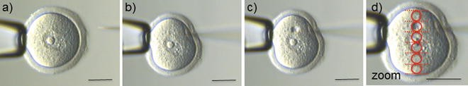
MO microinjection into the mouse GV oocyte. (a) Oocyte is immobilized using a holding pipette (left side) and set at the center of the field of view. Injection pipette is to the right. (b) The injection pipette is gently inserted into the cytoplasm through the zona pellucida , avoiding the nucleus. (c) Injection of MO into the cytoplasm. (d) A zoom of the image in (c) to illustrate estimation of injection size. Overlay illustrates that cytoplasmic displacement diameter is approximately 1/6 of the diameter of the oocyte. Scale bars 50 μm
-
20.
Experimental groups and controls depend upon the experimental design. However, since off-target effects have been occasionally reported with oligonucleotides [21], we strongly recommend that controls include specific MO-injected, water-injected, and random-sequence MO control-injected oocytes. MO-induced phenotypes should in most cases be reversible by expression of exogenous protein, and we consider corroboration of results by an alternative intervention (e.g., expression of a dominant-negative mutant version of the target) highly desirable to confirm specificity. For an example of such experimental design from our lab, see ref. [34]. For further notes on control experiments, see ref. [21].
-
21.
Required incubation time is highly dependent upon the rate of protein turnover. Cell cycle-related proteins often have quick turnover therefore making them good candidates for RNAi approaches. Some proteins are inappropriate targets due to slow turnover.
-
22.
The time required to complete in vitro oocyte maturation (IVM) depends upon the mouse strain but is 9–12 h in most cases. Completion of IVM is confirmed by the formation of a first polar body (see Fig. 2). In most laboratory mouse strains the conditions described herein should permit meiosis-I completion in 90 % or more of wild-type oocytes.
-
23.
We use a 96-well plate for immunofluorescence for examination of gene function in MO-injected oocytes. A bent Pasteur pipette with ~45–90 ° angle is required to use a 96-well plate. Add 50 μl of fixing/washing solution and 2–5 μl of antibody into the each well.
-
24.
Alexa-labeled secondary antibodies are light sensitive. Antibodies should be stored in nontranslucent tubes (or cover tubes in silver foil). Once oocytes have been exposed to fluorescent antibodies dishes, 96-well plates should be covered in silver foil.
-
25.
Immunolabeled oocytes can be stored in PBS + 1 % BSA in 96-well plates for many months and the fluorescence remain stable (depending upon the antibodies). Hoechst fluorescence is less stable, but can be reapplied on the day of imaging as required. Immunofluorescence of the target protein itself can provide a simple test of the effectiveness of depletion, and allows quantitative analysis of reduced abundance at a specific location (e.g., at the spindle). Western blotting provides an excellent quantitative alternative especially if the protein is predominantly cytoplasmic (see Chapter 15). For many cell division targets, analysis of spindle morphology (microtubule antibody, Sigma) can provide an informative first analysis of knockdown effect .

References
Jones KT, Lane SI (2013) Molecular causes of aneuploidy in mammalian eggs. Development 140:3719–3730
Hassold TJ, Hunt PA (2001) To err (meiotically) is human: the genesis of human aneuploidy. Nat Rev Genet 2:280–291
Nagaoka SI, Hassold TJ, Hunt PA (2012) Human aneuploidy: mechanisms and new insights into an age-old problem. Nat Rev Genet 18:493–504
Howe K, FitzHarris G (2013) Recent insights into spindle function in mammalian oocytes and early embryos. Biol Reprod 89:1–9
Li R, Albertini DF (2013) The road to maturation: somatic cell interaction and self-organization of the mammalian oocyte. Nat Rev Mol Cell Biol 14:141–152
Coticchio G, Dal Canto M, Mignini Renzini M, Guglielmo MC, Brambillasca F, Turchi D, Novara PV, Fadini R (2015) Oocyte maturation: gamete-somatic cells interactions, meiotic resumption, cytoskeletal dynamics and cytoplasmic reorganization. Hum Reprod Update 21:427–454
Kudo NR, Wassmann K, Anger M, Schuh M, Wirth KG, Xu H, Helmhart W, Kudo H, McKay M, Maro B, Ellenberg J, de Boer P, Nasmyth K (2006) Resolution of chiasmata in oocytes requires separase-mediated proteolysis. Cell 126:135–146
Sun QY, Liu K, Kikuchi K (2008) Oocyte-specific knockout: a novel in vivo approach for studying gene functions during folliculogenesis, oocyte maturation, fertilization, and embryogenesis. Biol Reprod 79:1014–1020
Singh P, Schimenti JC, Bolcun-Filas E (2015) A mouse geneticist’s practical guide to CRISPR applications. Genetics 199:1–15
Seruggia D, Montoliu L (2014) The new CRISPR-Cas system: RNA-guided genome engineering to efficiently produce any desired genetic alteration in animals. Transgenic Res 23:707–716
Yang H, Wang H, Jaenisch R (2014) Generating genetically modified mice using CRISPR/Cas-mediated genome engineering. Nat Protoc 9:1956–1968
Lane SI, Chang HY, Jennings PC, Jones KT (2010) The Aurora kinase inhibitor ZM447439 accelerates first meiosis in mouse oocytes by overriding the spindle assembly checkpoint. Reproduction 140:521–530
Nguyen A, Gentilello AS, Balboula AZ, Shirivastava V, Ohring J, Schindler K (2014) Phosphorylation of threonine 3 on histone H3 by haspin kinase is required for meiosis in mouse oocytes. J Cell Sci 127:5066–5078
FitzHarris G (2009) A shift from kinesin 5-dependent metaphase spindle function during preimplantation development in mouse. Development 136:2111–2119
Coelho PA, Bury L, Sharif B, Riparvelli MG, Fu J, Callaini G, Glover DM, Zernicka-Goetz M (2013) Spindle formation in the mouse embryo requires Plk4 in the absence of centrioles. Dev Cell 9:586–597
Yoshida S, Kaido M, Kitajima TS (2015) Inherent instability of correct kinetochore-microtubule attachments during meiosis I in oocytes. Dev Cell 33:589–602
Balboula AZ, Schindler K (2014) Selective disruption of Aurora C kinase reveals distinct functions from Aurora B kinase during meiosis in mouse oocytes. PLoS Genet 10:e1004194
Dalton CM, Carroll J (2013) Biased inheritance of mitochondria during asymmetric cell division in the mouse oocyte. J Cell Sci 126:2955–2964
Melkonian KA, Maier KC, Godfrey JE, Rodgers M, Schroer TA (2007) Mechanism of dynamitin-mediated disruption of dynactin. J Biol Chem 282:19355–19364
Summerton JE (2007) Morpholino, siRNA and S-DNA compared: impact of structure and mechanism of action on off-target effects and sequence specificity. Curr Top Med Chem 7:651–660
Eisen SE, Smith JC (2008) Controlling morpholino experiments: don’t stop making antisense. Development 135:1735–1743
Brunet S, Dumont J, Lee KW, Kinoshita K, Hikal P, Gruss OJ, Maro B, Verlhac M-H (2008) Meiotic regulation of TPX2 protein levels governs cell cycle progression in mouse oocytes. PLoS One 3:e3338
Pfender S, Kuznetsov V, Pasternak M, Tischer T, Santhanam B, Schuh M (2015) Live imaging RNAi screen reveals genes essential for meiosis in mammalian oocytes. Nature 524:239–242
Sharif B, Na J, Lykke-Harmann K, McLaughlin SH, Laue E, Glover DM, Zernicka-Goetz M (2010) The chromosome passenger complex is required for fidelity of chromosome transmission and cytokinesis in meiosis of mouse oocytes. J Cell Sci 123:4292–4300
Baumann C, Viveiros MM (2015) Meiotic spindle assessment in mouse oocytes by siRNA-mediated silencing. J Vis Exp. doi:10.3791/53586
Homer H, Gui L, Carroll J (2009) BubR1 is required for prophase I arrest and prometaphase progression during female meiosis I. Science 326:991–994
Homer H, McDougall A, Levaseur M, Yallop K, Murdoch AP, Herbert M (2005) Mad2 prevents aneuploidy and premature proteolysis and cyclin B and securin during meiosis I in mouse oocytes. Genes Dev 19:202–207
Marangos P, Stevense M, Niaka K, Lagoudaki M, Nabti I, Jessberger R, Carroll J (2015) DNA damage-induced metaphase I arrest is mediated by the spindle assembly checkpoint and maternal age. Nat Commun 6:8706
Christophe L, Terret ME, Djiane A, Rassinier P, Maro B, Verlhac M-H (2002) Meiotic spindle stability depends on MAPK-interacting and spindle-stabilizing protein (MISS), a new MAPK substrate. J Cell Biol 157:603–613
Madgwick S, Hansen DV, Levasseur M, Jackson P, Jones KT (2006) Mouse Emi2 is required to enter meiosis II by reestablishing cyclin B1 during interkinesis. J Cell Biol 174:791–801
Herbert M, Levasseur M, Homer H, Yallop K, Murdoch A, McDougall A (2003) Homologue disjunction in mouse oocytes requires proteolysis of securin and cyclin B1. Nat Cell Biol 5:1023–1025
Tsurumi C, Hoffmann S, Geley S, Graeser R, Polansky Z (2004) The spindle assembly checkpoint is not essential for CSF arrest of mouse oocytes. J Cell Biol 167:1037–1050
Balboula AZ, Stein P, Schultz RM, Schindler K (2014) Knockdown of RBBP7 unveils a requirement of histone deacetylation for CPC function in mouse oocytes. Cell Cycle 13:600–611
Illingworth C, Pirmadjid N, Serhal P, Howe K, FitzHarris G (2010) MCAK regulates chromosome alignment but is not necessary for preventing aneuploidy in mouse oocyte meiosis I. Development 137:2133–2138
Butler JE, Lechene C, Biggers JD (1988) Noninvasive measurement of glucose uptake by two populations of murine embryos. Biol Reprod 39:779–786
Fowler RE, Edwards RG (1957) Induction of superovulation and pregnancy in mature mice by gonadotrophins. J Endocrinol 15:374–384
Byers SL, Payson SJ, Taft RA (2006) Performance of ten inbred mouse strains following assisted reproductive technologies (ARTs). Theriogenology 65:1716–1726
Gosden RG, Telfer E (1987) Scaling of follicular sizes in mammalian ovaries. J Zool 211:157–168
Xiao S, Duncan FE, Bai L, Nguyen CT, Shea LD, Woodruff TK (2015) Size-specific follicle selection improves mouse oocyte reproductive outcomes. Reproduction 150:183–192
Schultz RM, Letourneau GE, Wassarman PM (1979) Program of early development in the mammal: changes in the patterns and absolute rates of tubulin and total protein synthesis during oocyte growth in the mouse. Dev Biol 73:120–133
Schultz RM, Letourneau GE, Wassarman PM (1978) Meiotic maturation of mouse oocytes in vitro: protein synthesis in nucleate and anucleate oocyte fragments. J Cell Sci 30:251–264
Baltz JM, Tartia AP (2010) Cell volume regulation in oocytes and early embryos: connecting physiology to successful culture media. Hum Reprod Update 16:166–176
Acknowledgements
Works in GFs lab is supported by CIHR, NSERC, CFI, and Fondation Jean-Louis Lévesque. Elements of the described procedure were learned from labs of John Carroll, Karl Swann, Jay Baltz, and Tomohiro Kono. We thank Jenna Haverfield, Cayetana Vázquez-Diez, and Angus MacCaulay for critical reading of the manuscript.
Author information
Authors and Affiliations
Corresponding author
Editor information
Editors and Affiliations
Rights and permissions
Copyright information
© 2016 Springer Science+Business Media New York
About this protocol
Cite this protocol
Nakagawa, S., FitzHarris, G. (2016). Quantitative Microinjection of Morpholino Antisense Oligonucleotides into Mouse Oocytes to Examine Gene Function in Meiosis-I. In: Nezis, I. (eds) Oogenesis. Methods in Molecular Biology, vol 1457. Humana Press, New York, NY. https://doi.org/10.1007/978-1-4939-3795-0_16
Download citation
DOI: https://doi.org/10.1007/978-1-4939-3795-0_16
Published:
Publisher Name: Humana Press, New York, NY
Print ISBN: 978-1-4939-3793-6
Online ISBN: 978-1-4939-3795-0
eBook Packages: Springer Protocols