
Abstract
Each kidney is supplied by a single renal artery in the majority of cases. These renal arteries generally arise from the aorta at L1–L2 interspace.
Access provided by Autonomous University of Puebla. Download chapter PDF
Keywords
- Renal Artery
- Renal Artery Stenosis
- Fibromuscular Dysplasia
- Atherosclerotic Renal Artery Stenosis
- Renal Artery Aneurysm
These keywords were added by machine and not by the authors. This process is experimental and the keywords may be updated as the learning algorithm improves.
1 Anatomy
Each kidney is supplied by a single renal artery in the majority of cases. These renal arteries generally arise from the aorta at L1–L2 inter space.
Extra renal arteries are present in about 30% of the population. The common variations being:
-
(a)
Extra renal arteries entering at the renal hilum arising directly from the aorta.
-
(b)
Polar branches directly to upper or lower pole from the aorta.
-
(c)
More rarely an extra renal artery may arise from an iliac artery.
-
(d)
Proximal bifurcation of the one or other of the main renal arteries is another common finding to be aware of.
2 Disease Definition
Flow limiting renal artery stenosis (RAS), so as to cause secondary hypertension or renal failure is thought to occur when the renal arteries demonstrate a > 50–75% reduction in luminal diameter.
3 Classification of Renal Artery Stenosis
-
1.
Atherosclerotic renovascular disease (ARVD) (90%)
-
2.
Fibromuscular dysplasia (FMD) 10%—A rare non-inflammatory condition that usually affects younger female patients.
-
3.
Other rare causes e.g. Takayasu’s disease—more common in India and China.
Atherosclerotic disease and FMD are two very different disease processes and respond differently to intervention. They should therefore be considered as two separate entities.
4 Diagnosis: Clinical and Laboratory
Renal artery stenosis is almost always asymptomatic; however, it should be suspected in certain patients:
-
1.
Uncontrollable hypertension
-
2.
Hypertension and asymmetric renal length on ultrasound
-
3.
20% or more reduction in eGFR or a 15% rise in serum creatinine at the time of starting an ACE inhibitor or angiotensin 11 receptor blocker
-
4.
Flash pulmonary oedema
-
5.
Kidney damage in the presence of known vascular disease elsewhere (especially if the legs are involved)
-
6.
Malignant or abrupt onset hypertension
-
7.
Hypertension onset when patient is <30 yrs old—consider FMD
Unfortunately there are no specific diagnostic laboratory investigations for renal artery stenosis, and the presence of a reduced eGFR or hypertension is not present in all cases. The diagnosis is usually made following appropriate imaging of the renal vasculature in patients where there is high clinical suspicion.
5 Diagnostic Imaging
5.1 Ultrasound
Assessment of kidney size is accurate and easily achieved, as is cortical thickness (kidneys <8 cm long are unlikely to benefit from revascularization in the treatment of ARVD). Colour flow duplex of the renal arteries is possible but the acoustic window is often limited by adverse body habitus and is also operator dependant. Other non-invasive modalities such as CT and MRI are more useful in diagnosis and planning treatment.
5.2 CT Scan
Multi-detector Computerized Tomographic Angiography (MDCTA) provides fast accurate interrogation of the renal arteries. Multi planar reformatting and 3D rendering allow for intuitive assessment and planning if intervention is required. In severely calcified ARVD, imaging may be degraded by artefact and confident assessment of the degree of stenosis difficult. Attention should also be made to any concurrent renal dysfunction, a GFR of <60 ml/min should prompt concern about possible iodinated contrast related nephrotoxicity and pre-hydration may be necessary.
5.3 Magnetic Resonance Angiography
Evaluation with gadolinium-enhanced magnetic resonance angiography (MRA) is the predominant modality in the assessment of renovascular disease due to its excellent sensitivity and specificity. Renal impairment (GFR < 60 ml/min) prompts careful consideration, as the administration of gadolinium contrast agents carry a risk of nephrogenic systemic fibrosis (NSF) which can result in significant morbidity and mortality. In these cases CTA may be preferable, as iodinated-related nephrotoxicity is usually mild and reversible and can be minimised by pre-hydration.
5.4 Diagnostic Angiography
This remains the gold standard in the assessment of renal artery disease although the above non-invasive modalities have largely replaced it. In severe renal failure, CO2 can be used instead of iodinated contrast as a contrast agent thus removing any risk of contrast nephropathy.
Steps involved in diagnostic angiography include:
-
1.
Femoral artery access
-
2.
Pigtail flush catheter placed into upper abdominal aorta at the level of L1 vertebrae.
-
3.
Power injection of 20 ml of iodinated contrast at a rate 10 ml/s.
-
4.
Oblique views may be required to accurately interrogate the renal artery origins. For example, the right renal artery often arises from the anterolateral aspect of the aorta requiring a 25 degree left anterior oblique projection. The key to alignment being that the image intensifier be perpendicular to the origin of the renal artery.
-
5.
In cases of non-atheromatous disease selective renal images are usually required. The renal arteries can be easily selected using either a femoro-visceral (Cobra) or a reverse curve (Shepherd’s crook) type shape. The reverse curve catheters need to be reformed in the aortic arch or over the iliac bifurcation but are very useful in caudally angulated vessels and very stable once in the renal artery. Hand injection of 10 ml of iodinated contrast is often sufficient for diagnostic needs.
6 Management
Management of patients with atherosclerotic renal artery stenosis remains controversial amongst radiologists and physicians. Individual reports and observational studies report successes from stenting, in terms of blood pressure control and stabilisation of renal function. However two recent randomised control trials, (ASTRAL and STAR) have failed to show any benefit of stenting over best medical treatment alone for a variety of outcomes including blood pressure, renal function and mortality. Therefore debate continues, especially in terms of the optimal time for intervention, the optimal patient on which to intervene and the precise benefits (if any) from stenting. There is however general agreement that stenting may be offered in cases of intractable hypertension, “flash” pulmonary oedema, rapidly deteriorating renal function and those who present with acute renal failure and occluded renal arteries.
Protection devices (filters) and platelet glycoprotein IIa/IIIb receptor inhibitors have been used either together or separately to try and improve the results of stenting by reducing the incidence of cholesterol embolization. There is some very weak anecdotal evidence in their favour which requires further investigation in larger trials.
In summary, careful consideration of the risk to benefit ratio is needed on a case by case basis for renal artery stenting in atheromatous disease.
The results of angioplasty in fibromuscular dysplasia are far more encouraging than with atheromatous disease. Following angioplasty, 10 year cumulative patency rates are as high as 87%. In addition blood pressure cure can be achieved in up to 50% and in the remainder improved blood pressure control with a reduction in the drug burden. Stents are only rarely required in non-atheromatous lesions.
6.1 List of Open Operative Choices
The minimally invasive nature of endovascular renal revascularisation has largely replaced conventional surgical reconstruction particularly, in elderly subjects with multiple co-morbidities. However there is still a need for open procedures particularly in cases of failed or complicated endovascular intervention. The principle options include:
-
(a)
Bypass (aortorenal, spleno/hepatorenal, iliorenal, aortic graft and renal bypass). Extra-anatomical bypass has the advantage of avoiding a heavily diseased aorta
-
(b)
Endarterectomy—seldom practised
-
(c)
Renal auto-transplantation to pelvis—occasionally used for complex FMD with bench surgery
-
(d)
Nephrectomy—occasionally useful with a shrunken kidney causing renin driven hypertension
7 Intervention: Techniques and Pitfalls
A.Ostial Atheromatous Stenosis
Primary stenting is advocated in the treatment of ostial ARVD. These lesions are due to aortic cushions of plaque and simply recoil and restenose if angioplastied alone.
All patients should have non-invasive imaging prior, to establish a diagnosis and plan the stenting. The numbers of renal arteries and the angle at which they arise from the aorta are both vital information. Non-invasive imaging will also dictate optimal C-arm positioning to allow for accurate stent placement ensuring the whole lesion is covered out into the aorta, for 2–3 mm.
Patients will normally be taking an antiplatelet agent (aspirin 75 mg daily or clopidogrel 75 mg daily) which should be continued for life. Pre-hydration with intravenous fluids for 12 h will help minimise the risk of contrast nephropathy. Some advocate the use of N-Acetyl cystine (to protect against contrast nephropathy) although the evidence for this is weak.
The small platform stent and angioplasty kits have largely replaced conventional 0.035 systems which will not be discussed further.
-
(a)
Femoral access although in approx 10% of cases the anatomy will dictate an arm approach. Flush aortogram (Fig. 20.1) via 4-French pigtail catheter to confirm anatomy.
Fig. 20.1 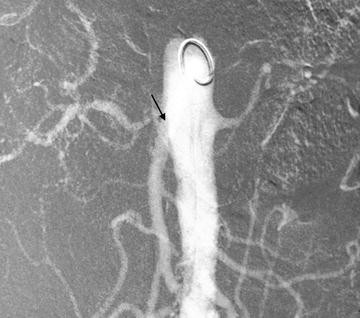
Flush aortogram via 4Fr pigtail catheter. A significant stenosis of the right renal artery is confirmed (arrow)
-
(b)
Place 7-Fr renal double curved introducer guide catheter leaving the tip adjacent to the renal artery ostium (Fig. 20.2).
Fig. 20.2 
A 7Fr double curved guide catheter is placed at the ostium of the right renal artery
-
(c)
Administer 3,000–5,000 units of heparin.
-
(d)
Cross the stenosis with a 0.014 or 0.018 guide wire alternatively if a protection device is going to be used the lesion is crossed with the filter at this stage (Fig. 20.3).
Fig. 20.3 
Renal protection device is advanced past the lesion (a 0.018 or 0.014 guide wire alone is an acceptable alternative)
-
(e)
Pre-dilate the lesion with a 3 mm small platform balloon.
-
(f)
Introduce the balloon mounted stent (usually 6 × 18 mm). Position checked by gentle contrast injection via the guide catheter.
-
(g)
Deploy stent allowing for 2–3 mm to project into the aorta (Fig. 20.4).
Fig. 20.4 
After pre-dilation the balloon mounted stent is deployed (arrow) note the open configuration of the renal protection device (arrowhead)
-
(h)
Check position with gentle contrast injection via the guide catheter (Fig. 20.5).
Fig. 20.5 
Check angiogram via the guide catheter. Guide wire or protection device must remain in the distal vessel so as to allow access to distal vessel if dissection or rupture occurs
-
(i)
Remove guide wire. Close and remove protection filter if used.
-
(j)
Manual compression to groin or closure device.


B.Non Ostial Atheromatous Lesions
These rare lesions may respond well to simple angioplasty and stents should be reserved for primary failures (e.g. recoil). Pre-dilation is not required and normally a 6 mm balloon is used.
C.Fibromuscular Dysplasia
Work up should include selective renal angiography as disease affecting the second or third order vessels may be missed by non-invasive methods. Technique is the same as above except that branch lesions may require more complex techniques using “kissing” balloons. Anti-platelets agents are not required.
8 Post-procedure Management
8.1
Immediately post-procedure patients should be monitored for bleeding/ haematoma around the arterial puncture site. The patient is required to lie flat in bed for a 2–6 h post-procedure (depending on local practice). During this time the nursing staff perform routine observations e.g. pulse and blood pressure.
A large diuresis can occur after revascularisation with a drop in blood pressure and careful attention to fluid replacement and anti-hypertensive treatment is required.
8.2 Follow-up Protocol
There is no consensus regarding follow up. Blood pressure and renal function should be monitored. Routine renal artery imaging is not required but may be indicated if there is a clinical deterioration. Re-stenosis is often asymptomatic but when associated with recurrent symptoms e.g. worsening renal function then re-intervention should be considered (angioplasty, stenting or surgery).
9 Complications and Management
-
1.
Puncture Site Haematoma. Careful pressure on the puncture site and the use of closure devices in selected patients will minimise this complication. Bleeding times are often prolonged in patients with impaired renal function.
-
2.
Renal Artery Dissection. Usually inconsequential if a stent is to be placed.
-
3.
Renal artery Rupture. A feared complication usually resulting in a nephrectomy in the past. Most can now be managed with either simple balloon tamponade or a covered stent.
-
4.
Acute Renal Failure. In the first few days this is usually contrast related and recovers with supportive management. A more insidious loss of renal function over several weeks often indicates cholesterol embolization for which the prognosis is more guarded.
10
When considering the management of ARVD it is worth considering that
-
Maximal medical management may well be as effective as revascularization.
-
Both endovascular and, open surgical revascularisation carry risks although these are often perceived to be more serious with surgery.
-
Surgical series have published good results but these are from centres of excellence, in highly selected patients.
-
For many elderly patients with multiple co-morbidities surgery is simply not an option.
It is our opinion that carefully selected patients may benefit from stenting and angioplasty. Surgery may have a place in complex disease or where stenting has failed, again careful patient selection on a case by case basis is recommended.
Primary angioplasty in FMD demonstrates good medium and long term patency rates and effective treatment of hypertension and renal failure. We would recommend this treatment over and above open surgery as primary treatment.
11 Renal Artery Dissections
The most common cause of renal artery dissection is secondary to endovascular treatment. Primary angioplasty of non ostial lesions and FMD may result in intimal dissection. This requires emergent treatment as the kidney will perish rapidly (60–90 min warm ischemic time).
Another scenario in which renal artery dissections are seen is as a part of aortic dissections. These are more complicated, but are treated in the same way, with the exception that the descending aortic dissection may need to be treated simultaneously.
As previously mentioned the guidewire should remain past the treated lesion until a satisfactory check angiogram has been performed. This wire will allow for access to the distal luminal vessel.
Management
-
1.
A repeat balloon angioplasty over the existing wire with a prolonged inflation time may “stick” the intima back into position restoring flow.
-
2.
If flow can be restored then stenting is recommended unless the angioplasty has resulted in a near-perfect angiogram. A suitable sized (6–8 mm diameter) balloon expandable stent will often provided the support necessary.
-
3.
A second stent projecting into the aortic lumen as for the treatment of ostial stenosis may be required.
12 Renal Artery Aneurysms
Renal artery aneurysms are rare and may be idiopathic (degenerative) or occur secondary to FMD. These are typically extra-parenchymal and usually occur at the first and second order bifurcations. More peripheral aneurysm formation can be seen in some of the vasculitides (e.g. PAN and Behcet’s disease), infection and trauma (including iatrogenic e.g. renal biopsy).
12.1 Diagnosis
MRA or MDCTA is usually sensitive enough to detect most aneurysms allowing for planning management and surveillance.
12.2 Management
Renal artery aneurysms are often asymptomatic. Extra parenchymal aneurysms >2 cm are at increased risk of rupture. These may be treated by either open surgical or more recently endovascular techniques. Options include coil embolization with and without stent scaffolding and stent grafting.
Flow chart summarising endovascular treatment for renal artery stenosis.

References
The ASTRAL Investigators. Revascularization versus medical therapy for renal-artery stenosis. N Engl J Med. 2009;361(20):1953–62.
Bax L, Woittiez AJ, Kouwenberg HJ, et al. Stent placement in patients with atherosclerotic renal artery stenosis and impaired renal function: a randomized trial. Ann Intern Med. 2009;150(12):840–8.
de Fraissinette B, Garcier JM, Dieu V, et al. Percutaneous transluminal angioplasty of dysplastic stenoses of the renal artery: results on 70 adults. Cardiovasc Intervent Radiol. 2003;26(1):46–51.
Hirsch AT, Haskal ZJ, Hertzer NR, et al. ACC/AHA 2005 Practice Guidelines for the management of patients with peripheral arterial disease (lower extremity, renal, mesenteric, and abdominal aortic): a collaborative report from the American Association for Vascular Surgery/Society for Vascular Surgery, Society for Cardiovascular Angiography and Interventions, Society for Vascular Medicine and Biology, Society of Interventional Radiology, and the ACC/AHA Task Force on Practice Guidelines. (Writing Committee to Develop Guidelines for the Management of Patients With Peripheral Arterial Disease): endorsed by the American Association of Cardiovascular and Pulmonary Rehabilitation; National Heart, Lung, and Blood Institute; Society for Vascular Nursing; TransAtlantic Inter-Society Consensus; and Vascular Disease Foundation. Circulation. 2006;113(11):e463–654.
Author information
Authors and Affiliations
Corresponding author
Editor information
Editors and Affiliations
Rights and permissions
Copyright information
© 2013 Springer Science+Business Media New York
About this chapter
Cite this chapter
Hay, C.S.M., Powell, J.R., Moss, J.G. (2013). Renal Artery Disease: Stenosis, Dissections and Aneurysms. In: Kumar, A., Ouriel, K. (eds) Handbook of Endovascular Interventions. Springer, New York, NY. https://doi.org/10.1007/978-1-4614-5013-9_20
Download citation
DOI: https://doi.org/10.1007/978-1-4614-5013-9_20
Published:
Publisher Name: Springer, New York, NY
Print ISBN: 978-1-4614-5012-2
Online ISBN: 978-1-4614-5013-9
eBook Packages: MedicineMedicine (R0)