
Abstract
CD49f+ CD34+ cells, a population rich in skin epithelial stem cells (EpSCs), were obtained from adult mouse skin and cultured with Wnt-3a for 10 days. On day 10, CD49f+ CD34+ cells were sorted and subjected to a second 10-day culture with Wnt-3a. The same procedures were repeated until fifteenth 10-day culture. CD49f+ CD34+ cells obtained from each 10-day culture retained the same EpSC-characteristics as seen in the original EpSCs from adult mouse skin. Here, wedescribe the culture protocol using Wnt-3a for successful maintenance of EpSCs.
Access provided by CONRICYT – Journals CONACYT. Download protocol PDF
Keywords
1 Introduction
Wnt signaling is critical for regulation of a number of basic cell functions, such as cell proliferation, fate, polarity, and differentiation [1–3], and is also considered to be deeply involved in maintenance of stem cells including embryonic, neural, hematopoietic, mammary, hepatic, and skin epithelial stem cells [4–8]. Recently, the cell surface protein CD34 was shown to be uniquely expressed in stem cells from the mouse hair follicle bulge region (HFSCs) and an isolation method for living skin epithelial stem cells (EpSCs) from adult mouse-derived primary skin epithelial cells (MPSECs) was established by fluorescent activated cell sorting (FACS) using two selective surface markers, CD49f (α6 integrin) and CD34 [9, 10]. The population of CD49f+ CD34+ cells is considered to be an EpSC-rich fraction containing HFSCs. In our previous study, we reported successful maintenance of EpSCs in vitro for a prolonged period (at least 150 days) using Wnt-3a and FACS [11]. In this chapter, we show our culture method protocol used to maintain EpSCs with sustained characteristics.
2 Materials
2.1 Isolation of EpSCs
-
1.
Inbred 8-week-old mice (Japan SLC, Shizuoka, Japan, C3H/HeN Slc).
-
2.
Dissection instruments (micro-scissors, fine forceps).
-
3.
Stereo microscope (Leica, model no. MS5).
-
4.
Saline (Otsuka, Tokyo, Japan, cat. no. D05352).
-
5.
Trypsin (0.25 %) (Invitrogen, Grand Island, NY, cat. no. 15050).
-
6.
Plastic petri dishes (Sigma, cat. no. SIAL506CC0SnV).
-
7.
Dulbecco’s modified Eagle’s medium (DMEM; Wako, Osaka, Japan, cat. no. 043-30085) containing 10 % fetal bovine serum and FBS (PAA Laboratory, Morningside QLD, cat. no. A15002).
-
8.
Phosphate-buffered saline (PBS), pH 7.4 (Wako, cat. no. 048-29805).
-
9.
Rat anti-α6 integrin (CD49f) directly coupled to PE (R&D Systems, Inc., Minneapolis, MN, cat. no. FAB13501P).
-
10.
Rat IgG2a isotype directly coupled to PE (R&D Systems, Inc, cat. no. IC006P).
-
11.
Rat anti-CD34 directly coupled to FITC (AbD Serotec, Oxford, UK, cat. no. MCA1825F).
-
12.
Rat IgG2a isotype directly coupled to FITC (eBioscience, San Diego, CA, cat. no. 11-4321-73).
-
13.
Polystyrene round-bottom tubes (BD, Bedford, MA, cat. no. 352235 or 352003).
-
14.
Cell strainer (40 μm) (BD, cat. no. 352340).
-
15.
Epilife™ serum-free culture medium kit (Thermo Fisher, Waltham, MA, cat. no. M-EPICF-500).
-
16.
FACSAria system equipped with FACS DiVa software (BD Biosciences, San Jose, CA).
-
17.
Trypan blue (Invitrogen, cat. no. 15250061).
-
18.
Hemocytometer (Waken-Btech, Kyoto, Japan, cat. no. WC2-100).
-
19.
Plastic cell culture dishes (6 cm) (BD, Bedford, MA, cat. no. 353002).
-
20.
Light microscope (Carl Zeiss; model no. Axiovert 40CFL).
2.2 EpSCs Sequential Culture
-
1.
Plastic cell culture dishes (6 cm).
-
2.
Collagen type I (Nippon Ham, Tsukuba, Japan).
-
3.
PBS.
-
4.
Trypsin–EDTA (0.25 %) (Invitrogen, Burlington, ON, Canada, cat. no. 25200-056).
-
5.
Rat anti-α6 integrin (CD49f) directly coupled to PE.
-
6.
Rat IgG2a isotype directly coupled to PE.
-
7.
Rat anti-CD34 directly coupled to FITC.
-
8.
Rat IgG2a isotype directly coupled to FITC.
-
9.
Polystyrene round-bottom tubes.
-
10.
Epilife™ serum-free culture medium kit.
-
11.
Wnt-3a recombinant protein (R&D Systems Inc., cat. no. 1324-WN/CF).
-
12.
FACSAria system equipped with FACS DiVa software.
-
13.
Trypan blue.
-
14.
Hemocytometer.
-
15.
Light microscope.
2.3 Characterization of EpSCs
2.3.1 Flow Cytometry
-
1.
PBS.
-
2.
Trypsin–EDTA (0.25 %).
-
3.
Rat anti-α6 integrin (CD49f) directly coupled to PE.
-
4.
Rat IgG2a isotype directly coupled to PE.
-
5.
Rat anti-CD34 directly coupled to FITC.
-
6.
Rat IgG2a isotype directly coupled to FITC.
-
7.
Polystyrene round-bottom tubes.
-
8.
PFA (1 %) solution (prepared by dilution of 4 % PFA solution, Wako, cat. no. 163-20145).
-
9.
FACSCalibur (BD Biosciences).
2.3.2 Hair Reconstitution Assay
-
1.
EpSCs (CD34+ CD49f+ cells sorted following serial cultivations) (see Sect. 3.2).
-
2.
Newborn (day 2) mice (from C3H/HeN Slc).
-
3.
Nude mice (BALB/c Slc-nu/nu, Japan SLC).
-
4.
PBS.
-
5.
Dispase (diluted in PBS, Godo Shusei Co. Ltd., Tokyo, Japan, cat. no. GD81020).
-
6.
Collagenase (diluted in PBS, Wako, cat. no. 034-10533).
-
7.
Cell strainer (40 μm).
-
8.
Isoflurane (final concentration 2–4 %, Wako, cat. no. 099-06571).
-
9.
Silicon transplantation chamber (kindly gifted by Dr. J. Kishimoto of Shiseido Co. Ltd.).
-
10.
Dissection instruments (micro-scissors, fine forceps).
-
11.
Animal clipper (Natsume Seisakusho, Tokyo, Japan, cat. no. C-29-8).
3 Methods
3.1 Isolation of EpSCs
-
1.
Shave dorsal skin areas of adult mice.
-
2.
Isolate dorsal skin tissues using forceps and scissors, and place in petri dishes.
-
3.
Wash isolated skin samples several times with PBS.
-
4.
Remove PBS and incubate skin tissues in 0.25 % trypsin at 4 °C overnight.
-
5.
Add same volume of DMEM containing 10 % FBS into dishes.
-
6.
Detach dermis from epidermis using fine forceps.
-
7.
Collect whole epithelial cells by scrubbing epidermis-facing surface of dermis using belly of the forceps. These cells were designated as mouse-derived primary skin epithelial cells (MPSECs).
-
8.
Filtrate whole cells using 40-μm cell strainer.
-
9.
Centrifuge cell suspension at 250 × g for 5 min, then wash cells three times with PBS.
-
10.
Prepare MPSECs as single cells by addition of PBS to pellets and determine cell number using trypan blue exclusion method.
-
11.
Add antibodies directly coupled with a fluorochrome and expose cells for 30 min on ice (see Note 1 ).
-
12.
After washing twice with PBS, resuspend cells in Epilife™ serum-free culture medium.
-
13.
Sort CD34+ CD49f+ cells (EpSCs) from MPSECs using FACSAria system (see Fig. 1).
Fig. 1 
Isolation of EpSCs. (a) MPSECs were stained with the isotype control, anti-CD34-FITC, or anti-CD49f-PE antibodies (isotype control Ab, CD34 Ab, CD49f Ab, respectively). EpSCs were collected from the MPSECs as CD34 and CD49f double-positive cells. (b) Bright microscopic field showing morphology of sorted EpSCs after attachment to culture plate. Scale bar = 50 μm


3.2 EpSC Sequential Cultures
Figure 2 shows the outline of the protocol for sequential culturing of EpSCs.


Experimental design and procedures. 0p-EpSCs (primary sorted cells) were cultured in the presence of Wnt-3a for 10 days. At the end of the first 10-day culture, CD49f+ CD34+ cells were collected as 1p-EpSCs and subjected to the second 10-day culture with Wnt-3a. By repeating this process, cultures of CD49f+ CD34+ cells were sequentially performed up to 15 passages
-
1.
Treat 60-mm dishes with collagen type I and wash with PBS three times.
-
2.
Resuspend CD49f+ CD34+ cells (EpSCs) sorted from MPSECs in Epilife™ serum-free culture medium and confirm their viability by trypan blue staining.
-
3.
Seed EpSCs at a density of 5 × 10⁴ cells per dish and culture in the presence of 200 ng/ml of Wnt-3a for 10 days.
-
4.
Change to fresh medium containing Wnt-3a every 2 days.
-
5.
On day 10, collect all cells by trypsin treatment and centrifuge cell suspensions at 250 × g for 5 min, then wash with PBS three times.
-
6.
Make single cell preparations into pellets by addition of PBS and determine the cell numbers using trypan blue exclusion method.
-
7.
Add antibodies with a fluorochrome and expose cells for 30 min on ice.
-
8.
After washing twice with PBS, resuspend cells in Epilife™ serum-free culture medium.
-
9.
Sort EpSCs using FACSAria system (see Note 2 ).
-
10.
Repeat steps 5–9 for each experiment (see Note 3 ).
3.3 Characterization of EpSCs
3.3.1 Flow Cytometry
-
1.
Culture EpSCs with or without Wnt-3a for 10 days (see Sect. 3.2.).
-
2.
Wash EpSCs with PBS and harvest with 0.25 % trypsin–EDTA.
-
3.
Centrifuge samples at 250 × g for 5 min, then wash with PBS and collect EpSCs.
-
4.
Make single cell preparations into pellets by addition of PBS and determine cell numbers using trypan blue exclusion method.
-
5.
Add antibodies with a fluorochrome and expose cells for 30 min on ice (see Sect. 3.2).
-
6.
After washing twice with PBS, resuspend cells in 1 % PFA solution.
-
7.
Analyze whole cells using FACSCalibur (see Note 4 ).
3.3.2 Hair Reconstitution Assay
A hair reconstitution experiment was performed as previously described by J. Kishimoto et al., with minor modifications.
3.3.2.1 Preparation of Dermal Cells
-
1.
Isolate dorsal skin tissues from dorsal skin areas of newborn (day 2) mice using forceps and scissors, and place into petri dishes.
-
2.
Wash isolated skin samples with PBS several times.
-
3.
Incubate skin tissues in 500 units/ml dispase at 4 °C overnight.
-
4.
Transfer skin tissues into dishes containing PBS and detach dermis samples from epidermis using fine forceps.
-
5.
Incubate skin dermis in 0.25 % collagenase at 37 °C for 1 h.
-
6.
Collect whole cells, then centrifuge and resuspend with PBS.
-
7.
Allow tubes to stand for 20 min (see Note 5 ).
-
8.
Collect all cells in supernatant and filtrate using 40-μm cell strainer.
-
9.
Centrifuge cell suspension at 250 × g for 5 min and wash cells with PBS three times.
-
10.
Add PBS to pellet samples and determine cell number using trypan blue exclusion method. Next, use dermal cell population containing dermal fibroblasts and papilla cells for transplantation (see Note 6 ).
3.3.2.2 Preparation of EpSCs as Epithelial Cells
On the day of transplantation, prepare cell population containing EpSCs cultured in serum-free medium with Wnt-3a.
-
1.
Collect all cells cultured in Epilife™ serum-free culture medium with Wnt-3a using trypsin treatment.
-
2.
Centrifuge cell suspensions at 250 × g for 5 min and wash with PBS three times.
-
3.
Check cell viability and determine numbers using trypan blue staining.
-
4.
Add antibodies with a fluorochrome and expose to cells for 30 min on ice (see Sect. 3.2.).
-
5.
After washing twice with PBS, resuspend cells in Epilife™ serum-free culture medium.
-
6.
Sort CD49f+ CD34+ cells as EpSCs using FACSAria system.
-
7.
After washing with PBS, use EpSCs as epithelial cells for transplantation.
3.3.2.3 Transplantation
-
1.
Prepare a mixed solution containing EpSCs (2.5 × 10⁷/ml) and dermal cells (2.5 × 10⁷/ml) to transplant into nude mice. Store on ice until transplantation.
-
2.
Anesthetize nude mice with an inhalation of isoflurane.
-
3.
Sterilize back skin of each mouse with 70 % ethanol.
-
4.
Remove a section of back skin tissue with a diameter of 10 mm using scissors.
-
5.
Implant silicon transplantation chambers into dorsal site of nude mice and fix using animal clipper.
-
6.
Inject 200-μl mixture of EpSCs (2.5 × 10⁶) and dermal cells (2.5 × 10⁶) into each chamber by pipette.
-
7.
Cut off the roof of the chamber at 1 week after transplantation.
-
8.
Remove chamber at 2 weeks after transplantation.
-
9.
Observe new hairs in graft sites at 3 weeks after removing chambers (see Fig. 4. and Note 7 ).
-
10.
Harvest skin tissues for assessment of reconstituted skin, if required.
4 Notes
-
1.
The following antibodies and dilutions were used: rat anti-α6 integrin (CD49f) labeled PE; 1:100; rat IgG2a isotype labeled PE; 1:100; rat anti-CD34 labeled FITC, 1:100; rat IgG2a isotype labeled FITC, 1:100.
-
2.
We termed CD49f+ CD34+ cells isolated from MPSECs as 0p-EpSCs, then those from the first culture as 1p-EpSCs and those from the second culture as 2p-EpSCs, with the same naming protocol used for CD49f+ CD34+ cells from each successive passaged culture.
-
3.
By repeating the procedure for CD49f+ CD34+ cell sorting on day 10 and subsequent passage to the next 10-day culture with Wnt-3a, sequential cultures were performed for up to 15 cultures.
-
4.
EpSCs were gated for single events and sorted according to their expressions of CD49f and CD34. The purity of sorted cells was determined using post-sort FACS analysis and typically exceeded 95 %. Representative results for 2p- and 15p-EpSCs are presented (see Fig. 3).
Fig. 3 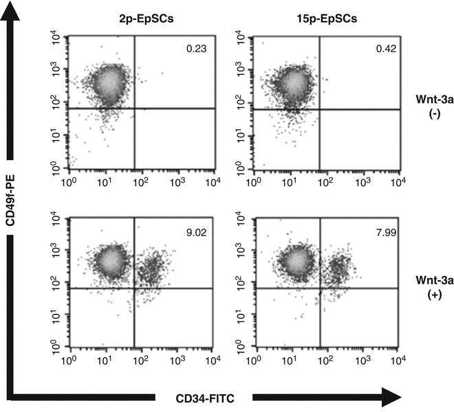
Long-term maintenance of EpSCs by Wnt-3a in sequential cultures. The CD49f+ CD34+ population comprised approximately 8–10 % of all cells on day 10 in cultures with Wnt-3a, while there were few CD49f+ CD34+ cells in cultures without Wnt-3a. Representative results for 2p- and 15p-EpSCs are shown
-
5.
The process utilized resulted in a sediment composed of large cells, debris, and follicles.
-
6.
The dermal fibroblast fraction contained unavoidable contamination by non-dermal cells such as melanocytes.
-
7.
The CD49f+ CD34+ population in sequential cultures with Wnt-3a showed an ability to reconstitute skin. Results from 2p- and 15p-EpSCs are presented (see Fig. 4).
Fig. 4 
EpSCs maintained in serial cultures showed sustained hair reconstitution ability. The CD49f+ CD34+ population in sequential cultures with Wnt-3a was able to reconstitute skin. Representative results from 2p- and 15p-EpSCs are presented


References
Peifer M, Polakis P (2000) Wnt signaling in oncogenesis and embryogenesis – a look outside the nucleus. Science 287(5458):1606–1609
Nusse R (2008) Wnt signaling and stem cell control. Cell Res 18(5):523–527. doi:10.1038/cr.2008.47
Moon RT, Bowerman B, Boutros M, Perrimon N (2002) The promise and perils of Wnt signaling through beta-catenin. Science 296(5573):1644–1646. doi:10.1126/science.1071549
Watt FM, Collins CA (2008) Role of beta-catenin in epidermal stem cell expansion, lineage selection, and cancer. Cold Spring Harb Symp Quant Biol 73:503–512. doi:10.1101/sqb.2008.73.011
Hu M, Kurobe M, Jeong YJ, Fuerer C, Ghole S, Nusse R, Sylvester KG (2007) Wnt/beta-catenin signaling in murine hepatic transit amplifying progenitor cells. Gastroenterology 133(5):1579–1591. doi:10.1053/j.gastro.2007.08.036
Singla DK, Schneider DJ, LeWinter MM, Sobel BE (2006) wnt3a but not wnt11 supports self-renewal of embryonic stem cells. Biochem Biophys Res Commun 345(2):789–795. doi:10.1016/j.bbrc.2006.04.125
Nguyen H, Merrill BJ, Polak L, Nikolova M, Rendl M, Shaver TM, Pasolli HA, Fuchs E (2009) Tcf3 and Tcf4 are essential for long-term homeostasis of skin epithelia. Nat Genet 41(10):1068–1075. doi:10.1038/ng.431
Kleber M, Lee HY, Wurdak H, Buchstaller J, Riccomagno MM, Ittner LM, Suter U, Epstein DJ, Sommer L (2005) Neural crest stem cell maintenance by combinatorial Wnt and BMP signaling. J Cell Biol 169(2):309–320. doi:10.1083/jcb.200411095
Trempus CS, Morris RJ, Bortner CD, Cotsarelis G, Faircloth RS, Reece JM, Tennant RW (2003) Enrichment for living murine keratinocytes from the hair follicle bulge with the cell surface marker CD34. J Invest Dermatol 120(4):501–511. doi:10.1046/j.1523-1747.2003.12088.x
Blanpain C, Lowry WE, Geoghegan A, Polak L, Fuchs E (2004) Self-renewal, multipotency, and the existence of two cell populations within an epithelial stem cell niche. Cell 118(5):635–648. doi:10.1016/j.cell.2004.08.012
Ouji Y, Ishizaka S, Nakamura-Uchiyama F, Okuzaki D, Yoshikawa M (2015) Partial maintenance and long-term expansion of murine skin epithelial stem cells by Wnt-3a in vitro. J Invest Dermatol 135(6):1598–1608. doi:10.1038/jid.2014.510
Author information
Authors and Affiliations
Corresponding authors
Editor information
Editors and Affiliations
Rights and permissions
Copyright information
© 2016 Springer Science+Business Media New York
About this protocol
Cite this protocol
Ouji, Y., Yoshikawa, M. (2016). Maintenance of Skin Epithelial Stem Cells by Wnt-3a In Vitro. In: Turksen, K. (eds) Stem Cell Heterogeneity. Methods in Molecular Biology, vol 1516. Humana Press, New York, NY. https://doi.org/10.1007/7651_2016_320
Download citation
DOI: https://doi.org/10.1007/7651_2016_320
Published:
Publisher Name: Humana Press, New York, NY
Print ISBN: 978-1-4939-6549-6
Online ISBN: 978-1-4939-6550-2
eBook Packages: Springer Protocols