
Opinion statement
The management of infrapopliteal peripheral arterial occlusive disease (PAD) is challenging. For patients with asymptomatic disease or claudication, exercise and optimal medical management, including antiplatelet agents, blood pressure control, statin therapy and tight glucose control for patients with diabetes mellitus, are the mainstays of therapy. However, patients with isolated tibial artery occlusive disease often have diabetes mellitus or renal insufficiency and present with critical limb ischemia (CLI). CLI is advanced occlusive disease marked by the development of rest pain, ischemic ulceration, or gangrene and is associated with a high mortality rate. Limb salvage requires an intervention in cases of CLI, but careful operative planning is required as patients often have multilevel disease and limited options for revascularization. A surgical bypass with a vein graft remains the best treatment for infrapopliteal PAD, especially in patients with a life expectancy of over 2 years. Balloon angioplasty can play an important role in limb salvage, especially for patients lacking adequate vein for bypass, at high operative risk, or with a life expectancy of less than 2 years. However, a lack of rigorous trials has left unanswered questions as to the efficacy of infrapopliteal angioplasty with or without stents compared to bypass surgery. As such, endovascular therapy is currently not a proven treatment for intermittent claudication. Patients who are unable to undergo a revascularization procedure for infrapopliteal CLI have few options besides amputation or palliation. New therapies, such as drug-eluting stents, drug-coated balloons, and stem cell therapy are under development, but their efficacy and effectiveness remain unproven.
Introduction
Below the popliteal artery, blood flow to the calf and foot is supplied by three arteries: the anterior tibial (which becomes the dorsalis pedis at the ankle), the posterior tibial, and the peroneal arteries. When affected by peripheral arterial disease (PAD), the tibial and pedal arteries are typically heavily calcified and, because of extensive collateral vessels, chronic total occlusions (CTO) may occur in one or more infrapopliteal vessel before symptoms arise. In fact, most patients with symptomatic PAD of the infrapopliteal segment have multi-level disease with atherosclerosis in the aortoiliac and femoropopliteal segments as well [1]. Fewer patients, usually those with diabetes mellitus or renal failure, develop symptomatic disease from isolated infrapopliteal PAD [2].
Although a diminished pedal pulse suggests infrapopliteal PAD, the diagnosis is generally established with an ankle-brachial index (ABI) of <0.9. Alternatively, a toe-brachial index (TBI) of <0.7 can be used for the diagnosis in cases where incompressible, heavily calcified tibial arteries give a falsely elevated ABI value (eg, >1.40) [3, 4•]. In patients with an abnormal ABI, symptoms classify patients into three categories: asymptomatic, intermittent claudication, and critical limb ischemia (CLI). Importantly, intermittent claudication, which is calf or foot pain that occurs with exercise and is relieved by rest, affects lifestyle, whereas CLI, which is defined as rest pain and/or ischemic tissue loss, carries a high risk of limb loss and mortality.
The distinction in severity of symptoms and disease plays an important role in determining therapy. Most patients have asymptomatic infrapopliteal disease or intermittent claudication and are best managed with exercise and medical therapy according to the ACC/AHA and TASC II guidelines for patients with PAD [1, 3]. The anatomic challenges of intervening on the small and often heavily calcified tibial and pedal arteries have led to the generally inferior results of infrapopliteal revascularization compared to revascularization of larger proximal arterial segments. As a result, intervention in patients with infrapopliteal PAD is generally reserved for patients with CLI, as these patients have the highest risk of limb loss and death.
Until recently, much of the data for infrapopliteal PAD therapies have come from small trials and case series studying a heterogeneous group of PAD patients. Much of the data for non-interventional therapies still come from studies of patients with multi-level PAD. However, several trials have published (and several more announced) that focus on the specific treatment of infrapopliteal disease, and more rigorous trials comparing treatment approaches would be welcomed. This article discusses the current recommendations for medical and surgical therapies for infrapopliteal PAD as well as treatments on the horizon.
Treatment
Diet and lifestyle
-
Although no recommendations exist specifically for infrapopliteal PAD, there is consensus that a low-fat, heart-healthy diet, smoking cessation, and tight glucose control (for patients with diabetes mellitus) are recommended for all individuals with PAD [3, 4•].
-
Exercise therapy, both at home and as a structured, supervised program, has been shown to increase the distance for claudication symptoms and quality of life measures. Exercise is generally recommended for a minimum of 30 to 45 min, 3 times per week for 12 weeks. Because intermittent claudication frequently limits the mobility and thus general physical fitness of patients, exercise programs may also provide protection from future cardiovascular events [5].
Pharmacologic treatment
-
The presence of PAD is considered a coronary artery disease equivalent; therefore, all patients should be treated according to cardiovascular disease risk-reduction strategies [6]. There are no medical or pharmacologic treatments specifically for infrapopliteal PAD.
-
Aspirin has been shown to decrease the rate of cardiovascular events (myocardial infarction, stroke, or vascular death) as well as incidence of lower extremity ischemic events but does not improve claudication symptoms [4•, 7]. Clopidogrel is also effective in reducing the risk of cardiovascular events in patients with PAD [1, 4•].
-
Blood pressure medications, particularly beta-blockers and angiotensin-converting enzyme inhibitors (ACE-I), help reduce overall cardiovascular events [3]. Several small studies have also suggested that the ACE-I (lisinopril), beta-blockers (nebivolol, metoprolol), and thiazide diuretics (hydrochlorothiazide) are associated with improvements in intermittent claudication symptoms [8–10]. Larger clinical trials are needed to confirm these results.
-
Statin drugs are recommended for all patients with PAD as they have beneficial cardiovascular effects and have been shown to increase walking time for those with claudication [3, 7, 11]. Patients with lower extremity PAD are in the “high risk” or “very high risk” categories according to the National Cholesterol Education Program Adult Treatment Panel III guidelines and thus should have a goal low-density lipoprotein (LDL) level of <100 mg/dL or <70 mg/dL, respectively [6]. In addition, evidence is emerging that statins reduce ultrasensitive C-reactive protein (CRP) levels, which are a marker of overall inflammation and cardiovascular risk. The recently published Justification for the Use of statins in Primary prevention: an Intervention Trial Evaluating Rosuvastatin (JUPITER) randomized controlled trial of over 17,000 patients with LDL <130 mg/dL and CRP >2 mg/L demonstrated that patients randomized to rosuvastatin treatment had a significantly lower rate of cardiovascular events [12].
-
Cilostazol, a phosphodiesterase-3 inhibitor, is currently approved for use in the United States for improving claudication symptoms and walking distance in patients with PAD without heart failure. However, it does not protect against ischemic events [3].
-
Other medications have been used for claudication symptoms, such as pentoxifylline, dipyridamole/aspirin, iloprost, and L-arginine, but there is insufficient evidence to recommend these therapies [3].
Interventional procedures
-
Endovascular revascularization for PAD is currently a method for limb salvage revascularization in patients with CLI according to Inter-Society Consensus (TASC II) guidelines [1]. A recent review of Medicare claims found a trend toward increasing endovascular procedures for CLI and declining major amputation rates for CLI over the past decade [13], but there are no data to establish a causal relationship between the two trends. In addition, there are no data to recommend infrapopliteal endovascular therapy for intermittent claudication.
-
Because most patients with infrapopliteal PAD have multilevel disease, evaluation and treatment of arterial insufficiency in the aortoiliac and femoropopliteal segments is usually necessary for successful treatment of infrapopliteal PAD [14]. Furthermore, there is an inverse correlation between the lesion length and rate of successful outcomes after endovascular infrapopliteal interventions [15, 16].
-
The mainstay of infrapopliteal intervention has been angioplasty (Fig. 1), with stenting reserved for flow-limiting dissections in patients with CLI [17]. Initial reports documenting technical feasibility and safety have been followed by recent medium-term reports on outcomes after balloon angioplasty [18], bare metal stent (BMS) [19], and drug-eluting stent (DES) treatment [20, 21]. Data comparing angioplasty to angioplasty plus stenting (BMS to DES) are so far limited to two randomized trials, although several trials involving BMS, DES, and drug-coated balloons (DCB) are now underway (Table 1).
Figure 1 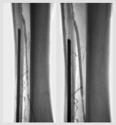
Angioplasty of a heavily calcified anterior tibial artery in a patient with critical limb ischemia. A 76-year-old diabetic man had an open hallux fracture with subsequent osteomyelitis and deep space infection of the foot. He had no significant stenosis of the aortoiliac or femoropopliteal segments but had severe infrapopliteal atherosclerosis, including complete occlusions of the peroneal and posterior tibial arteries. The anterior tibial artery had diffuse stenosis and a chronic total occlusion in the distal calf, which was treated with plain balloon angioplasty to 3 mm. His dorsalis pedis ankle-brachial index improved from 0.68 to 1.2 and his foot wound healed after debridement and toe amputation.
Table 1 Randomized controlled trials of endovascular therapy for infrapopliteal peripheral arterial occlusive disease -
Randon et al. [22] published a single-center randomized trial comparing PTA to PTA plus a single BMS in 38 limbs of 35 patients with CLI (Rutherford class 4–6) and a >70% stenosis of a tibial vessel. The length of the treated lesions ranged from <2 cm to >15 cm, of which 64% were occlusions and 36% were stenoses. The mean length of angioplasty was 3.9 cm (range, 2–10 cm) and the mean stent length was 2.15 cm (range, 1.2–4 cm). There was no significant difference in 12-month primary patency rates (66% PTA vs 56% PTA + BMS), secondary patency rates (79.5% vs 64%), limb salvage rates (90% vs 91.7%), or survival rates (69.3% vs 74.1%) [22].
-
In 2011, Rastan, et al. [23] published a randomized multicenter trial of BMS versus sirolimus DES in 161 patients with >70% stenosis of a tibial artery on duplex ultrasonography (DUS) or digital subtraction angiography (DSA) and target lesion length <4.5 cm. Of the study patients, 86 (53%) had intermittent claudication (Rutherford class 2 or 3) and 75 (47%) had CLI (Rutherford class 4 or 5). For the BMS vs DES groups, the mean lesion length (3.1 ± 0.9 cm vs 3.0 ± 0.8 cm, P = not significant), number of total occlusions (21.5% vs 23.2%, P = not significant), and number of stents used (overall 1.3 per patient) were similar. The primary outcome was binary restenosis rate at 12 months, defined as >50% in-stent stenosis on DSA or peak systolic velocity ratio >2.4 on DUS. Although the authors reported a significant difference in 12-month in-stent restenosis rates (44.4% BMS vs 19.4% DES, P = 0.004), only results for the 77.6% of patients completing the 12-month follow-up were reported, rather than the results of a right-censored Kaplan-Meier survival analysis. A Kaplan-Meier survival analysis for the composite end-point of “event-free survival” (freedom from death, minor or major amputation or target lesion revascularization) showed no difference between the groups [23].
-
There have been a number of series published on the technical feasibility and short-term results of a variety of additional endovascular methods, including laser atherectomy [24], mechanical atherectomy [25], cryoplasty [26, 27], drug-coated balloons (DCB) [28], and subintimal angioplasty from the antegrade and retrograde direction [29]. In general, the success rates in these reports have been highest for short lesions, but the series have been small with short follow-up periods and limited comparisons between techniques.
-
There has been one randomized controlled trial (RCT) comparing bypass surgery to angioplasty for the treatment of severe limb ischemia. The Bypass versus Angioplasty in Severe Ischemia of the Leg (BASIL) trial enrolled 452 patients with at least 2 weeks of ischemic rest or night pain and/or ischemic tissue loss. Study subjects at 27 centers in the United Kingdom were randomized to bypass surgery or balloon angioplasty with a median follow-up of 5 years (range 3 to >7 years). In the final intention-to-treat analysis of the trial, there was no difference in amputation free survival (AFS) or overall survival between treatment groups [30]. Although only one third of patients had an infrapopliteal revascularization, a number of results are worth considering: 1) For patients who survived at least 2 years after randomization, there was a significant improvement in overall survival and a trend towards improved AFS for patients undergoing bypass surgery [30]; 2) in a by-treatment-received analysis, AFS was significantly worse for patients undergoing bypass surgery after failed angioplasty [31•]; and 3) there was no difference in quality-of-life measures or overall cost between the two treatment groups [32].

Endovascular infrapopliteal revascularization
- Standard procedure :
-
Angioplasty is recommended for patients with CLI with a life expectancy <2 years or with no suitable bypass graft conduit [4•]. Stenting is recommended only for cases of severe recoil or dissection after angioplasty. There is insufficient evidence to recommend routine infrapopliteal stenting with either BMS or DES.
- Contraindications :
-
Intermittent claudication, unsuitable anatomy. Relative: renal insufficiency.
- Complications :
-
Arterial dissection, thrombosis, distal embolization, blue toe syndrome, hematoma from puncture site, contrast nephropathy, and renal failure.
- Special points :
-
Given the distance involved, an antegrade femoral puncture often provides superior pushability and wire control, whereas access from the contralateral femoral artery often requires long guide sheaths to establish a treatment platform. The use of 0.014-inch or 0.018-inch platforms may facilitate lesion crossing. Angioplasty is generally performed with 1:1 sized balloons (typically 2.5–4 mm) using low-pressure and long inflation times.
- Cost/cost-effectiveness :
-
The BASIL trial cost-analysis found that at 1 year after randomization, the costs for bypass surgery ($34,378 to $28,701 hospital stay and $5677 procedure costs) was significantly higher than for angioplasty ($25,909 to $22,605 hospital stay and $3305 procedure costs). However, angioplasty was associated with an increased need for subsequent interventions, thus by 3 years, there was no longer a significant difference in cost ($45,322 bypass surgery vs $39,801 angioplasty) [32].
Surgery
-
Lower extremity bypass surgery is considered the gold standard for treating infrapopliteal disease, especially among patients with CLI [33•].
-
Lower extremity bypass with autogenous vein offers the highest rate of successful revascularization and is the most durable procedure with proven long-term benefit. Based on data from the PREVENT III trial, single-segment vein conduit, vein diameter >3 mm, and shorter bypass graft length are factors associated with higher rates of vein graft patency [34]. The challenge of treating infrapopliteal PAD is often related to the long graft length required for a distal target if the proximal anastomosis is to the common femoral artery. However, graft inflow from the superficial femoral artery or popliteal artery to a tibial or pedal target does not affect long-term graft outcome [34, 35].
-
Prosthetic grafts of polyethylene terephthalate (Dacron©) or polytetrafluoroethylene (PTFE) have been shown to have inferior results compared to autogenous vein when used in the infrapopliteal position and are not recommended [36]. For this reason, use of arm vein or a composite vein graft is preferred to a prosthetic graft. Cryopreserved femoral or saphenous veins have also been used as bypass graft conduit, but graft outcomes from large series are not better than for prosthetic grafts [37].
-
Based on the available data from three large multi-center RCTs of lower extremity bypass (n = 838), recent Objective Performance Goals (OPG) were published for evaluating catheter-based treatment of CLI [33•]. Infrapopliteal bypass (n = 505) was defined as “high anatomic risk” with the following characteristics: 1) 30-day safety benchmarks: major adverse cardiovascular event (MACE) rate 7.3% (95% CI, 5.2%–10.0%), major adverse limb event (above ankle amputation or major bypass reintervention [MALE]) rate 6.1% (95% CI, 4.2%–8.6%), and amputation rate 2.2% (95% CI, 1.1%–3.9%); 2) 1-year efficacy benchmarks (as percent free of event at 1 year): amputation-free survival (AFS) 74.4% (95% CI, 70.6%–78.3%), any reintervention or above ankle amputation 58.3% (95% CI, 54.0%–63.0%), limb salvage 86.6% (95% CI, 83.6%–89.8%), and survival 85.9% (95% CI, 82.9%–89.0%).
Lower extremity bypass surgery
- Standard procedure :
-
Lower extremity bypass surgery with autogenous vein. The preferred conduit is ipsilateral saphenous vein of good quality and diameter >3 mm. The vein configuration (reversed, non-reversed, or in situ) and graft inflow site play little role in graft outcomes.
- Contraindications :
-
Lack of suitable vein, high operative risk, unsuitable anatomy (no distal target). Relative contraindication: life expectancy <2 years
- Complications :
-
Perioperative cardiovascular events, wound infection, long recovery time.
- Special points :
-
Most authors recommend re-establishing inline blood flow to the foot for patients with CLI, which may require technically demanding procedures such as pedal artery bypass for patients with infrapopliteal PAD.
- Cost/cost-effectiveness :
-
The BASIL trial cost-analysis found that at 1 year after randomization, the costs for bypass surgery ($34,378 to $28,701 hospital stay and $5677 procedure costs) was significantly higher than for angioplasty ($25,909 to $22,605 hospital stay and $3305 procedure costs). However, angioplasty was associated with an increased need for subsequent interventions, thus by 3 years, there was no longer a significant difference in cost ($45,322 bypass surgery vs $39,801 angioplasty) [32].
Assistive devices
-
For those patients with CLI who have no options for revascularization, major amputation is often required. However, there have been recent studies in the use of intermittent pneumatic compression (IPC) devices or spinal cord stimulation to reduce amputation rates. Chemical or surgical lumbar sympathectomy may relieve pain in this group of patients, but the efficacy of this treatment has not been studied. Although these assistive therapies may avoid amputation by arterial flow augmentation or reduced rest pain, they are not a substitute for revascularization and the risk of limb-loss remains high.
Intermittent pneumatic compression
- Usage :
-
IPC of the foot, ankle, and calf aids venous return and lowers venous pressure in the lower leg and foot. The resulting arteriovenous pressure gradient augments arterial flow and may improve collateral circulation. In a case series of 171 patients with CLI and non-reconstructible PAD, IPC was performed for 3 h twice daily for 3 months. At 12 months, there was an increase in toe pressure and popliteal blood flow and the limb salvage rate was 94% at 3.5 years [38]. In a non-randomized study comparing IPC and wound care (n = 24) to wound care alone (n = 24), IPC was used for 2 h three times daily for 18 months. During the 18-month study period, 42% of the IPC plus wound care group underwent a below-knee amputation compared to 83% of wound care-only group [39].
- Special points :
-
Therapy can be performed at home.
- Cost/cost-effectiveness :
-
The cost of IPC therapy was approximately $5350 per patient whereas the cost of a major amputation was approximately $39,994 per patient in the study by Sultan et al. [38]. Although IPC therapy appears to be a cost-effective solution compared to amputation, larger RCTs are needed to confirm this finding.
Spinal cord stimulation
- Usage :
-
An implanted subcutaneous pulse generator stimulates an electrode in the epidural space, leading to analgesia of the ischemic leg. Several studies have suggested that spinal cord stimulation leads to decreased pain, improved ulcer healing, and improved skin microcirculation as measured with transcutaneous oximetry (TcpO2) and laser Doppler perfusion. A meta-analysis by Ubbink et al. [40] of six controlled trials involving 444 patients also showed an 11% lower amputation rate and significantly lower analgesic use after 12 months compared to medical treatment alone.
- Special points :
-
An analysis of the published trials suggested that patients with a TcpO2 measurement between 10 and 30 mm Hg had the greatest benefit from spinal cord stimulation. Patients with a TcpO2 <10% usually required an amputation regardless of treatment [40].
- Cost/cost-effectiveness :
-
There are few data to support the cost-effectiveness of spinal cord stimulation. One study found no difference in limb salvage rates, whereas the cost of spinal cord stimulation was 28% higher than medical treatment alone [41].
Lumbar sympathectomy
- Usage :
-
Chemical or surgical ablation of the lumbar sympathetic chain can provide pain relief and an increase in blood flow in the skin, foot, and calf of patients with CLI and non-reconstructible PAD. The exact mechanism, however, is unclear and may be due to blocking both efferent vasoconstrictive sympathetic nerves and afferent cutaneous neuropeptide sensory nerves [42]. Although modern techniques have a vastly improved safety profile compared to traditional open surgical sympathectomy, there have been no contemporary studies examining the efficacy of the chemical or surgical ablation in palliation or limb salvage.
- Special points :
-
Chemical ablation with local anesthetic, 6% phenol, or 100% ethanol is typically performed under fluoroscopic or CT guidance. Surgical sympathectomy can be performed with open or laparoscopic techniques [43].
- Cost/cost-effectiveness :
-
Not studied.
Emerging therapies
-
Several trials of DES in the tibial arteries have been published or announced (Table 1). Most of these trials use antiproliferative agents such as sirolimus (and derivatives) or paclitaxel to inhibit restenosis. Although the allure of DES technology has been fueled by a reduction of in-stent restenosis rates in the coronary arteries, there are important differences between coronary and tibial arteries that may need to be addressed. Namely, the tibial vessels are anatomically different (muscular vs elastic arteries), frequently have long-segment, heavily-calcified occlusions, and are subject to mechanical strain transmitted by the movement of the lower leg. Alternative methods for delivering antiproliferative agents without a permanent stent such as drug-coated balloons (DCB) [28] and absorbable metal stents [44] have been demonstrated, but their efficacy has yet to be proven.
-
Gene therapy with growth factors has been explored as a way to augment angiogenesis and initiate vasculogenesis for patients with claudication or CLI. The challenge of gene delivery hampered development of widespread gene therapy, but the results of initial studies have been promising. Intramuscular injection of a hepatocyte growth factor (HGF) encoding plasmid was examined in a randomized trial of 27 CLI patients with tissue loss. After 6 months, patients treated with HGF plasmid had a significant improvement in TBI and pain, but there was no difference in complete ulcer healing, major amputation rate, or survival [45]. Vascular endothelial growth factor (VEGF) has been investigated in two separate trials. In an RCT in 105 patients with claudication, unilateral intramuscular injection with a VEGF-encoding adenovirus did not improve exercise performance or quality-of-life measures [46]. However, an RCT in 54 adults with CLI, a significant improvement in skin ulcers and pain was observed with intramuscular injection of a VEGF-encoding plasmid, but there was no difference in amputation rate [47]. Cell therapy for CLI using intramuscular injection of autologous bone marrow-derived stem cells into an ischemic limb showed promise in several initial series [48, 49]. Recently, the interim results of the multicenter, randomized, double-blinded, placebo-controlled RESTORE-CLI trial were published and demonstrated a significant increase in time to treatment failure and amputation-free survival with stem cell therapy [50].
References and Recommended Reading
Papers of particular interest, published recently, have been highlighted as: • Of importance
Norgren L, Hiatt W, Dormandy J, et al. Inter-society consensus for the management of peripheral arterial disease (TASC II). J Vasc Surg. 2007;45:S5–S67.
Gray BH, Grant AA, Kalbaugh CA, et al. The impact of isolated tibial disease on outcomes in the critical limb ischemic population. Ann Vasc Surg. 2010;24:349–59.
Hirsch AT, Haskal ZJ, Hertzer NR, et al. ACC/AHA 2005 practice guidelines for the management of patients with peripheral arterial disease (lower extremity, renal, mesenteric, and abdominal aortic): a collaborative report from the american association for vascular surgery/society for vascular surgery, society for cardiovascular angiography and interventions, society for vascular medicine and biology, society of interventional radiology, and the ACC/AHA task force on practice guidelines (writing committee to develop guidelines for the management of patients with peripheral arterial disease): endorsed by the american association of cardiovascular and pulmonary rehabilitation; national heart, lung, and blood institute; society for vascular nursing; TransAtlantic inter-society consensus; and vascular disease foundation. Circulation. 2006;113:e463–654.
Rooke TW, Hirsch AT, Misra S, et al. 2011 ACCF/AHA Focused Update of the Guideline for the Management of Patients With Peripheral Artery Disease (Updating the 2005 Guideline): A Report of the American College of Cardiology Foundation/American Heart Association Task Force on Practice Guidelines. Circulation 2011, 124:2020–45.
Hamburg NM, Balady GJ. Exercise rehabilitation in peripheral artery disease: functional impact and mechanisms of benefits. Circulation. 2011;123:87–97.
Grundy SM. Implications of recent clinical trials for the national cholesterol education program adult treatment panel III guidelines. Circulation. 2004;110:227–39.
Momsen AH, Jensen MB, Norager CB, et al. Drug therapy for improving walking distance in intermittent claudication: a systematic review and meta-analysis of robust randomised controlled studies. Eur J Vasc Endovasc Surg. 2009;38:463–74.
Lane DA, Lip GY. Treatment of hypertension in peripheral arterial disease. Cochrane database of systematic reviews 2009:CD003075.
Diehm C, Pittrow D, Lawall H. Effect of nebivolol vs. hydrochlorothiazide on the walking capacity in hypertensive patients with intermittent claudication. J Hypertens. 2011;29:1448–56.
Espinola-Klein C, Weisser G, Jagodzinski A, et al. beta-Blockers in patients with intermittent claudication and arterial hypertension: results from the nebivolol or metoprolol in arterial occlusive disease trial. Hypertension. 2011;58:148–54.
Mohler ER. Cholesterol reduction with atorvastatin improves walking distance in patients with peripheral arterial disease. Circulation. 2003;108:1481–6.
Ridker PM, Danielson E, Fonseca FA, et al. Rosuvastatin to prevent vascular events in men and women with elevated C-reactive protein. N Engl J Med. 2008;359:2195–207.
Goodney PP, Beck AW, Nagle J, et al. National trends in lower extremity bypass surgery, endovascular interventions, and major amputations. J Vasc Surg. 2009;50:54–60.
Sadek M, Ellozy SH, Turnbull IC, et al. Improved outcomes are associated with multilevel endovascular intervention involving the tibial vessels compared with isolated tibial intervention. J Vasc Surg. 2009;49:638–44.
Fernandez N, McEnaney R, Marone LK, et al. Predictors of failure and success of tibial interventions for critical limb ischemia. J Vasc Surg 2010:1–9.
Giles KA, Pomposelli FB, Hamdan AD, et al. Infrapopliteal angioplasty for critical limb ischemia: relation of TransAtlantic InterSociety consensus class to outcome in 176 limbs. J Vasc Surg. 2008;48:128–36.
Romiti M, Albers M, Brochado-Neto FC, et al. Meta-analysis of infrapopliteal angioplasty for chronic critical limb ischemia. J Vasc Surg. 2008;47:975–81.e1.
Conrad MF, Kang J, Cambria RP, et al. Infrapopliteal balloon angioplasty for the treatment of chronic occlusive disease. J Vasc Surg. 2009;50:799–805.e4.
Donas KP, Torsello G, Schwindt A, et al. Below knee bare nitinol stent placement in high-risk patients with critical limb ischemia is still durable after 24 months of follow-up. J Vasc Surg. 2010;52:356–61.
Siablis D, Karnabatidis D, Katsanos K, et al. Infrapopliteal application of paclitaxel-eluting stents for critical limb ischemia: midterm angiographic and clinical results. J Vasc Interv Radiol. 2007;18:1351–61.
Siablis D, Karnabatidis D, Katsanos K, et al. Infrapopliteal application of sirolimus-eluting versus bare metal stents for critical limb ischemia: analysis of long-term angiographic and clinical outcome. J Vasc Interv Radiol. 2009;20:1141–50.
Randon C, Jacobs B, De Ryck F, Vermassen F. Angioplasty or primary stenting for infrapopliteal lesions: results of a prospective randomized trial. Cardiovasc Intervent Radiol. 2010;33:260–9.
Rastan A, Tepe G, Krankenberg H, et al. Sirolimus-eluting stents vs. bare-metal stents for treatment of focal lesions in infrapopliteal arteries: a double-blind, multi-centre, randomized clinical trial. Eur Heart J. 2011;32:2274–81.
Stoner MC, deFreitas DJ, Phade SV, et al. Mid-term results with laser atherectomy in the treatment of infrainguinal occlusive disease. J Vasc Surg. 2007;46:289–95.
Zeller T, Sixt S, Schwarzwälder U, et al. Two-year results after directional atherectomy of infrapopliteal arteries with the SilverHawk device. J Endovasc Ther. 2007;14:232–40.
Bosiers M, Deloose K, Vermassen F, et al. The use of the cryoplasty technique in the treatment of infrapopliteal lesions for critical limb ischemia patients in a routine hospital setting: one-year outcome of the cryoplasty CLIMB registry. J Cardiovasc Surg (Torino). 2010;51:193–202.
Das TS, McNamara T, Gray B, et al. Primary cryoplasty therapy provides durable support for limb salvage in critical limb ischemia patients with infrapopliteal lesions: 12-month follow-up results from the BTK Chill Trial. J Endovasc Ther. 2009;16:II19–30.
Schmidt A, Piorkowski M, Werner M, et al. First experience with drug-eluting balloons in infrapopliteal arteries restenosis rate and clinical outcome. J Am Coll Cardiol. 2011;58:1105–9.
Yeh K-H, Tsai Y-J, Huang H-L, et al. Dual vascular access for critical limb ischemia: immediate and follow-up results. Catheter Cardiovasc Interv. 2011;77:296–302.
Bradbury AW, Adam DJ, Bell J, et al. Bypass versus Angioplasty in Severe Ischaemia of the Leg (BASIL) trial: an intention-to-treat analysis of amputation-free and overall survival in patients randomized to a bypass surgery-first or a balloon angioplasty-first revascularization strategy. J Vasc Surg. 2010;51:5S–17S.
Bradbury AW, Adam DJ, Bell J, et al. Bypass versus Angioplasty in Severe Ischaemia of the Leg (BASIL) trial: Analysis of amputation free and overall survival by treatment received. J Vasc Surg 2010, 51:18S–31S.
Forbes JF, Adam DJ, Bell J, et al. Bypass versus Angioplasty in Severe Ischaemia of the Leg (BASIL) trial: health-related quality of life outcomes, resource utilization, and cost-effectiveness analysis. J Vasc Surg. 2010;51:43S–51S.
Conte MS, Geraghty PJ, Bradbury AW, et al. Suggested objective performance goals and clinical trial design for evaluating catheter-based treatment of critical limb ischemia. J Vasc Surg 2009, 50:1462–73.e3.
Schanzer A, Hevelone N, Owens CD, et al. Technical factors affecting autogenous vein graft failure: observations from a large multicenter trial. J Vasc Surg. 2007;46:1180–90. discussion 90.
Reed AB, Conte MS, Belkin M, et al. Usefulness of autogenous bypass grafts originating distal to the groin. J Vasc Surg. 2002;35:48–54. discussion, -5.
Albers M, Battistella VM, Romiti M, et al. Meta-analysis of polytetrafluoroethylene bypass grafts to infrapopliteal arteries. J Vasc Surg. 2003;37:1263–9.
Randon C, Jacobs B, De Ryck F, et al. Fifteen years of infrapopliteal arterial reconstructions with cryopreserved venous allografts for limb salvage. J Vasc Surg. 2010;51:869–77.
Sultan S, Hamada N, Soylu E, et al. Sequential compression biomechanical device in patients with critical limb ischemia and nonreconstructible peripheral vascular disease. J Vasc Surg. 2011;54:440–7.
Kavros SJ, Delis KT, Turner NS, et al. Improving limb salvage in critical ischemia with intermittent pneumatic compression: a controlled study with 18-month follow-up. J Vasc Surg. 2008;47:543–9.
Ubbink DT, Vermeulen H. Spinal cord stimulation for critical leg ischemia: a review of effectiveness and optimal patient selection. J Pain Symptom Manage. 2006;31:S30–5.
Klomp HM, Steyerberg EW, van Urk H, Habbema JD. Spinal cord stimulation is not cost-effective for non-surgical management of critical limb ischaemia. Eur J Vasc Endovasc Surg. 2006;31:500–8.
Coventry BJ, Walsh JA. Cutaneous innervation in man before and after lumbar sympathectomy: evidence for interruption of both sensory and vasomotor nerve fibres. ANZ J Surg. 2003;73:14–8.
Nemes R, Surlin V, Chiutu L, et al. Retroperitoneoscopic lumbar sympathectomy: prospective study upon a series of 50 consecutive patients. Surg Endosc. 2011;25:3066–70.
Investigators O, Bot AI, Bosiers M. AMS INSIGHT—absorbable metal stent implantation for treatment of below-the-knee critical limb ischemia: 6-month analysis. Cardiovasc Intervent Radiol. 2008;32:424–35.
Powell RJ, Goodney P, Mendelsohn FO, et al. Safety and efficacy of patient specific intramuscular injection of HGF plasmid gene therapy on limb perfusion and wound healing in patients with ischemic lower extremity ulceration: results of the HGF-0205 trial. J Vasc Surg. 2010;52:1525–30.
Rajagopalan S, Mohler 3rd ER, Lederman RJ, et al. Regional angiogenesis with vascular endothelial growth factor in peripheral arterial disease: a phase II randomized, double-blind, controlled study of adenoviral delivery of vascular endothelial growth factor 121 in patients with disabling intermittent claudication. Circulation. 2003;108:1933–8.
Kusumanto YH, van Weel V, Mulder NH, et al. Treatment with intramuscular vascular endothelial growth factor gene compared with placebo for patients with diabetes mellitus and critical limb ischemia: a double-blind randomized trial. Hum Gen Ther. 2006;17:683–91.
Amann B, Luedemann C, Ratei R, Schmidt-Lucke JA. Autologous bone marrow cell transplantation increases leg perfusion and reduces amputations in patients with advanced critical limb ischemia due to peripheral artery disease. Cell Transplant. 2009;18:371–80.
Tateishi-Yuyama E, Matsubara H, Murohara T, et al. Therapeutic angiogenesis for patients with limb ischaemia by autologous transplantation of bone-marrow cells: a pilot study and a randomised controlled trial. Lancet. 2002;360:427–35.
Powell RJ, Comerota AJ, Berceli SA, et al. Interim analysis results from the RESTORE-CLI, a randomized, double-blind multicenter phase II trial comparing expanded autologous bone marrow-derived tissue repair cells and placebo in patients with critical limb ischemia. J Vasc Surg. 2011;54:1032–41.
ClinicalTrials.gov. Available at: http://clinicaltrials.gov/. Accessed August 2011.
Martens JM, Knippenberg B, Vos JA, et al. Update on PADI trial: percutaneous transluminal angioplasty and drug-eluting stents for infrapopliteal lesions in critical limb ischemia. J Vasc Surg. 2009;50:687–9.
Bosiers M, Deloose K, Callaert J, et al. Drug-eluting stents below the knee. J Cardiovasc Surg (Torino). 2011;52:231–4.
Zeller T, Schmitmeier S, Tepe G, Rastan A. Drug-coated balloons in the lower limb. J Cardiovasc Surg (Torino). 2011;52:235–43.
Disclosure
No conflicts of interest relevant to this article were reported.
Author information
Authors and Affiliations
Corresponding author
Rights and permissions
About this article
Cite this article
Gasper, W.J., Runge, S.J. & Owens, C.D. Management of Infrapopliteal Peripheral Arterial Occlusive Disease. Curr Treat Options Cardio Med 14, 136–148 (2012). https://doi.org/10.1007/s11936-012-0164-y
Published:
Issue Date:
DOI: https://doi.org/10.1007/s11936-012-0164-y