Abstract
Trichosporon asahii and Rhodotorula mucilaginosa are important fungal species causing disseminated disease in immunocompromised patients. Onychomycosis prevalence rate ranges from 2 to 30%, which were 50% of nail diseases and 30% of superficial mycosis, respectively. Although important, little is known about the co-habitation of T. asahii and R. mucilaginosa in the causation of onychomycosis. Here, we present the co-habitation of T. asahii and R. mucilaginosa as causative agents of onychomycosis in a healthy 41-year-old male in China. Direct microscopic examination, fungal culture and MALDI-TOF MS were employed in isolated pathogens; hence, antifungal susceptibility test was evaluated. T. asahii was sensitive to posaconazole, voriconazole and itraconazole, whereas R. mucilaginosa was sensitive to both 5-flucytosine and amphotericin B. This report highlights the co-habitation of T. asahii and R. mucilaginosa in the causation of onychomycosis and to raise the awareness of this infection among dermatologists.
Avoid common mistakes on your manuscript.
Introduction
Onychomycosis is a common fungal infection in dermatologic clinics, caused by dermatophytes, Candida spp., or a non-dermatophyte mold [1]. Humidity, occlusive footwear, repeated nail trauma, genetic predisposition and concurrent diseases such as diabetes are risk factors for onychomycosis [2]. Worldwide, the prevalence ranges from 2 to 30%. It accounts for 30% of superficial mycoses and 50% of nail diseases and is common among men of all ages and in women above 60 years of age [3]. Most cases of onychomycosis are caused by dermatophytes that belong to three genera Trichophyton, Microsporum and Epidermophyton. Distal lateral subungual onychomycosis and total dystrophy onychomycosis are the prevalent forms of onychomycosis in China, followed by superficial white onychomycosis and proximal subungual onychomycosis [4]. Trichosporon spp. and Rhodotorula spp. are Basidiomycetous yeasts that are ubiquitously found in soil, decomposing wood and air [5, 6]. Trichosporon sp. is an opportunistic pathogen that resides commensally on the skin of their hosts. Alteration of the surrounding environment of this fungus may activate its pathogenic potential [7]. Nonetheless, Rhodotorula sp. is an uncommon agent in the etiology of onychomycosis with only few cases reported [8,9,10]. Onychomycosis is frequently diagnosed using direct microscopic examination with 10% potassium hydroxide and fungal culture [1]. In this study, we report identification and in vitro antifungal susceptibility test of two different species of Basidiomycetous in a toe nail of 41-year-old Chinese patient with onychomycosis by direct microscopic examination of toe nail fragments sample, fungal culture on Sabouraud dextrose agar and MALDI-TOF MS identification system.
Case Report
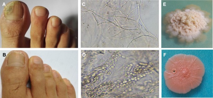
A 41-year-old Chinese male patient was attended at the dermatology department of the First Affiliated Hospital of the Chongqing Medical University in China with a history of 3-year deformity and thickening of the right toe nail. Prior to his presentation, he had not been on any medication and upon direct questioning; he had no known history of chronic disease, malignancy, or familial genetic disorder except poor hygiene management, humidity and occlusive footwear. On examination, he presented with lateral discoloration of toe nail and provisional diagnosis of lateral subungual onychomycosis was made (Fig. 1a). In contrast, nails of his fingers and left feet were normal. Preliminary laboratory reports of diabetes were negative. Hence, Fig. 1b shows a picture of recovered toe nail after treated with itraconazole (100-mg capsules) orally twice a day and naftifine hydrochloride (100 mg/g-cream) applied twice a day for 7 months.
a Picture of a 41-year-old male’s right foot showing lateral discoloration of toe nail, and provisional diagnosis of lateral subungual onychomycosis was made. b Picture of recovered toe nail showing the nail plate that grew out completely. c Observation of direct microscopic examination of toe nail fragments sample previously treated with 10% potassium hydroxide under compound microscope (Olympus) with 100 magnification yielding wide and true mycelia of T. asahii. d Spores of R. mucilaginosa.e Colony morphology of T. asahii.f Colony morphology of R. mucilaginosa after 15 days on SDA, cultivated at 25 °C
Materials and Methods
Three nail fragment samples were collected with sterile scalpel for direct microscopic examination and mycology laboratory examination immediately [11]. Direct microscopic examination of samples previously treated with 10% potassium hydroxide (Guanghua Sci-Tech Co. Ltd, China) yielded wide and septate hyphae and round to oval single-budding blastoconidia (Fig. 1c, d). Samples cultured on Sabouraud dextrose agar (SDA; Oxoid Ltd, England) were supplemented with 1% ampicillin (Genview, China) and incubated for 15 days at 25 °C. Mixed growth was presented on the culture medium after 15 days which showed an irregular folded or verrucosed whitish-creamy colonies about 80 mm (Fig. 1e) and a salmon-orange-pink, moist and smooth surface about 90 mm after 15 days (Fig. 1f). VITEK® MSMALDI-TOF mass spectrometry system (bioMérieux) was used for identification of the fungi and the value expressed as logscore (LS) range between 1.7 and ≥ 2.0. Purity plating was done by touching the surface of a single colony of each growth and sub-culturing on SDA supplemented with 1% ampicillin. The morphologies of fungi were characterized using a compound microscope (Olympus) with 100 magnification.
In vitro antifungal susceptibility test was performed against isolates from clinical sample with pure powders provided by manufacturers using standard broth micro-dilution method according to the Clinical and Laboratory Standards Institute (CLSI) guideline M27-A2 [12]. The antifungal agents, amphotericin B, caspofungin, micafungin, anidulafungin, 5-flucytosine, voriconazole, fluconazole, itraconazole and posaconazole, were all purchased from Sigma-Aldrich (St. Louis, MO, USA). The stock solutions of itraconazole (100 mg/mL), voriconazole (100 mg/mL) and amphotericin B (100 mg/mL) were freshly prepared in dimethyl sulfoxide (DMSO, Sigma). Fluconazole (100 mg/mL), 5-flucytosine (100 mg/mL), caspofungin (100 mg/mL), micafungin (100 mg/mL) and anidulafungin (100 mg/mL) were dissolved in sterile distilled water. Candida parapsilosis ATCC 22019 and C. krusei ATCC 6258 were used as the quality control strains. Final drug dilutions were prepared in standard RPMI-1640 medium (Oxoid Ltd., England).
Results and Discussion
Trichosporon spp. and Rhodotorula spp. are Basidiomycetous yeasts that are particularly found in deep liquid substrates or moist and uneven surfaces that are rich in simple soluble nutrients such as sugars and amino acids and establish their habitat in water bodies and animals [13, 14]. Observation of direct microscopic examination of toe nail fragments sample under the compound microscope (Olympus) with 100 magnification revealed the presence of definite regular septate hyaline hyphae of Trichsoporon spp. (Figure 1c) and small round to oval single-budding blastoconidia of Rhodotorula spp. from the same nail fragment repeatedly (Fig. 1d). The structure of hyphae observed under microscope pointed to the genus Trichosporon spp. [14] which is parallel to a study conducted by Mariné et al. [6].
Some criteria were evaluated to determine whether these two isolates were causative agents of onychomycosis not colonizer such as no dermatophyte or other known bacteria were presented, the definite structure presented repeatedly on the same nail fragments sample, well-taken nail samples, and the growth of colonies on culture were associated with the direct microscopy examination. The morphology of colonies growth on SDA was evaluated by microscopy examination, and the result showed the same structures as on the direct microscopy examination of nail fragments sample after several rounds of pure isolation. The species of isolates were identified as T. asahii and R. mucilaginosa by using MALDI-TOF MS system.
In vitro antifungal susceptibility test and minimum inhibitory concentration (MIC50) results showed T. asahii was sensitive to posaconazole (MIC ≤ 0.5 µg/mL), voriconazole (MIC ≤ 0.25 µg/mL) and itraconazole (MIC > 0.5 µg/mL) but less sensitive to amphotericin B (4 µg/mL),while R. mucilaginosa was sensitive to 5-flucytosine (MIC ≤ 0.06 µg/mL) and amphotericin B (1.0 µg/mL). Combining our results and other studies published before, fluconazole, itraconazole and voriconazole appeared as effective antifungal drugs against the majority of clinical isolates of Rhodotorula sp. in vitro, but amphotericin B exhibits good activity and is a reasonable alternative for empirical treatment. Moreover, since azoles and allylamines prove effective for onychomycosis infection [15,16,17], the patient is treated with itraconazole (100-mg capsules) orally twice a day and naftifine hydrochloride (100 mg/g-cream) applied twice a day. Clinical cure criteria showed a completely normal-appearing toe nail at study endpoint as shown in Fig. 1b. In conclusion, our report revealed the presence of T. asahii and R. mucilaginosa in the infected toe nail onychomycosis. Further surveillance and investigation into the mechanism of co-infection of fungal species is recommended.
References
Yue X, Wang A. The role of scanning electron microscopy in the direct diagnosis of onychomycosis. Scanning. 2018. https://doi.org/10.1155/2018/1581495.
Oliveira CD, Rodrigues F, Gonçalves S, et al. The cell biology of the Trichosporon-host interaction. Front Cell Infect Microbiol. 2017. https://doi.org/10.3389/fcimb.2017.00118.
Velasquez-agudelo V, Cardona-arias JA. Meta-analysis of the utility of culture, biopsy, and direct KOH examination for the diagnosis of onychomycosis. BMC Infect Dis. 2017. https://doi.org/10.1186/s12879-017-2258-3.
Wang AP, Yu J, Wan Z, Li FQ, et al. Multi-center epidemiological survey of pathogenic fungi of onychomycosis in China. Chin J Mycol. 2015. https://doi.org/10.1007/s11684-017-0601-0(in Chinese).
Colombo AL, Padovan ACB, Chaves GM. Current knowledge of Trichosporon spp. and trichosporonosis. Clin Microbiol Rev. 2011;24:682–700.
Mariné M, Brown NA, Riaño-Pachón DM, et al. On and under the skin: emerging basidiomycetous yeast infections caused by Trichosporon species. PLoS Pathog. 2015. https://doi.org/10.1371/journal.ppat.1004982.
Zhang E, Sugita T, Tsuboi R, et al. The opportunistic yeast pathogen Trichosporon asahii colonizes the skin of healthy individuals: analysis of 380 healthy individuals by age and gender using a nested polymerase chain reaction assay. Microbiol Immunol. 2011. https://doi.org/10.1371/journal.ppat.1004982.
Uludag Altun H, Meral T, Turk Aribas E, Gorpelioglu C, et al. A case of onychomycosis caused by Rhodotorula glutinis. Case Rep Dermatol Med. 2014. https://doi.org/10.1155/2014/563261.
da Cunha MML, Dos Santos LPB, Dornelas Ribeiro M, et al. Identification, antifungal susceptibility and scanning electron microscopy of a keratinolytic strain of Rhodotorula mucilaginosa: a primary causative agent of onychomycosis. FEMS Immunol Med Microbiol. 2009. https://doi.org/10.1111/j.1574-695X.2009.00534.x.
Zhou J, Chen M, Chen H, Pan W, et al. Rhodotorula minuta as onychomycosis agent in a Chinese patient: first report and literature review. Mycoses. 2014. https://doi.org/10.1111/myc.12143.
Azambuja CVDA, Pimmel LA, Klafke GB, et al. Onychomycosis: clinical, mycological and in vitro susceptibility testing of isolates of Trichophyton rubrum. An Bras Dermatol. 2014. https://doi.org/10.1590/abd1806-4841.20142630.
Canton E, Pemán J, Espinel-Ingroff A, et al. Comparison of disc diffusion assay with the CLSI reference method (M27-A2) for testing in vitro posaconazole activity against common and uncommon yeasts. J Antimicrob Chem. 2008. https://doi.org/10.1093/jac/dkm442.
Choudhary DK, Johri BN. Basidiomycetous yeasts: current status. Yeast Biotechnol Divers Appl. 2009. https://doi.org/10.1007/978-1-4020-8292-4-2.
Biegańska MJ, Rzewuska M, Dąbrowska I, Malewska-Biel B, et al. Mixed infection of respiratory tract in a dog caused by Rhodotorula mucilaginosa and Trichosporon jirovecii: a case report. Mycopathologia. 2018. https://doi.org/10.1007/s11046-017-0227-4.
Dyanne P, Westerberg DO, Michael J, Yoyack DO. Onychomycosis: current trends in diagnosis and treatment. Am Fam Physician. 2013;88:762–70.
Gupta AK, Ryder JE, Johson AM. Cumulative meta-analysis of systemic antifungal agents for the treatment of onychomycosis. Br J Dermatol. 2004. https://doi.org/10.1046/j.1365-2133.2003.05728.
Taj-Aldeen SJ, Al-Ansari HI, Boekhout T, Theelen B. Co-isolation of Trichosporon inkin and Candida parapsilosis from a scalp white piedra case. Med Mycol. 2004. https://doi.org/10.1080/1369378032000-4-453.
Acknowledgements
We are thankful for the funds provided by Basic Medical College of Chongqing Medical University (Grant No. 4101070003] and Chongqing Research Program of Basic Research and Frontier Technology (No. cstc2015jcyjA10006). We also thank the patient and Professor Xia Yun from Clinical Department, First Affiliated Hospital of Chongqing Medical University, for their technical support of fungi identification.
Author information
Authors and Affiliations
Corresponding author
Ethics declarations
Conflict of interest
All authors declare that they have no conflict of interest.
Additional information
Publisher's Note
Springer Nature remains neutral with regard to jurisdictional claims in published maps and institutional affiliations.
Handling Editor: Cunwei Cao.
Rights and permissions
About this article
Cite this article
Idris, N.F.B., Huang, G., Jia, Q. et al. Mixed Infection of Toe Nail Caused by Trichosporon asahii and Rhodotorula mucilaginosa. Mycopathologia 185, 373–376 (2020). https://doi.org/10.1007/s11046-019-00406-y
Received:
Accepted:
Published:
Issue Date:
DOI: https://doi.org/10.1007/s11046-019-00406-y


