
Abstract
Previously, we produced two groups of gnotobiotic mice, GB-3 and GB-4, which showed different responses to Escherichia coli O157:H7 challenge. E. coli O157:H7 was eliminated from GB-3, whereas GB-4 mice became carriers. It has been reported that the lag time of E. coli O157:H7 growth in 50% GB-3 caecal suspension was extended when compared to GB-4 caecal suspension. In this study, competition for nutrients between intestinal microbiota of GB-3 and GB-4 mice and E. coli O157:H7 was examined. Amino acid concentrations in the caecal contents of GB-3 and GB-4 differed, especially the concentration of proline. The supplementation of proline into GB-3 caecal suspension decreased the lag time of E. coli O157:H7 growth in vitro. When E. coli O157:H7 was cultured with each of the strains used to produce GB-3 mice in vitro, 2 strains of E. coli (proline consumers) out of 5 enterobacteriaceae strains strongly suppressed E. coli O157:H7 growth and the suppression was attenuated by the addition of proline into the medium. These results indicate that competition for proline with indigenous E. coli affected the growth of E. coli O157:H7 in vivo and may contribute to E. coli O157:H7 elimination from the intestine.
Introduction
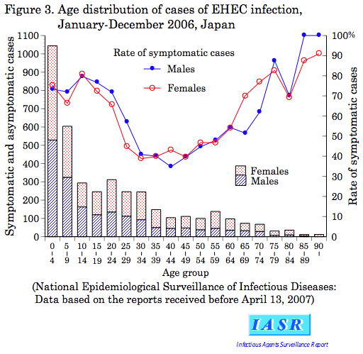
Shiga toxin-producing Escherichia coli such as E. coli O157:H7 are recognized as food-borne pathogens of great concern. The susceptibility to infection is higher in infants, children, and the elderly than in healthy adults (http://idsc.nih.go.jp/iasr/28/327/graph/f3273.gif). There have been many reports showing that gastrointestinal microbiota have antagonistic activity against bacterial infections (Carman et al. 2004; Donskey et al. 2001; Que et al. 1986; Silva et al. 2001). This means that intestinal microbiota play an essential role in protecting hosts from enteric infection. The composition of the intestinal microbiota of high risk groups such as infants and elderly people is different from that of adults and not stable (Benno et al. 1984, 1989; Gavini et al. 2001; Matsuki et al. 1999; Mitsuoka 2000). Therefore, it is likely that intestinal microbiota are involved in susceptibility to E. coli O157:H7 infection.
In our previous study (Momose et al. 2005), we inoculated 0.5 ml of 10−3 human infant faecal dilutions into germfree (GF) mice and produced baby-flora-associated (BFA) mice. Two groups of BFA mice, BFA-3 and BFA-4, were selected from the viewpoint of their different responses against E. coli O157:H7 challenge. Orally administered E. coli O157:H7 was eliminated from BFA-3 mice, whereas BFA-4 mice became carriers. The composition and species of BFA-3 faecal bacteria were similar to those often observed in healthy infants. In contrast, the microbiota of BFA-4 were not typical of healthy infants. We produced various types of gnotobiotic (GB) mice associated with intestinal bacteria isolated from BFA-3 and BFA-4. Among these GB mice, GB-3 and GB-4 were shown to most closely reflect the original characteristics of BFA-3 and BFA-4, respectively, with regard to their capacity for E. coli O157:H7 elimination.
We already reported that one of the mechanisms of E. coli O157:H7 elimination from the GB-3 intestine was a reduction of E. coli O157:H7 motility due to exposure to a mixture of acetate plus lactate under anaerobic conditions (Momose et al. 2008). We also proposed that in addition to these organic acids, other mechanisms involving inhibitory substances or nutritional competition that might affect E. coli O157:H7 growth itself may exist.
In this study, we focused on the competition for nutrients between intestinal microbiota of GB-3 and GB-4 mice and E. coli O157:H7, and also on inhibitors.
Materials and methods
Animals
GF BALB/c mice (female, 8–14 weeks old), GB-3 and GB-4 mouse groups (Momose et al. 2005) were used in this study. The mice were given a pelletted commercial diet (CMF, Oriental Yeast Co. Ltd., Japan) sterilized by γ-irradiation at 50 kGy and autoclaved water ad libitum. They were housed in a room on a 12:12 light cycle at 24 ± 1°C with a relative humidity of 55 ± 5%. All experiments were performed in accordance with the guidelines for care and use of laboratory animals of The University of Tokyo.
Bacteria
E. coli O157:H7 strain 44Rf (Stx 2) is a rifampicin-resistant mutant originally derived from bovine faeces. Other strains used in this study are listed in Table 1. They were isolated from faeces of BFA-3 and BFA-4 mice and identified in our previous study (Momose et al. 2005).
Preparation of culture supernatants of BFA isolates
Bacterial strains isolated from BFA-3 and BFA-4 were used. Each isolate was inoculated into 50% GF caecal suspension and incubated in an anaerobic chamber (N2: 85%, CO2: 5%, H2: 10%) for 1 or 2 days. A 50% GF caecal suspension was prepared by suspending GF mouse caecal contents in equal volume (w/v) of distilled water and supplemented with 0.84 g Na2CO3 and 0.5 g L-cysteine·HCl·H2O/l (pH 7.0). The suspension was kept in an anaerobic chamber for at least 2 days before use to remove dissolved oxygen. After incubation, the culture supernatants were obtained by centrifugation (3,000 × g, 10 min) and filtration (pore size: 0.22 μm) and used for the diffusion and overlay assays.
Diffusion assay
EG agar (Nissui, Japan) containing 106.0 colony forming units (CFU)/ml of E. coli O157:H7 was used for the diffusion assay. The agar plates were bored using a cork borer and the caecal contents of GB-3 and GB-4 mice and the culture supernatant of each BFA isolate were inoculated into the hole. The plates were incubated under anaerobic conditions using mixed gas (N2: 85%, CO2: 5%, H2: 10%) at 4°C for 3 h to diffuse the tested samples, and then at 37°C overnight. Clear zones around the holes were observed after incubation.
Overlay assay
The overlay assay was carried out according to the method of Pugsley and Oudega (1987) with some modifications. The caecal contents of GB-3 and GB-4 mice were inoculated onto EG agar without E. coli O157:H7. The BFA isolates were also inoculated. The plates were incubated under anaerobic conditions using mixed gas (N2: 85%, CO2: 5%, H2: 10%) overnight. A piece of filter paper was introduced into the lid and impregnated with 0.5 ml of chloroform. The plates were kept closed for 30 min. Soft agar seeded with 106.0 CFU/ml E. coli O157:H7 was overlaid and plates were re-incubated under anaerobic conditions using mixed gas. Clear zones around the colonies were observed.
Detection of amino acids
The caecal contents of GF, GB-3, and GB-4 mice were collected and 50% caecal suspensions were prepared in distilled water. These suspensions were mixed with an equal volume of 5% trichloroacetic acid and centrifuged (3,000 × g, 10 min). The supernatants were filtered (pore size: 0.45 μm) and used for the detection of amino acids. These prepared samples were sent to Hitachi Science Systems and amino acids were detected by HPLC (Hitachi: L-8800A Amino Acid Analyzer).
Proline utilization assay
Peptone water (1%, w/v) supplemented with 0.84 g of Na2CO3 and 0.5 g of L-cysteine·HCl·H2O/l (pH 7.0) was used. l-proline (Wako, Japan) was added to the medium after filtration (pore size: 0.22 μm) at a final concentration of 1.5 mM. E. coli O157:H7 and each enterobacteriaceae strain isolated from BFA-3 and BFA-4 was inoculated into the medium and incubated in an anaerobic chamber for 2 days. The culture supernatants were used for the proline utilization assay. The measurement of proline was carried out according to the method of Grainger and Aitken (2004) with some modifications. Briefly, 0.6 ml of each supernatant was dried overnight at 75°C and dissolved in 0.6 ml of 0.5 M sodium citrate buffer (pH 4.1). A 588 μl of each solution and 12 μl of 10% (w/v) solution of isatin (Wako, Japan) in dimethyl sulfoxide was mixed and incubated at 95°C for 6 h. After incubation, 200 μl of dimethyl sulfoxide was added and further incubated at room temperature for 15 min. The absorbance at 595 nm was measured.
Culture conditions for studying nutritional competition
The media used to examine competition for nutrients were 50% caecal suspensions of either GF or GB mice prepared in distilled water. Each caecal suspension was supplemented with 0.84 g of Na2CO3 and 0.5 g of l-cysteine·HCl·H2O/l, adjusted to pH 7.0.
E. coli O157:H7 was inoculated into GB-3 and GB-4 caecal suspensions at the initial concentration of 104.0 CFU/ml and incubated in an anaerobic chamber. l-proline was added to the GB-3 caecal suspension at the beginning of incubation. Aliquots (50 μl) of the culture were taken at 0, 1, 3, 5, 7, and 24 h after inoculation and the number of E. coli O157:H7 was enumerated on Trypticase Soy Agar (Becton Dickinson) and Sorbitol MacConkey Agar (Oxoid) plates.
Screening of enterobacteriaceae strains that compete for proline with E. coli O157:H7
We incubated E. coli O157:H7 in the GF caecal suspensions pre-cultured with each BFA enterobacteriaceae isolate, and the growth kinetics of E. coli O157:H7 were examined. Each enterobacteriaceae strain was inoculated at the initial concentration of 104.0 CFU/ml and incubated in an anaerobic chamber. E. coli O157:H7 was inoculated 5 h after the inoculation of each isolate at the concentration of 105.5–5.7 CFU/ml. For the proline competition test, l-proline was added to the medium after 5 h of incubation; application occurred simultaneously with the inoculation of E. coli O157:H7. A 50 μl aliquot was taken after 5, 7, 9, and 24 h of incubation, and the number of E. coli O157:H7 was enumerated on Sorbitol MacConkey agar plates with 50 μg/ml rifampicin (Sigma, USA). The number of each BFA enterobacteriaceae isolate was enumerated on Sorbitol MacConkey agar plates without rifampicin.
Statistical analysis
Data were analysed by Student’s t-test. A significant difference was defined as a P < 0.05.
Results
Inhibitory substances against E. coli O157:H7
We screened BFA isolates to select strains possessing antagonistic activity against E. coli O157:H7 by means of diffusion and overlay assays. We could not find any clear zones around the holes or colonies by either method.
Amino acids
Amino acid concentrations in the caecal suspensions differed between GB-3 and GB-4 (Table 2). Among the 17 amino acids measured, the concentration of proline was lower in the GB-3 caecal suspension when compared to GB-4.
E. coli O157:H7 growth in caecal suspensions and the effect of proline supplementation
The lag time of E. coli O157:H7 growth was extended in the GB-3 caecal suspension. When E. coli O157:H7 was incubated anaerobically in GB-3 caecal suspension supplemented with proline adjusted to the same concentration as present in GB-4 (1.5 mM), the lag time of E. coli O157:H7 growth decreased. The number of E. coli O157:H7 did not significantly vary from GF and GB-4 caecal suspensions at any time point (Fig. 1).

The growth kinetics of E. coli O157:H7 in the caecal suspensions of GF, GB-3 and GB-4 mice and the effect of supplementation with proline. Data are the means and standard deviations of duplicated experiments (n = 2 for each replicate and n = 4 in total). Closed circles, control (GF caecal suspension); open circles, GB-3 caecal suspension; closed triangles, GB-4 caecal suspension; open triangles, GB-3 caecal suspension supplemented with proline. Significant differences were observed compared to GB-4 mouse groups (** P < 0.01; *** P < 0.001)
Proline utilization
The ability of enterobacteriaceae strains of BFA isolates and E. coli O157:H7 to utilize proline was examined (Fig. 2). Among five strains from BFA-3 and six strains from BFA-4, two strains of E. coli from BFA-3 reduced the proline concentration in the test medium lower than that observed for the other strains. Although E. coli O157:H7 also reduced the proline concentration, the residual concentration was still higher than that observed for the two E. coli strains.

Proline utilization of enterobacteriaceae strains isolated from BFA-3 and BFA-4 mice and E. coli O157:H7. Data are the means and standard deviations of duplicated experiments (n = 2 for each replicate and n = 4 in total). Significant differences were observed compared to control cultures without bacterial inoculation (* P < 0.05; *** P < 0.001)
E. coli O157:H7 growth suppression by enterobacteriaceae and the effect of proline supplementation
When E. coli O157:H7 was incubated in GF caecal suspensions pre-cultured with each enterobacteriaceae isolate from BFA-3 and BFA-4, suppressive effects on E. coli O157:H7 growth varied with the isolate, whereas all the enterobacteriaceae isolates from both BFA-3 and BFA-4 reached to 108.8–9.0 CFU/ml after 24 h of incubation. When comparing numbers of E. coli O157:H7 at the end of incubation, four out of five enterobacteriaceae strains from BFA-3 suppressed E. coli O157:H7 growth and E. coli 1 exhibited the strongest suppressive effect, whereas only one out of six strains from BFA-4 showed growth suppression against E. coli O157:H7. Among BFA-3 enterobacteriaceae strains, the number of E. coli O157:H7 at the end of incubation was suppressed to 106.5 CFU/ml by E. coli 1, 107.3 CFU/ml by E. coli 1′, 107.9 CFU/ml by Citrobacter freundii 1, 108.7 CFU/ml by Pantoea sp., whereas the number in the cultures of E. coli O157:H7 alone achieved to 108.9 CFU/ml. By contrast, among BFA-4 enterobacteriaceae strains, only Klebsiella oxytoca 4 suppressed the number of E. coli O157:H7 to 108.6 CFU/ml.
The E. coli 1 strain was further tested to determine whether the growth kinetics of E. coli O157:H7 change after the addition of proline to the GF caecal suspension, thereby examining whether or not E. coli 1 and E. coli O157:H7 compete for proline as a nutritional source. In results, the numbers of E. coli O157:H7 after 9 and 24 h of incubation were significantly higher when compared to cultures without proline. The number of E. coli O157:H7 cultured without proline was 105.9 and 106.5 CFU/ml after 9 and 24 h of incubation, respectively. The number of E. coli O157:H7 cultured with proline was 106.2 and 107.6 CFU/ml, respectively.
Discussion
It has been known for several decades that intestinal microbiota play a major role in colonization resistance, and several mechanisms have been proposed including the production of inhibitory substances (Que et al. 1986; Silva et al. 2001), competition for limiting nutrients (Guiot 1982; Wilson and Perini 1988; Ushijima and Seto 1991; Yamamoto-Osaki et al. 1994), and competition for available adhesion sites (Snoeyenbos 1979).
We reported in a previous paper that when E. coli O157:H7 was incubated anaerobically in a 50% suspension of GB-3 or GB-4 caecal contents, the lag time of E. coli O157:H7 growth was significantly extended in the GB-3 caecal suspension when compared to GB-4 (Momose et al. 2008). Because the intestinal contents are continuously flowing downstream, an extended lag time for intestinal growth could disadvantage the colonization of exogenous bacteria. Besides organic acids, bile acids (Itoh et al. 1999) and hydrogen sulfide (Freter et al. 1983) have been proposed to be inhibitory factors that suppress the growth of exogenous bacteria in the intestine. Inhibitory activity of colicins against E. coli O157:H7 has also been demonstrated (Murinda et al. 1996) and colicinogenic bacteria that inhibit E. coli O157 have been isolated from human stool specimens (Toshima et al. 2007). To elucidate the mechanisms that generate extended lag time, we first analysed bacterial isolates from BFA-3 and BFA-4 mice to determine whether they could produce inhibitory substances against E. coli O157:H7. However, we could not detect inhibitory substances or their producers in our study.
We examined whether amino acid concentrations differed between caecal suspensions of GB-3 and GB-4 because there have been some reports demonstrating that indigenous microbiota compete for amino acids with exogenous pathogens and contribute to colonization resistance (Ushijima and Seto 1991; Yamamoto-Osaki et al. 1994). Among the 17 amino acids measured, the proline concentration in the caecal suspension was the most different between GB-3 and GB-4. The proline concentration was lower in the GB-3 caecal suspension than in the GB-4 suspension. When proline was added to the GB-3 caecal suspension to an equivalent concentration as that found in the GB-4 suspension, the initial growth of E. coli O157:H7 was accelerated; the lag time significantly decreased until it was not different from the lag times observed for GF and GB-4. These results suggest that proline is a nutritional source competed for by indigenous GB-3 microbiota and E. coli O157:H7, thereby resulting in extension of the lag time of E. coli O157:H7 growth in the GB-3 caecum. Reinders et al. (2001) reported that E. coli O157:H7 ATCC 43895 survived better in a model apple juice medium with proline than without it. However, the effect was observed only for log-phase-grown cells at low pH. In our previous study (Momose et al. 2008), we showed that caecal pH of gnotobiotic mice was neutral, and under neutral pH, organic acids did not inhibit E. coli O157:H7 growth itself, but its motility. Therefore, proline is thought not to be relevant to the survival of E. coli O157:H7 in the intestine.
Because E. coli O157:H7 is a member of the enterobacteriaceae and thought to have similar biological properties, we hypothesized that competition for nutritional sources might particularly occur between indigenous enterobacteriaceae and E. coli O157:H7.With the aim of finding strains that compete for proline with E. coli O157:H7, we examined the proline utilization of enterobacteriaceae strains of BFA isolates and E. coli O157:H7. E. coli O157:H7 was also cultured in GF caecal suspension pre-cultured with each enterobacteriaceae strain, and the effect on E. coli O157:H7 growth was examined. Two E. coli strains isolated from BFA-3, but no strain isolated from BFA-4, consumed more proline than E. coli O157:H7. When E. coli O157:H7 was cultured with each enterobacteriaceae strain from BFA-3, E. coli 1, one of the E. coli strains that consumed proline at a high level, exerted the strongest suppressive effect against E. coli O157:H7 growth. This growth suppression was attenuated by the addition of proline to the medium. These results suggest that proline competition with indigenous E. coli contributes to extending the lag time of E. coli O157:H7 growth in the caecum.
To our knowledge, the data have not yet been reported on the difference in proline requirements, metabolism and biosynthesis between E. coli O157:H7 and other E. coli strains. As the additional examination, we used several other E. coli O157:H7 strains isolated from patients in outbreak cases and carried out the proline utilization and competition assays. In results, two out of six strains of E. coli O157:H7 reduced the proline concentration in the test medium. The growth of these two strains was suppressed in the medium pre-cultured with E. coli 1, and the growth suppression was attenuated by the supplementation with proline to the medium (data not shown), as well as the case of strain 44Rf. These data indicated that competition for proline contributes to the colonization resistance against E. coli O157:H7 but it is not the case for all E. coli O157:H7 strains. There could be several competitive nutrients and in this study, we defined one of them. The other competitive nutrients should be demonstrated in the further studies.
In conclusion, extension of the lag time of growth by competition for at least one nutritional source with indigenous microbiota contributed to the elimination of E. coli O157:H7 from the intestine. We already demonstrated that enterobacteriaceae are indispensable in the elimination of E. coli O157:H7 and that enterococci and bifidobacteria play a supportive role (Momose et al. 2005). And also, motility of E. coli O157:H7 was suppressed by a combination of acetate plus lactate under anaerobic conditions (Momose et al. 2008). In conjunction with these previous reports, we conclude the following: the colonization resistance exerted by the infant indigenous microbiota against E. coli O157:H7 colonization is a consequence of at least two mechanisms. These mechanisms exert relatively mild suppression by themselves, rather than a consequence of only one crucial mechanism. One is that enterobacteriaceae, especially E. coli, compete for proline and extend the lag time of E. coli O157:H7 growth in the caecum. The other is that enterococci and bifidobacteria may generate caecal conditions that suppress the motility of E. coli O157:H7 under anaerobic conditions, i.e. the combination of acetate plus lactate. These two mechanisms likely contribute to the elimination of E. coli O157:H7 from the intestine by infant indigenous microbiota.
Abbreviations
- BFA:
-
Baby-flora-associated
- CFU:
-
Colony forming units
- GB:
-
Gnotobiotic
- GF:
-
Germfree
References
Benno Y, Endo K, Mizutani T, Namba Y, Komori T, Mitsuoka T (1989) Comparison of fecal microflora of elderly persons in rural and urban areas of Japan. Appl Environ Microbiol 55:1100–1105
Benno Y, Sawada K, Mitsuoka T (1984) The intestinal microflora of infants: composition of fecal flora in breast-fed and bottle-fed infants. Microbiol Immunol 28:975–986
Carman RJ, Simon MA, Fernández H, Miller MA, Bartholomew MJ (2004) Ciprofloxacin at low levels disrupts colonization resistance of human fecal microflora growing in chemostats. Reg Toxicol Pharmacol 40:319–326
Donskey CJ, Hume ME, Callaway TR, Das SM, Hoyen CK, Rice LB (2001) Inhibition of vancomycin-resistant enterococci by an in vitro continuous-flow competitive exclusion culture containing human stool flora. J Infect Dis 184:1624–1627
Freter R, Brickner H, Botney M, Cleven D, Aranki A (1983) Mechanisms that control bacterial populations in continuous-flow culture models of mouse large intestinal flora. Infect Immun 39:676–685
Gavini F, Cayuela C, Antoine JM, Lecoq C, Lefebvre B, Membre JM, Neut C (2001) Differences in the distribution of bifidobacterial and enterobacterial species in human faecal microflora of three different (children, adults, elderly) age groups. Microbial Ecol Health Dis 13:40–45
Grainger DJ, Aitken S (2004) A microtitre format assay for proline in human serum or plasma. Clin Chim Acta 343:113–118
Guiot HFL (1982) Role of competition for substrate in bacterial antagonism in the gut. Infect Immun 38:887–892
Itoh M, Wada K, Tan S, Kitano Y, Kai J, Makino I (1999) Antibacterial action of bile acids against Helicobacter pylori and changes in its ultrastructural morphology: effect of unconjugated dihydroxy bile acid. J Gastroenterol 34:571–576
Matsuki T, Watanabe K, Tanaka R, Fukuda M, Oyaizu H (1999) Distribution of bifidobacterial species in human intestinal microflora examined with 16S rRNA-gene-targeted species-specific primers. Appl Environ Microbiol 65:4506–4512
Mitsuoka T (2000) Significance of dietary modulation of intestinal flora and intestinal environment. Biosci Microflora 19:15–25
Momose Y, Hirayama K, Itoh K (2005) Antagonism of intestinal bacteria isolated from human infants against Escherichia coli O157:H7 infection in gnotobiotic mice. Microbial Ecol Health Dis 17:9–14
Momose Y, Hirayama K, Itoh K (2008) Effect of organic acids on inhibition of Escherichia coli O157:H7 colonization in gnotobiotic mice associated with infant intestinal microbiota. Antonie van Leeuwenhoek 93:141–149
Murinda SE, Roberts RF, Wilson RA (1996) Evaluation of colicins for inhibitory activity against diarrheagenic Escherichia coli strains, including serotype O157:H7. Appl Environ Microbiol 62:3196–3202
Pugsley AP, Oudega B (1987) Methods for studying colicins and their plasmids. In: Hardy KG (ed) Plasmids: a practical approach. IRL Press, Oxford, pp 105–161
Que JU, Casey SW, Hentges DJ (1986) Factors responsible for increased susceptibility of mice to intestinal colonization after treatment with streptomycin. Infect Immun 53:116–123
Reinders RD, Biesterveld S, Bijker PGH (2001) Survival of Escherichia coli O157:H7 ATCC 43895 in a model apple juice medium with different concentrations of proline and caffeic acid. Appl Environ Microbiol 67:2863–2866
Silva SH, Vieira EC, Dias RS, Nicoli JR (2001) Antagonism against Vibrio cholerae by diffusible substances produced by bacterial components of the human faecal microbiota. J Med Microbiol 50:161–164
Snoeyenbos GH (1979) Role of native intestinal microflora in protection against pathogens. Proc Annu Meet US Anim Health Assoc 83:388–393
Toshima H, Hachio M, Ikemoto Y, Ogasawara J, Hase A, Takahashi K, Masaki H, Nishikawa Y (2007) Prevalence of enteric bacteria that inhibit growth of enterohaemorrhagic Escherichia coli O157 in humans. Epidemiol Infect 135:110–117
Ushijima T, Seto A (1991) Selected faecal bacteria and nutrients essential for antagonism of Salmonella typhimurium in anaerobic continuous flow cultures. J Med Microbiol 35:111–117
Wilson KH, Perini F (1988) Role of competition for nutrients in suppression of Clostridium difficile by the colonic microflora. Infect Immun 56:2610–2614
Yamamoto-Osaki T, Kamiya S, Sawamura S, Kai M, Ozawa A (1994) Growth inhibition of Clostridium difficile by intestinal flora of infant feces in continuous flow culture. J Med Microbiol 40:179–187
Acknowledgements
We wish to thank Ms. Masako Fujiwara for technical support in studies using germfree mice. This work was supported by grants from the Yakult Bio-Science Foundation and the Morinaga Foundation for Health & Nutrition.
Author information
Authors and Affiliations
Corresponding author
Rights and permissions
About this article
Cite this article
Momose, Y., Hirayama, K. & Itoh, K. Competition for proline between indigenous Escherichia coli and E. coli O157:H7 in gnotobiotic mice associated with infant intestinal microbiota and its contribution to the colonization resistance against E. coli O157:H7. Antonie van Leeuwenhoek 94, 165–171 (2008). https://doi.org/10.1007/s10482-008-9222-6
Received:
Accepted:
Published:
Issue Date:
DOI: https://doi.org/10.1007/s10482-008-9222-6