Abstract
Objectives
The purpose of this study was to assess the reproducibility of substantia nigra pars compacta (SNpc) and locus coeruleus (LC) delineation and measurement with neuromelanin-sensitive MRI.
Materials and methods
Eleven subjects underwent two neuromelanin-sensitive MRI scans. SNpc and LC volumes were extracted for each scan. Reproducibility of volume and magnetization transfer contrast measurements in SNpc and LC was assessed using intraclass correlation coefficients (ICC) and dice similarity coefficients (DSC).
Results
SNpc and LC volume measurements showed excellent reproducibility (SNpc-ICC: 0.94, p < 0.001; LC-ICC: 0.96, p < 0.001). SNpc and LC were accurately delineated between scans (SNpc-DSC: 0.80 ± 0.03; LC-DSC: 0.63 ± 0.07).
Conclusion
Neuromelanin-sensitive MRI can consistently delineate SNpc and LC.
Explore related subjects
Discover the latest articles, news and stories from top researchers in related subjects.Avoid common mistakes on your manuscript.
Introduction
The catecholamine nuclei, locus coeruleus (LC) and substantia nigra pars compacta (SNpc), consist of neuromelanin containing catecholaminergic neurons. Neuronal loss in one or both nuclei is known to occur in Alzheimer’s disease (AD) and Parkinson’s disease (PD) with LC hypothesized to degenerate in the prodromal stages of both diseases [1, 2]. Thus, a safe, cost-effective and reproducible method for in vivo imaging of LC and SNpc is an important unmet need in translational biomarker development for both diseases.
Neuromelanin-sensitive MRI offers a noninvasive way to image the catecholamine nuclei and probe their integrity. Neuromelanin-sensitive MRI approaches generate neuromelanin sensitive contrast using incidental magnetization transfer (MT) effects, in the case of T1-weighted turbo spin echo (TSE) pulse sequences [3, 4], or explicit MT effects, for magnetization transfer prepared gradient echo pulse sequence (GRE) [5–8]. Both approaches found group differences in volume or contrast in the SNpc or LC occurring after onset of PD or AD [5, 9–13].
To date, no study has examined the reproducibility of LC or SNpc volume, composition, and spatial location in MRI. In this work, we examine the reproducibility of volume and magnetization transfer contrast (MTC) of SNpc and LC between two separate scans using a recently developed MT prepared GRE-based approach [6].
Materials and methods
Subjects
A cohort of 11 subjects (ten male and one female; aged: 28.0 ± 3.9 years) participated in this study. All participants gave written, IRB-approved, informed consent in accordance with the Declaration of Helsinki in its currently applicable form.
Image acquisition
All imaging data were acquired with a 3T scanner (Prisma, Siemens Medical Solutions, Malvern, PA, USA) using a 20-channel receive-only coil. Neuromelanin-sensitive data were acquired using a 2-D GRE sequence with a modified MT preparation pulse using the following parameters: TE/TR = 3.10/354 ms, 15 contiguous slices, 416 × 512 imaging matrix, 162 × 200 mm (0.39 × 0.39 × 3 mm3), seven measurements, flip angle (FA) = 40°, MTC preparation pulse (300°, 1.2 kHz off-resonance, 10 ms duration), and 470 Hz/pixel receiver bandwidth with a scan time of 17 min 12 s. The seven measurements were saved individually for offline registration and averaging.
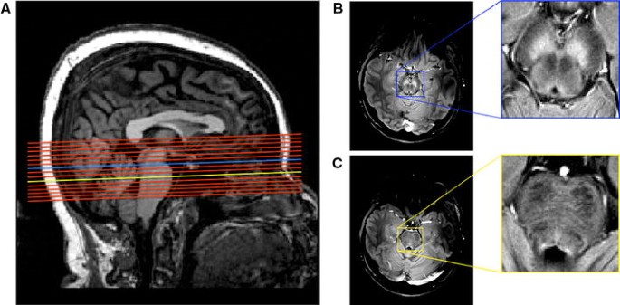
Structural images were acquired for slice placement and registration with an MP-RAGE sequence: TE/TR = 2.46/1900 ms, inversion time = 900 ms, 192 slices, FA = 9°, voxel size = 0.8 × 0.8 × 0.8 mm3, scan time = 5 min 42 s. On the MP-RAGE images, GRE slices were prescribed perpendicular to the dorsal edge of the brain stem, covering the SNpc and LC (see Fig. 1). Each subject was scanned twice using the modified GRE sequence. To emulate multiple scanning sessions, subjects were removed from the scanner after the first session, repositioned on the table, and scanned again with the MP-RAGE and GRE sequences with slices prescribed identically to the first session.
Image processing
Imaging data were analyzed with FMRIB Software Library (FSL) and MATLAB (The Mathworks, Natick, MA). First, images from the seven GRE measurements were registered to the first image using a linear transformation in FMRIB’s Linear Image Registration Tool (FLIRT) tool and averaged. The averaged image was used in the subsequent analysis.
Segmentation of neuromelanin-sensitive GRE images was performed in MATLAB. SNpc and LC volumes were delineated using a semi-automated thresholding method based on anatomical landmarks as detailed previously [6]. Thresholds for each structure were calculated using the mean, denoted μ REF, and standard deviation, denoted σ REF, of a reference region placed in the cerebral peduncle. For segmentation, voxels with signal intensity greater than I > μ REF + 3σ REF or than I > μ REF + 4σ REF were considered part of SNpc or LC, respectively.
SNpc and LC volumes were transformed into Montreal Neurological Institute (MNI)-152 space using FLIRT and FMRIB’s Nonlinear Image Registration Tool (FNIRT) tools in FSL as follows. First, brain extracted T1-weighted images were aligned with the MNI brain extracted image using an affine transformation. Second, a nonlinear transformation was used to generate a transformation from individual subject space to common space. Finally, individual SNpc and LC masks were transformed to their respective T1-weighted images using FLIRT and then transformed to common space using FNIRT. After transformation to common space, the center of mass for the LC and SN masks were recorded for each subject.
MTC of a voxel is defined as
where I(x, y) and I ref denote the signal intensity of a voxel located at (x, y) and the mean signal intensity of a reference region, respectively. To reduce variability in the MTC calculation, a reference region in the cerebral peduncle was drawn in MNI space and transformed to individual subject space using the inverse of the nonlinear transform described in the previous paragraph.
Reproducibility between SNpc and LC volumes were calculated using the dice similarity coefficient (DSC) and is defined as
where A and B denote catecholamine nuclei volumes for scans 1 and 2, respectively, and ∩ represents the intersection operator. DSC was calculated in MNI space.
Statistical analysis
Scan-rescan reproducibility of volume and MTC was assessed using intraclass correlation coefficients (ICC). Reproducibility of interscan volume and MTC measurements was tested with a two-way random ICC evaluating absolute agreement. ICC is a measure of agreement between two groups, and ICC values were interpreted according to the criteria set by Landis and Koch: (0.8, 1] = almost perfect agreement and (0.6, 0.8] = substantial agreement [14]. All statistical analyses were performed using IBM SPSS Statistics software version 22 (IBM Corporation, Somers, NY, USA).
Results
Both volume measurements showed excellent reproducibility with high ICC (SNpc: 0.94, p < 0.001; LC: 0.96, p < 0.001). Figure 2 a, b show the interscan reproducibility for SNpc and LC volumes, respectively. MTC measurements showed substantial agreement between scans. SN and LC ICC values were 0.81 and 0.76, respectively. Interscan reproducibility of SN and LC MTC measurements are shown in Fig. 3a, b, respectively.
The segmented SNpc volumes from the two scans were found to be highly reproducible with significant overlap between the two scans (SNpc DSC: 0.80 ± 0.03). Furthermore, no difference was seen in SNpc center of mass between scans 1 and 2. The average distance in center of mass between scan 1 and scan 2 was 0.8 ± 0.7 and 1.0 ± 0.5 mm for the left and right hemispheres, respectively. The LC was reproducibly delineated in the two scans. The mean DCS for LC was 0.63 ± 0.07. The LC centers of mass for the two scans were found to be in virtually the same location with an average deviation being 1.6 ± 1.2 mm for the left LC and 1.2 ± 0.9 mm for the right LC.
Discussion
Neuromelanin-sensitive MRI has been used to examine differences cross-sectional volumetric, slice area, or contrast differences occurring in SNpc from PD [4, 5, 9–12]. However, a longitudinal study examining changes in SNpc volume or MTC after onset of PD has not been reported in the literature and, the reproducibility of SNpc volume or MTC measurements had not been examined prior to this work. We found SNpc volume to show excellent reproducibility between sessions with high ICC and DSC values. In addition, MTC in SNpc was found to have high reproducibility between scanning sessions. The results presented here indicate the neuromelanin-sensitive approach used in this work is well suited for examining longitudinal changes in SNpc.
We found minor differences in location and volume of SNpc between scanning sessions. The discrepancies in SNpc volume between scans may be due to differences in partial volume effects and slice profiles between scans. Since the subject was physically removed from the scanner after the first session, it is likely that there were slight differences in slice orientation between sessions 1 and 2. These slight differences in slice orientation will be manifested as slight changes in SNpc morphometry in each scanning session. These morphological differences will be present after transformation to common space, and the discrepancy in SN DSC may be due to differences in slice orientation between scans.
LC volume showed excellent reproducibility as indicated by a high ICC. The mean absolute difference in LC volume from the two sessions was approximately 1.8 mm3, which represents a difference of approximately 2.5 % as compared to the bilateral LC volume reported in the literature [15]. Neuronal loss in LC from AD and PD range from 38 to 88 and 21 to 93 % as compared to controls, respectively [15, 16]. Thus, differences in LC volume from PD or AD should be measurable using the presented approach.
The spatial location of LC exhibited more variation than SNpc as evidenced by the smaller LC DSC value. The differences in spatial location of LC may be attributed to a combination of factors, including partial volume effects from different slice orientations and pulsation in the 4th ventricle. In addition, the small size of LC may lead to greater registration errors than those seen in SNpc. The registration errors may be due to the loss of resolution associated with the transformation from GRE space to T1-space, and these errors may be mitigated in future studies by increasing the spatial resolution of MP-RAGE images.
There are some caveats in the present study. First, the population used in this study consisted of young, healthy subjects. Neuromelanin concentration in SN has been found to increase throughout life [17] while neuromelanin concentration in LC has been found to peak around age 50 [18]. In aged populations, the decrease in NM concentration in LC may reduce the reproducibility of LC volume measurements. In clinical populations, the reproducibility of LC and SNpc volume may be decreased since clinical symptoms (such as PD tremor) may introduce artefacts in neuromelanin-sensitive MRI images. Furthermore, degradation of these structures from disease will decrease MTC and could reduce reproducibility in clinical populations. Second, since the reproducibility experiment was conducted on the same day, scanner performance and subject physiological status were similar for the two scans. Reproducibility for scans conducted on different days may be somewhat lower.
Conclusion
We found that measurement of LC and SNpc volume and MTC, performed with an MT prepared GRE sequence and automated thresholding method, has excellent test–retest reproducibility. Demonstrating test–retest reproducibility is a necessary step in the establishment of possible biomarkers for clinical and translational application.
References
Braak H, Thal DR, Ghebremedhin E, Del Tredici K (2011) Stages of the pathologic process in Alzheimer disease: age categories from 1 to 100 years. J Neuropathol Exp Neurol 70(11):960–969
Braak H, Tredici KD, Rüb U, de Vos RAI, Jansen Steur ENH, Braak E (2003) Staging of brain pathology related to sporadic Parkinson’s disease. Neurobiol Aging 24(2):197–211
Sasaki M, Shibata E, Kudo K, Tohyama K (2008) Neuromelanin-sensitive MRI basics, technique, and clinical applications. Clin Neuroradiol 18(3):147–153
Schwarz ST, Rittman T, Gontu V, Morgan PS, Bajaj N, Auer DP (2011) T1-weighted MRI shows stage-dependent substantia nigra signal loss in Parkinson’s disease. Mov Disord 26(9):1633–1638
Ogisu K, Kudo K, Sasaki M, Sakushima K, Yabe I, Sasaki H, Terae S, Nakanishi M, Shirato H (2013) 3D neuromelanin-sensitive magnetic resonance imaging with semi-automated volume measurement of the substantia nigra pars compacta for diagnosis of Parkinson’s disease. Neuroradiology 55(6):719–724
Chen X, Huddleston DE, Langley J, Ahn S, Barnum CJ, Factor SA, Levey AI, Hu X (2014) Simultaneous imaging of locus coeruleus and substantia nigra with a quantitative neuromelanin MRI approach. Magn Reson Imaging 32(10):1301–1306
Langley J, Huddleston D, Chen X, Sedlacik J, Zachariah N, Hu X (2015) A multicontrast approach for comprehensive imaging of substantia nigra. NeuroImage 112(1):7–13
Trujillo P, Smith AK, Summers PE, Mainardi LM, Cerutti S, Smith SA, Costa A (2015) High-resolution quantitative imaging of the substantia nigra. Conf Proc IEEE Eng Med Biol Soc 2015:5428–5431
Nakane T, Nihashi T, Kawai H, Naganawa S (2008) Visualization of neuromelanin in the Substantia nigra and locus ceruleus at 1.5T using a 3D-gradient echo sequence with magnetization transfer contrast. Magn Reson Med Sci 7(4):205–210
Castellanos G, Fernandez-Seara MA, Lorenzo-Betancor O, Ortega-Cubero S, Puigvert M, Uranga J, Vidorreta M, Irigoyen J, Lorenzo E, Munoz-Barrutia A, Ortiz-de-Solorzano C, Pastor P, Pastor MA (2015) Automated neuromelanin imaging as a diagnostic biomarker for Parkinson’s disease. Mov Disord 30(7):945–952
Ohtsuka C, Sasaki M, Konno K, Koide M, Kato K, Takahashi J, Takahashi S, Kudo K, Yamashita F, Terayama Y (2013) Changes in substantia nigra and locus coeruleus in patients with early-stage Parkinson’s disease using neuromelanin-sensitive MR imaging. Neurosci Lett 541:93–98
Reimao S, Pita Lobo P, Neutel D, Correia Guedes L, Coelho M, Rosa MM, Ferreira J, Abreu D, Goncalves N, Morgado C, Nunes RG, Campos J, Ferreira JJ (2015) Substantia nigra neuromelanin magnetic resonance imaging in de novo Parkinson’s disease patients. Eur J Neurol 22(3):540–546
Sasaki M, Shibata E, Tohyama K, Takahashi J, Otsuka K, Tsuchiya K, Takahashi S, Ehara S, Terayama Y, Sakai A (2006) Neuromelanin magnetic resonance imaging of locus ceruleus and substantia nigra in Parkinson’s disease. Neuroreport 17(11):1215–1218
Landis JR, Koch GG (1977) The measurement of observer agreement for categorical data. Biometrics 33(1):159–174
German DC, Manaye KF, White CL 3rd, Woodward DJ, McIntire DD, Smith WK, Kalaria RN, Mann DM (1992) Disease-specific patterns of locus coeruleus cell loss. Ann Neurol 32(5):667–676
Hoogendijk WJ, Pool CW, Troost D, van Zwieten E, Swaab DF (1995) Image analyser-assisted morphometry of the locus coeruleus in Alzheimer’s disease, Parkinson’s disease and amyotrophic lateral sclerosis. Brain 118(Pt 1):131–143
Zecca L, Gallorini M, SchuÈnemann V, Trautwein AX, Gerlach M, Riederer P, Vezzoni P, Tampellini D (2001) Iron, neuromelanin and ferritin content in the substantia nigra of normal subjects at different ages: consequences for iron storage and neurodegenerative processes. J Neurochem 76(6):1766–1773
Manaye KF, McIntire DD, Mann DM, German DC (1995) Locus coeruleus cell loss in the aging human brain: a non-random process. J Comp Neurol 358(1):79–87
Acknowledgments
This work was partially supported by the Michael J. Fox Foundation (MJF 10854) and by Siemens Healthcare.
Author information
Authors and Affiliations
Corresponding author
Ethics declarations
Conflict of interest
The authors declare that they have no conflict of interest.
Ethical standards
All procedures performed in studies involving human participants were in accordance with the ethical standards of the institutional and/or national research committee and with the 1964 Helsinki declaration and its later amendments or comparable ethical standards.
Informed consent
Informed consent was obtained from all individual participants included in the study.
Rights and permissions
About this article
Cite this article
Langley, J., Huddleston, D.E., Liu, C.J. et al. Reproducibility of locus coeruleus and substantia nigra imaging with neuromelanin sensitive MRI. Magn Reson Mater Phy 30, 121–125 (2017). https://doi.org/10.1007/s10334-016-0590-z
Received:
Revised:
Accepted:
Published:
Issue Date:
DOI: https://doi.org/10.1007/s10334-016-0590-z


