
Abstract
Purpose
To evaluate the results of a consecutive series of patients affected by lumbar discogenic pain associated with facet pain and canal stenosis surgically treated with the PercuDyn device.
Methods
From 2009, 129 consecutive patients (96 M, 33 F, mean age 62) were treated with posterior dynamic stabilization screws (PercuDyn). Inclusion criteria were minimum follow-up of 24 months; pain localized at the lumbar spine column alone or in association to lower limb radicular pain; magnetic resonance evidence of disc degeneration associated with facet degeneration and canal stenosis. Patients were clinically studied using VAS scale and Oswestry Disability Index (ODI); CT assessment of the neuroforamina and spinal canal areas was done at 1 month of follow-up.
Results
At 24 months of follow-up, 96 patients fulfilled the inclusion criteria. 96 intervertebral spaces were treated (85 levels L5–S1, 11 levels L4–L5). The VAS scale showed a statistically significant difference at 1 month, 6 months and 2 years with respect to the pre-operative value (p < 0.001). The ODI score registered a significant difference with the same fashion (p < 0.001 both at 1- and 6-month, and 2-year follow-up with respect to the pre-operatory). At 1-month follow-up, neuroforamina and spinal canal areas were considerably wider (p < 0.05). 70 (72.5 %) patients were satisfied of the procedure.
Conclusions
In this wide cohort study, the PercuDyn ensured good clinical and radiological results, with more than 70 % of patients satisfied of the procedure. Very few complications were noted, with an immediate return to daily activities. At longer follow-ups, 10 % of patients received revision surgery.
Introduction
Painful conditions of the aging spine include degenerative disc disease, facet arthropathy, central foraminal stenosis and spondylolisthesis. The degenerating lumbar spine is a major source of low back pain and disability in western industrial societies. Recently introduced dynamic stabilization devices have been designed to alleviate pain by purportedly stiffening or supporting the motion segment in attempt to restore the native biomechanical neutral zone. Potential advantages of these devices include retention and protection of the intervertebral disc, earlier surgical intervention, and minimally invasive techniques. The most notable advantage is the ability to maintain or restore controlled motion at the treated level. This not only contributes to increased total range of motion and natural anatomic alignment but may also reduce the risk of accelerated degeneration at adjacent levels, a major concern with fusion. A category of posterior dynamic stabilization devices comprises the pedicle-based systems. A recently introduced pedicle-based design is the PercuDyn (PercuDyn; Interventional Spine Inc.; Irvine, CA) that serves to stabilize the middle column of the spine. This system is delivered percutaneously through a paramedian Wiltse type approach into the bilateral superior articular processes of the inferior vertebral body where it acts as a mechanical stop to the inferior articulating process coming down from the level above thereby directly augmenting the stiffness of the facet column. Among posterior dynamic stabilization devices, the PercuDyn has demonstrated to be most effective in decreasing hyperextension without hindering motion substantially in the other planes [1]. In 2011, Sangiorgio et al. [1] studied the biomechanics of the Percudyn using human cadaveric lumbar spinal motion segments, and they concluded that it reduces extension by more than 50 %, with a minimally-invasive and motion-sparing procedure, preventing facet joints hyperextension without fusion.
Literature about the PercuDyn is limited to few case series of patients affected by lumbar spine stenosis [2], degenerative lumbar low back pain [3], and patients treated with microdiscectomy associated to stabilization with the PercuDyn [4]. In our department, in the last 7 years, we have used the PercuDyn to treat low back pain caused by severe degeneration of intervertebral disc, lumbar spine vertebrae instability, facet arthropathy and central foraminal stenosis for a total of 129 patients. The aim of the present prospective cohort study was to evaluate the clinical and radiographic results of dynamic posterior stabilization of the lumbar column using the PercuDyn device.
Materials and methods
In this prospective cohort study, we hypothesized that the PercuDyn device is an effective and long lasting minimally invasive device for the treatment of chronic discogenic lumbar spine pain. From March 2008, 129 consecutive patients [96 M, 33 F, mean age 62 (range 35–81, SD 27)], with lumbar pain and/or neuralgia-claudication were treated with posterior dynamic stabilization screws (PercuDyn, Interventional Spine Inc., USA). All patients were affected by lumbar discogenic pain associated with facet pain and canal stenosis. The source of these patients is our Private Practice Offices and Orthopedic Spine Ambulatory, directed by the senior author. Demographic data were collected on all the subjects. Data included age, height, weight, body mass index (BMI), and sport activity. Inclusion criteria were age between 30 and 90 years old; minimum follow-up of 24 months; pain localized at the lumbar spine column alone or in association to lower limb radicular pain; magnetic resonance evidence of disc degeneration (from 3 to 5 according to Pfirrmann’s classification) [5] associated with facet degeneration. In addition, to be included in the series, patients had a clear correlation between disc degeneration and symptoms, no evidence of focused neurological signs. All the patients were non responders to at least 8 months of non-operative treatment including steroids, NSAIDs, muscle relaxants, local injective therapies and rehabilitation. Exclusion criteria were: patients younger 30 or older 90 years old; insulin dependent diabetes, rheumatoid or serum-negative arthritis; previous traumatic injuries to the column spine; cauda syndrome and/or limb motor paralysis; the presence of severe spondylolisthesis; severe osteoporosis; migrated disc herniation. In summary, clinical and medical history evaluation had to be confirmed by recent lumbosacral X-ray, CT scan, and MRI with axial scan T1W and sagittal scan T1W, T2W e STIR, 4 mm of thickness.
All patients were informed of the purpose and content of the project and signed a written consent to participate in the study, according to the Declaration of Helsinki.
Surgical procedure
2 g cefazolin was used as antibiotic prophylaxis in all patients. Preoperatively, the patients received light anesthesia with sedation using midazolam 0.05 mg/kg e.v., and local analgesia into the skin, subcutaneous tissues and the periosteum, with lidocaine 2 % (20 ml). The surgical procedure has already been described elsewhere [6]. Following, we report only the main steps that we believe are the basis of a standardized surgical technique.
-
The patient is positioned prone on a radiolucent operative table. We have found that a slight flexion of lumbar spine helps to decrease the articular facets overlapping (Fig. 1);
Fig. 1 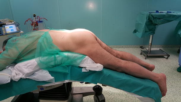
Patient’s prone position on a radiolucent operative table. A slight flexion of lumbar spine helps to decrease the articular facets overlapping
-
Latero-lateral (LL)\antero-posterior (AP) fluoroscopic images are acquired, focusing on the interdisc spaces to be treated;
-
The bilateral paramedian 1 cm incision (Fig. 2) sites are localized with antero-posterior fluoroscopy with the amplioscope at the level to the top of pedicle where the device will be implanted. The skin and fascia are opened, and an access needle is introduced and under fluoroscopy it is placed over the pedicle, ensuring that its tip is exactly positioned at the bottom of the articular facet.
Fig. 2 
The bilateral paramedian 1 cm incision localized using the amplioscope
-
Once the position is verified, the proximal end of the needle is moved 10° medially and 10° caudally to be introduced through the superior articulating process into the pedicle;
-
On AP view, check the needle is exactly positioned inside the pedicle without overcoming medial wall, in order to exclude failure of the nerve root or neural sac.
-
On lateral view, be aware not to advance beyond the somatic anterior wall. (Sequences of the procedure under fluoroscopic guidance are shown on Fig. 3).
Fig. 3 
The sequences of the procedure under fluoroscopic guidance


In summary, the procedure provides a tunnel screwed hole through the pedicle and somatic trabecular bone for placement of the Anchor device, that is similar to a transpedicular screw, on which the Stabilizer device will be anchored. Before placing the Stabilizer device, that is the working component of the system that prevents articular facets overlapping, the tip of the inferior articular process of the above soma and the tip of the superior articular process of the below soma are drilled with 5-mm boor gaining room for the Stabilizer itself. The same steps are performed on the contralateral pedicle.
Post-operative antero-posterior and lateral views are consistently taken to check the final position of the implanted PercuDyn (Fig. 4). Surgeries are performed on an outpatient surgery basis, with patients’ discharge the same day. Prescription of total rest for 48 h and physical therapy for at least 2 months is consistently done. Return to sport activity is allowed at 6 months of follow-up.

Anteroposterior and lateral post-operative X-ray of a L5–S1 PercuDyn implant
Follow-up
Clinical examination of patients was done before surgery and at 1 month, 6 and 24 months of follow-up using the visual analogic scale (VAS) [7] and the Oswestry Disability Index (ODI) [8] by an independent researcher not involved in the study. A CT assessment of the neuroforamina and spinal canal areas was done at pre-operatively and at 1 month of follow-up by the two authors. All images were examined on diagnostic quality liquid crystal display monitors using DICOM (Digital Imaging and Communication in Medicine) compliant grading software (IMPACS Web 1000; Agfa, Mortsel, Belgium). The measurements were performed using multiplanar rendering (MPR) CT imaging in axial planes.
Ability to return to sport activities was evaluated at 6 months of follow-up.
Statistical analysis
Power calculation detected a significant difference in total ODI score as 19 ± 14 at the last follow-up. From this difference, assuming a two-tailed α value of 0.05 (sensitivity 95 %) and a β value of 0.95 (study power 95 %); we determined that at least 40 patients were required at follow-up evaluation (G power 3 power analysis program). Intra- and inter-tester reliability of the CT assessment of the neuroforamina and spinal canal areas was determined calculating intra-class correlation coefficients. Interpretation of the κ statistic was performed as described by Landis and Koch [9] in 1977. Agreement was considered excellent if κ fell between 0.81 and 1.0, high if κ was between 0.61 and 0.80, moderate if κ was 0.41–0.60, fair if κ was 0.21–0.40, and poor if κ was 0.20 or less. VAS and ODI score data are presented as means (with standard deviations) and differences were analyzed with T Student test, as well as for neuroforamina and spinal canal areas. p < 0.05 was considered statistically significant.
Results
Of the total 129 patients treated with the PercuDyn, 96 (74.4 %) were available at the final follow-up of 24 months. Of the 33 who were not, 15 had a follow-up shorter than 24 months; 8 were not complained with the post-operative restrictions and rehabilitation; and 5 were lost to clinical follow-up. In the remaining 5 excluded cases, the PercuDyn device was used to prevent instability in the intervertebral levels adjacent to traditional posterior intervertebral arthrodesis. The mean age of the 96 studied patients was 60 (80 M, 16 F; age range 35–76, SD 22); 71 patients were sedentary and 25 practiced recreational sport activities. Morphometric data are synthesized in Table 1.
96 intervertebral spaces were treated (85 levels L5–S1, 11 levels L4–L5). The VAS scale showed a statistically significant difference at 1 month, 6 months and 2 years with respect to the pre-operative value (p < 0.001). No statistically significant difference was present between the VAS values comparing 1 month versus 2 years (p = 0.83). The ODI score registered a significant difference with the same fashion (p < 0.001 both at 1- and 6-month, and 2-year follow-up respect to the pre-operatory), and no statistically significant difference was detected comparing 1 month versus 2 years (p = 0.75). The mean values with range and standard deviation of VAS scale and ODI score are summarized on Table 2.
Intra- and inter-tester reliability of the CT assessment of the neuroforamina and spinal canal areas was high (κ values—neuroforamina canal area: intra-tester 0.79, inter-tester 0.78; spinal canal area: intra-tester 0.8, inter-tester 0.71). At 1 month of follow-up, we detected an increase of the area of 15 and 16.5 % for the right and left neuroforamina, respectively [pre-op right foramina area: 0.91, post-op: 1.05 mm2 (p = 0.016); pre-op left foramina area: 0.92, post-op: 1.08 mm2 (p = 0.014)]. Accordingly, the spinal canal area increased from 1.92 to 2.19 mm2 (p = 0.0021).
Three major complications were noted: one too medial positioning of one of the two screws of the device (Fig. 5) that caused severe irritation of the sciatic nerve root, with prompt removal after 40 days and successful revision of the implant; one screw mobilization in a recreational weight-lifter; and one superficial infection that was successfully treated with oral antibiotics.

MRI axial scan showing too medial positioning of one of the two screws of the device, with severe irritation of the sciatic nerve root
Of the 25 patients (26 % of the study group) who practiced recreational sport activities, 16 (64 %) were able to resume training at 6 months of follow-up, with restriction of heavy activities, in particular weight-lifting and contact sports.
At the final follow-up, 70 (72.5 %) patients were satisfied of the procedure.
Of the 21 patients who had a follow-up longer than 5 years, 3 (14.3 %) received revision surgery with posterior static stabilization using bars and screws because of mobilization of the device with worsening of symptoms.
Discussion
This study deals with the overall widest cohort study of patients treated with the PercuDyn device with a considerably long follow-up. After 2 years, more than 70 % of patients were satisfied of the procedure, with a good improvement of subjective and objective scores and of considered radiologic parameters. In addition, even with the difficulties to isolate a single specific pathogenic entity for each patient due to the complex nature of the structures involved and their reciprocal interactions, we have studied a relatively homogeneous group of patients, both for pre-operative characteristics and for received treatment, in all cases with one intervertebral level stabilized with the PercuDyn. Patients who had additional surgeries, as the 5 patients in whom the PercuDyn device was used to prevent instability in the intervertebral levels adjacent to traditional posterior intervertebral arthrodesis, were not considered, even if we always use the device in these cases and we recognize it as an indication to implant the PercuDyn. Similarly, Kafer et al. have found that posterior dynamic stabilization in combination with total disc replacement reduces flexion/extension ROM and segmental lordosis in a monosegmental biomechanical model [10].
Our results are comparable to those emerging from previous studies. In 2014, Marcia and colleagues studied a cohort of 38 patients and found VAS pain reduction as well as ODI improvement, with a statistically significant widening of the neuroforaminal area; their indications for surgery were lumbar discogenic pain (disc degeneration from 3 to 5 according to Pfirrmann’s classification) [5] associated with facet pain and canal stenosis [3]. According to the results of Chen et al. [11], they concluded that the PercuDyn is able to restore neuroforamina and spinal canal area, acting as a dynamic stabilization, because it is placed on the facet joint bases and leads to natural full range of motion and rotation, increasing interspinous ligament motility in order to reduce the lumbar low back pain source. Masala et al. [2] treated 20 patients with symptoms of lumbar spine stenosis and obtained 16 cases with satisfactory results, suggesting that the PercuDyn may at least delay the time for classic decompression surgeries. Maida et al. [4] in 2014 used the device in association with microdiscectomy in a selected cohort of patients with severe disc degeneration, good range of motion of the lumbar spine and integrity of posterior intervertebral processes. Even with the limits of their study, they confirmed that dynamic stabilization produces a good relief from back pain preserving the range of motion [4]. The common link between these studies [2–4], is the very low morbidity of the procedure, with a very fast recovery and return to daily activities, and the quite absence of intra- or post-operative complications, with very good results in term of pain resolution. This dynamic stabilization system seems to be a very good alternative to the standard surgical fusion in patients with severe disc degeneration, especially because potential surgical complications as irreversible spine deformity and invalidation of quality of life are not reported [8, 12]. The PercuDyn significantly prevents narrowing of the spinal canal and neural foramina in extension, decompressing the individual degenerative spinal levels that cause symptoms. PercuDyn is designed to relieve the patients’ symptoms while standing and walking, which allows the patient to resume their normal posture. In summary, indications for the implant of the PercuDyn device are severe degeneration of intervertebral disc, lumbar spine vertebrae instability (I–II degree), facet arthropathy with central foraminal stenosis and to prevent instability in the intervertebral levels adjacent to traditional posterior intervertebral arthrodesis; contraindications are represented by severe osteoporosis, cauda syndrome, migrated disc herniation and severe spondylolisthesis.
In our experience, the improvement of clinical scores reaches a plateau in the first 6 months of follow-up. This evidences that patients that do not improve in the first months do not improve later, and, so, it is recommended to have serial close follow-ups to monitor the results. This is absolutely true especially for high-demanding patients that practice heavy sport (weight-lifting) or contact sport activities (rugby, American football, and wrestling). Beyond the biomechanical characteristics of the device that have been widely described [1, 13], we have noted that the device cannot ensure its effect when patients do not respect post-operative restrictions with excessive loads on the lumbar spine. In addition, we have to highlight that more than 10 % of patients with a follow-up of 5 years or longer received traditional stabilization with bars and screws for mobilization and failure of the PercuDyn. We discourage patients to continue with heavy or contact sport activities, but, at the same time, we strongly encourage them to avoid sedentary lifestyle with possible weight gain. The BMI of patients treated with this device should be monitored at each follow-up, and any eventual significant increase of the value should be carefully valued.
In materials and methods section, the main steps for a correct surgical procedure are reported. The device can be easily used at L5/S1 as well as other lumbar segments. In fact, at L5/S1, the device may be easier to apply given the orientation of the facet joint at this level is almost perpendicular to the trajectory of device application. At higher levels, the facet joints are oriented in a more parallel fashion, with a more difficult trajectory angle. Respect to other devices, due to the poorly developed S1 spinous process, the PercuDyn may be easily used at L5–S1 level, while other minimally invasive stabilization devices are not [1, 14]. Respecting the over-reported steps, complications like too medial positioning of the screw may be easily avoided (Fig. 5). In this study group, we have consistently obtained a CT assessment at 1 month of follow-up for radiologic evaluation; in any case, especially when the surgeon is not too familiar with the implant, it is mandatory to obtain the post-operative CT or MRI scan to assess the position of the device.
This study has a potential limitation that need to be addressed. This is not a randomized study and so there is a lack of a comparison group receiving an alternative procedure for the disease. On the other hand, this study has the widest cohort in literature with a considerably long follow-up, up to 7 years.
References
Sangiorgio SN, Sheikh H, Borkowski SL, Khoo L, Warren CR, Ebramzadeh E (2011) Comparison of three posterior dynamic stabilization devices. Spine (Phila Pa 1976) 36:E1251–E1258. doi:10.1097/BRS.0b013e318206cd84
Masala S, Tarantino U, Nano G, Iundusi R, Fiori R, Da Ros V, Simonetti G (2013) Lumbar spinal stenosis minimally invasive treatment with bilateral transpedicular facet augmentation system. Cardiovasc Intervent Radiol 36:738–747. doi:10.1007/s00270-012-0478-x
Marcia S, Saba L, Anselmetti GC, Marini S, Piras E, Marras M, Masala S, Georgy B (2014) Effectiveness of percutaneous screws for treatment of degenerative lumbar low back pain. Cardiovasc Intervent Radiol 37:1329–1335. doi:10.1007/s00270-013-0786-9
Maida G, Altruda C, Gatti M, Saletti A, Borrelli M, Sarubbo S (2014) Two-year follow-up after microsurgical discectomy and dynamic percutaneous stabilization in degenerate and herniated lumbar disc: clinical and neuroradiological outcome. J Neurosurg Sci 58:95–102
Pfirrmann CW, Metzdorf A, Zanetti M et al (2001) Magnetic resonance classification of lumbar intervertebral disc degeneration. Spine (Phila Pa 1976) 26:1873–1878
Smith ZA, Armin S, Raphael D, Khoo LT (2011) A minimally invasive technique for percutaneous lumbar facet augmentation: technical description of a novel device. Surg Neurol Int 2:165. doi:10.4103/2152-7806.90026
Grönblad M, Lukinmaa A, Konttinen YT (1990) Chronic lowback pain: intercorrelation of repeated measures for pain and disability. Scand J Rehabil Med 22:73–77
Little DG, MacDonald D (1994) The use of the percentage change in Oswestry Disability Index score as an outcome measure in lumbar spinal surgery. Spine (Phila Pa 1976) 19:2139–2143
Landis JR, Koch GC (1977) The measurement of observer agreement for categorical data. Biometrics 33:159–174
Käfer W, Cakir B, Midderhoff S, Reichel H, Wilke HJ (2014) Circumferential dynamic stabilization of the lumbar spine: a biomechanical analysis. Eur Spine J 23:2330–2339. doi:10.1007/s00586-014-3286-y
Chen D, Fay LA, Lok J, Yuan P, Edwards WT, Yuan HA (1995) Increasing neuroforaminal volume by anterior interbody distraction in degenerative lumbar spine. Spine (Phila Pa 1976) 20:74–79
Arbit E, Pannullo S (2001) Lumbar stenosis: a clinical review. Clin Orthop 384:137–143
Palmer S, Mahar A, Oka R (2007) Biomechanical and radiographic analysis of a novel, minimally invasive, extension-limiting device for the lumbar spine. Neurosurg Focus 22:E4
Fei H, Xu J, Wang S, Xie Y, Ji F, Xu Y (2015) Comparison between posterior dynamic stabilization and posterior lumbar interbody fusion in the treatment of degenerative disc disease: a prospective cohort study. J Orthop Surg Res 10:87. doi:10.1186/s13018-015-0231-7
Author information
Authors and Affiliations
Corresponding author
Ethics declarations
Conflict of interest
No conflict of interest to declare.
Rights and permissions
About this article

Cite this article
Canero, G., Carbone, S. The results of a consecutive series of dynamic posterior stabilizations using the PercuDyn device. Eur Spine J 24 (Suppl 7), 865–871 (2015). https://doi.org/10.1007/s00586-015-4268-4
Received:
Revised:
Accepted:
Published:
Issue Date:
DOI: https://doi.org/10.1007/s00586-015-4268-4