
Abstract
Background
Breast augmentation surgery with implants is one of the most common aesthetic surgical procedures. Round and anatomical textured implants are employed very often, and fat grafting has proven to be a very useful complementary procedure in breast augmentation. Many authors report a more natural result with anatomical compared to round implants. Nevertheless, anatomical implants can be associated with complications such as implant rotation with subsequent shape distortion. In this article, we propose a combination of high-profile round implants and fat grafting to obtain a natural result analyzing its impact on the aesthetic outcome and patient satisfaction.
Methods
In this study, we report our personal approach on 31 consecutive patients undergoing primary aesthetic breast augmentation with high-profile round implants and fat grafting. We describe our personal technique of breast augmentation via the periareolar approach and fat grafting. We evaluated short- and medium-term aesthetic outcomes and patient satisfaction using a 10-point VAS scale.
Results
We achieved in all cases high patient satisfaction and good aesthetic outcomes with a “natural” breast shape and a “smoothened” upper pole with low complication rates. The technique is safe, simple, fast, and it leads to high levels of patient satisfaction.
Conclusions
Our observations show that the combination of high-profile round implants and fat grafting in aesthetic breast augmentation can improve the aesthetic outcome and patient satisfaction as with anatomical implants eliminating the risk of implant rotation.
Level of Evidence IV
This journal requires that authors assign a level of evidence to each article. For a full description of these Evidence-Based Medicine ratings, please refer to the Table of Contents or the online Instructions to Authors www.springer.com/00266.
Introduction
Breast augmentation surgery with implants is one of the most common aesthetic surgical procedures since the 1960s [1, 2]. Breast shapes and features may vary, and some conditions, such as asymmetric, tuberous or constricted breasts, are very common; thus, a combination of different procedures is often required to achieve satisfying results [3]. The safety of breast augmentation has been extensively reported in the literature [4,5,6,7,8,9], and no interference on oncological surveillance by physical examination and imaging has been described. Breast augmentation aims at providing greater volume to hypoplasic breasts and fuller contours in hypotrophic breasts, ensuring a “natural” shape and durability of the result over time. The use of fat grafting has been extensively reported in both cosmetic and reconstructive breast surgery, and it represents nowadays a fundamental, easy, fast and effective complementary procedure integrating the traditional surgical approach to breast augmentation [10]. Round textured implants are currently employed in breast augmentation. Even though round implants represent the best option in selected patients, they can lead to a less natural result with a convex appearance of the upper pole associated with implant palpability and/or visibility, especially in thin patients with severe glandular hypotrophy. Moreover, some authors reported more natural results with anatomical implants in this kind of patients [11,12,13,14,15,16]. On the other hand, anatomical implant malposition and rotation can cause shape distortion with an incidence ranging from 0.9 to 14 percent [17,18,19,20,21,22,23,24,25]. Fat grafting can help to overcome those issues leading to a better aesthetic outcome. Nevertheless, the combination of high-profile round implants and fat grafting and its impact on the aesthetic outcome and patient satisfaction have been poorly investigated. In this study, we report our experience with breast augmentation with high-profile round implants and fat grafting.
Materials and Methods
We prospectively considered 31 female patients who underwent primary aesthetic breast augmentation with round implants and fat grafting for hypomastia from January 2016 to June 2017. Breast reconstruction, secondary breast augmentation, severe constricted/tuberous breast deformity, Poland’s syndrome and transgender patients were not included in this study. The follow-up, ranging from 3 to 12 months, consisted of monthly clinical examination. The preoperative workup included clinical history and physical examination, routine blood tests, EKG and cardiologist consultation (if over 55 years old), mammary ultrasound (US) imaging in all patients and mammography in patients over 40 years old. Preoperative standard photographs were taken (anterior/posterior view, lateral views and oblique views). The intervention was performed under general anesthesia. Preoperative antibiotic prophylaxis with cefazolin (2 g, i.v.) was administered 30 mins’ before surgery, cefazolin (1 g every 12 h, i.v.) during hospitalization and cefixime (400 mg/24 h, oral) for 5 days after the patient was discharged.
Operative Technique and Perioperative Care
-
Preoperative measurements: The desired “horizontal” width of the breast is planned; the surgeon considers the corresponding round implant and verifies whether its size matches the desired “vertical” height, planning the lowering of the inframammary fold; on this basis and with regard to the overall thoracic size, the round implant with that diameter and the desired projection and volume is chosen during surgery
-
Inferior periareolar incision (3–4 cm), trans-glandular electrocautery dissection, identification of the outer pectoral fascia and suprafascial glandular undermining. From the lateral pectoralis border separation creation of a wide subpectoral (partial submuscular) pocket by blunt finger dissection associated with a precise monopolar dissection. After accurate hemostasis, a 19 French lateral closed suction drain is positioned
-
High-profile round textured implant placement and subsequent reconstruction of the glandular parenchyma followed by intradermal cutaneous reabsorbable suture
-
The adipose tissue was harvested through liposuction from abdominal and hip subcutaneous fat. We used to infiltrate only physiologic solution with epinephrine without local anesthetic. We harvest the fat using blunt cannulas of 2 to 3 mm in diameter of variable length (between 15 and 23 cm). The cannula used for sampling is connected with a 10-ml Luer-lock syringe. The syringe plunger is pulled at the top and secured by hand. This creates inside the syringe a slight negative pressure, which allows the levy of adipose tissue while the cannula is advanced and retracted with radial movements inside the donor area and processed, according to Coleman’s technique, by centrifugation at 3000 rpm for 5 min. The adipocyte cell fraction was purified in the ultracentrifuged adipose material by removing the pellet and the oil fraction. The fat graft was injected using an 18-gauge angiographic needle with a snap-on wing (Cordis, a Johnson & Johnson Company, N. V, Roden, the Netherlands) with a retrograde technique to avoid intravascular injection [26,27,28]. The mean amount of the injected adipose tissue was 134 cc. Fat was injected into the subcutaneous fat layer to obtain a breast augmentation as with an anatomical implant (Figs. 1, 2).
Fig. 1 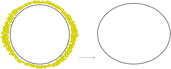
Front view of fat grafting
Fig. 2 
Lateral view of fat grafting


We evaluated patients at the 5th day post-op, weekly for the first month, every 15 days for the first 3 months and then monthly until the first year. The short- and mid-term aesthetic outcome was evaluated independently by five plastic surgeons with expertise in breast cosmetic surgery. We considered the following parameters: shape, symmetry (volume symmetry and NAC symmetry in terms of diameter and position), presence/absence of rippling, implant visibility and/or palpability, capsular contracture, upper pole contour (Table 1). Patient short-term and mid-term satisfaction was evaluated according to a 10-point VAS scale, and the questionnaire was administered to the patients every 4 months on the first year after surgery (Table 2).
Results
The average patient age was 34.3, ranging from 16.5 to 66.1. The average operative time was 67 min, ranging from 45 to 87 min. All patients were discharged on the first day after surgery, and drains were removed before discharge.
Transient areolar hypoesthesia was quite common, no permanent hypoesthesia at 1 year after surgery occurred. We did not experience any poor scarring needing revision surgery. We did not observe any capsular contracture and/or implant displacement. No double-bubble deformity was observed. We did not have any fat grafting-related complications, and the percentage of fat take on the breast was satisfying and stable over time in all patients.
Aesthetic outcome in terms of shape and symmetry was satisfying and stable over time. We achieved a breast natural appearance without implant palpability in the infero-lateral pole and with a “smoothened” upper pole contour and a gentle takeoff from the chest preventing and hiding the convex shape of the round implant (Figs. 3, 4, 5, 6; Table 3).

Pre–post-operative frontal view (case 1)

Pre–post-operative oblique view (case 1)

Pre–post-operative frontal view (case 2)

Pre–post-operative oblique view (case 2)
Most patients were satisfied with both breast size and symmetry. No secondary surgery to increase the volume was required with high patient satisfaction according to the 10-point VAS scale (mean patients aesthetic value of 9.2 SD 1.1) (Table 4.)
Discussion
Areolar, axillary, inframammary and umbilical approaches as well as subglandular, totally or partially submuscular, subfascial and dual-plane pockets have been described and thoroughly evaluated in the past years [29,30,31,32,33,34,35,36]. In our experience, the inferior periareolar incision provides adequate exposure and allows us controlled suprafascial undermining and separation of pectoralis major muscle fibers, whereas the resulting scar is in most cases favorable and well concealed. In addition, the inframammary fold can be lowered as requested after the implant placement and the formation of a new mound [36]. Indications include constricted basis/inferior pole, areolar pseudo-herniation of the parenchyma, but this approach is suitable in most cases, and it can be easily combined with periareolar mastopexy. However, if the areola is very small or its margins are indistinct [37], this access can be more difficult.
Glandular dissection by finger detachment is simple and safe; glandular function is provided by the upper pole, and the nipple-areola complex sensitivity is preserved in all cases. We create a subglandular pocket by electrocautery and blunt finger dissection in a suprafascial plane, whose extension is related to the result we want to achieve. This plane allows very “natural” repositioning of the glandular parenchyma over the submuscular implant and does not definitively compromise vascularity or innervation.
Submuscular implant positioning is particularly indicated if the glandular tissue is poorly developed at the superior pole, but it is suitable in most cases. Separation of the pectoralis major muscle fibers is performed over a rib, preventing chest wall injury and pneumothorax. In our opinion, if high-profile round implant placement has been planned, the submuscular pocket should be wide. Blunt finger dissection does not increase the risk of bleeding in the submuscular plane and allows to accurately control the extension of the undermining even though we always complete the pocket by monopolar electrocautery dissection, especially in the lower and medial quadrants. The pectoralis major muscle lower sternal and rib insertions are often divided [37]. This also allows an easy lowering of the inframammary crease, adapting the mound contour to the new volume.
Cohesive gel implants are slightly firmer than responsive gel, and the firmness of the implant can sometimes result in palpable edges [38]. In our experience, this may occur more often laterally and inferiorly, because at this level the partially submuscular placement does not ensure coverage.
With these technical refinements and choices, round textured implants are suitable in most cases and provide long-term favorable contours, particularly if we imagine that mild to severe ptosis may subsequently occur.
In our experience, mammary implants do not have an “expiration date” and each choice should be made considering long-term outcomes. Fat grafting was performed mostly in the upper and medial pole smoothening its contour, hiding the implant edges and preventing its visibility and palpability. The fat “camouflage” in the upper, lateral, medial, central and lower pole was stable over time leading to a more natural breast shape even in low-BMI patients with thin tissues and/or a severe hypotrophic gland. The use of needles for fat injection allows lower pole and inframammary fold release with a decreased risk of double-bubble deformity as we described in stenotic breast treatment in our previous article [39] similarly to Rigotti [40].
Finally, many authors reported rotation and malposition with shape distortion associated with anatomical implants with an incidence ranging from 0.9 to 14 percent. The combination of high-profile round implants with fat grafting, as investigated in this study, can provide natural results similarly to anatomical implants with no risk of implant rotation.
Conclusions
In this study, we resume our experience with breast augmentation combining high-profile round textured implants and fat grafting. We achieved in all cases high patient satisfaction and favorable, “natural” results in primary aesthetic breast augmentation. The technique is safe, simple and fast, and we advocate the use of fat grafting every time there is an indication to place a round implant. Therefore, we firmly believe that in a modern approach to breast augmentation, fat grafting is becoming an essential procedure for breast reshaping when associated with round implants.
References
Brown MH, Shenker R, Silver SA (2005) Cohesive silicone gel breast implants in aesthetic and reconstructive breast surgery. Plast Reconstr Surg 116(3):768–779
Cronin TD, Gerow FJ (1964) Augmentation mammoplasty: a new “natural feel” prosthesis. In: Transactions of the 3rd international congress of plastic surgery. Excerpta Medica International Congress Series, #66, Amsterdam, p 41
Bostwich J II (1983) Aesthetic and reconstructive breast surgery. C.V. Mosby, St Louis, p 29
Mc Grath M, Burkhardt BR (1984) The safety and efficacy of breast implants for augmentation mammoplasty. Past Reconstr Surg 74:550
Berkel H, Birdsell DC, Jenkin S (1992) Breast augmentation: a risk factor for breast cancer? N Engl J Med 326:1649
Deapen D, Hamilton A, Bernstein L, Brody GS (1992) The relationship between breast cancer and augmentation mammaplasty: an epidemiologic study. Plast Reconstr Surg 89:660
The American Society of Plastic and Reconstructive Surgeons. Institute of Medicine Report released: breast implants get clean bill of health. Plast Surg. News, July 1999
Sanchez-Guerrero J, Schnur PH, Sergent IS, Liang MH (1994) Silicone breast implants and rheumatic disease: clinical, immunologic, and epidemiologic studies. Arthritis Rheumatol 37:158
Deapen DM, Brody GS (1995) Augmentation mammaplasty and breast cancer: a five year update of the Los Angeles study. J Clin Epidemiol 48:551
Autologous Fat Grafting in Cosmetic Breast Augmentation (2016) A systematic review on radiological safety, complications, volume retention, and patient/surgeon satisfaction. Aesthet Surg J 36:993–1007
Hedén P, Montemurro P, Adams WP Jr, Germann G, Scheflan M, Maxwell GP (2015) Anatomical and round breast implants: how to select and indications for use. Plast Reconstr Surg 136:263–272
Adams WP Jr, Mallucci P (2012) Breast augmentation. Plast Reconstr Surg 130:597e–611e
Adams WP Jr, Small KH (2015) The process of breast augmentation with special focus on patient education, patient selection and implant selection. Clin Plast Surg 42:413–426
Hedén P, Brown MH, Luan J, Maxwell GP, Munhoz AM, Carter M (2015) Delphi study consensus recommendations: patient selection and preoperative planning measurements for Natrelle 410. Plast Reconstr Surg Glob Open 3:e556
Caplin DA (2014) Indications for the use of MemoryShape breast implants in aesthetic and reconstructive breast surgery: longterm clinical outcomes of shaped versus round silicone breast implants. Plast Reconstr Surg 134(Suppl):27S–37S
Hammond DC (2014) Technique and results using MemoryShape implants in aesthetic and reconstructive breast surgery. Plast Reconstr Surg 134(Suppl):16S–26S
Hedén P, Jernbeck J, Hober M (2001) Breast augmentation with anatomical cohesive gel implants: the world’s largest current experience. Clin Plast Surg 28:531–552
Baeke JL (2002) Breast deformity caused by anatomical or teardrop implant rotation. Plast Reconstr Surg 109:2555–2564 (discussion 2568)
Tebbetts JB. Dual plane breast augmentation: Optimizing implant-soft-tissue relationships in a wide range of breast types. Plast Reconstr Surg. 2006;118(Suppl):81S–98S; discussion 99S–102S
Hahn M, Kuner RP, Scheler P et al (2008) Sonographic criteria for the confirmation of implant rotation and the development of an implant-capsule-interaction (“interface”) in anatomically formed textured breast implants with texturised Biocell-surface. Ultraschall Med 29:399–404
Hammond DC, Migliori MM, Caplin DA, Garcia ME, Phillips CA (2012) Mentor contour profile gel implants: clinical outcomes at 6 years. Plast Reconstr Surg 129:1381–1391
Maxwell GP, Van Natta BW, Murphy DK, Slicton A, Bengtson BP (2012) Natrelle style 410 form-stable silicone breast implants: core study results at 6 years. Aesthet Surg J 32:709–717
Lista F, Tutino R, Khan A, Ahmad J (2013) Subglandular breast augmentation with textured, anatomic, cohesive silicone implants: a review of 440 consecutive patients. Plast Reconstr Surg 132:295–303
Maxwell GP, Scheflan M, Spear S, Nava MB, Hedén P (2014) Benefits and limitations of macrotextured breast implants and consensus recommendations for optimizing their effectiveness. Aesthet Surg J 34:876–881
Maxwell GP, Van Natta BW, Bengtson BP, Murphy DK (2015) Ten-year results from the Natrelle 410 anatomical form-stable silicone breast implant core study. Aesthet Surg J 35:145–155
Maione L, Vinci V, Klinger M, Klinger FM, Caviggioli F (2015) Autologous fat graft by needle: analysis of complications after 1000 patients. Ann Plast Surg 74(3):277–280
Maione L, Vinci V, Caviggioli F, Klinger F, Banzatti B, Catania B, Lisa A, Klinger M (2014) Autologous fat graft in postmastectomy pain syndrome following breast conservative surgery and radiotherapy. Aesthet Plast Surg 38(3):528–532. https://doi.org/10.1007/s00266-014-0311-9
Caviggioli F, Forcellini D, Vinci V, Cornegliani G, Klinger F, Klinger M (2012) Employment of needles: a different technique for fat placement. Plast Reconstr Surg 130(2):373e–374e
Groen JW, Negenborn VL, Twisk JW, Ket JC, Mullender MG, Smit JM (2016) Autologous fat grafting in cosmetic breast augmentation: a systematic review on radiological safety, complications, volume retention, and patient/surgeon satisfaction. Aesthet Surg J 36:993–1007
Lewis JR Jr (1965) The augmentation mammoplasty. Plast Reconstr Surg 35:51
McKinney P, Shedbalker A (1974) Augmentation mammoplasty using a non-inflatable prosthesis through circum-areolar incision. Br J Plast Surg 27:35
Hoehler H (1973) Breast Augmentation: the Axillary Approach. Br J Plast Surg 26:373
Johnson GW, Christ JE (1993) The endoscopic breast augmentation: the transumbelical insertion of saline-filled breast implants. Plast Reconstr Surg 92:801
Griffiths CO (1967) The submuscular implant in augmentation mammaplasty. In: Translation of the fourth international congress of plastic surgery. Excepta Medica Foundation, Amsterdam, p 1009
Truppman ES, Ellenby JD (1978) A13-year evaluation of subpectoral augmentation mammaplasty. In: Owsley JQ, Peterson RA (eds) Symposium on aesthetic surgery of the breast. Mosby, St Luis
Hidalgo AD (2000) Breast augmentation: choosing the optimal incision, implant, and pocket plane. Plast Reconstr Surg 105(6):2202–2216
Fruhstorfer BH, Hodgson ELB, Malata CM (2004) Early experience with an anatomical soft cohesive silicone gel prosthesis in cosmetic and reconstructive breast implant surgery. Ann Plast Surg 53(6):536–542
Hamas RS (1999) The postoperative shape of round and teardrop saline-filled breast implants. Aesthet Surg J 19:369–374
Klinger M, Klinger F, Giannasi S, Veronesi A, Bandi V, Banzatti B, Catania B, Vinci V, Lisa A, Cornegliani G, Giaccone M, Caviggioli F, Maione L (2017) Stenotic breast malformation and its reconstructive surgical correction: a new concept from minor deformity to tuberous breast. Aesthet Plast Surg 41(5):1068–1077
Khouri RK, Rigotti G, Cardoso E, Khouri RK Jr, Biggs TM (2014) Megavolume autologous fat transfer: part II. Practice and techniques. Plast Reconstr Surg 133(6):1369–1377
Acknowledgements
No financial support or benefits have been received by any author. We do not have any relationship with any commercial source related directly or indirectly to this scientific paper. The principles outlined in the Declaration of Helsinki have been followed in this study.
Author information
Authors and Affiliations
Corresponding author
Rights and permissions
About this article

Cite this article
Maione, L., Caviggioli, F., Vinci, V. et al. Fat Graft in Composite Breast Augmentation with Round Implants: A New Concept for Breast Reshaping. Aesth Plast Surg 42, 1465–1471 (2018). https://doi.org/10.1007/s00266-018-1240-9
Received:
Accepted:
Published:
Issue Date:
DOI: https://doi.org/10.1007/s00266-018-1240-9