
Abstract
In recent years, requests for breast implant surgery have occurred for several reasons. First, the number of diagnosed breast cancer cases has increased, and the number of reconstructive surgeries consequently has multiplied. Second, the number of patients who constantly try to achieve a better physical shape, corresponding in Western countries to the common image of prosperous and tonic breasts, has proliferated. These circumstances have led to an increasingly frequent need for more accurate and sophisticated imaging methods to study prosthetic breast implants and their integrity. Diagnostic imaging for the study of patients with suspected breast implant ruptures uses different techniques of radiologic investigation such as mammography and ultrasonography, even if the current gold standard is magnetic resonance imaging (MRI).This study aimed to draw attention to the main MRI signs capable of highlighting contractures or ruptures of the implants that are not always clinically detectable and thus to provide plastic surgeons with an adequate instrument for discerning any possible alterations in prosthetic implants. Furthermore, it was necessary to stress the importance of teamwork. In fact, proper cooperation and coordination between radiologists and dedicated plastic surgeons are fundamental for the proper management of patients and the complications they may experience.
In recent years, the concomitant occurrence of two phenomena have made breast plastic surgery increasingly frequent: the increase in the number of diagnosed breast cancers due to better diagnostic technology and the increase in the number of women desiring tonic and prosperous breasts as an indication of better physical shape. These events have led to the inevitable proliferation of reconstructive surgery. All this resulted in the necessity of relying increasingly on methods of diagnostic imaging to study prosthetic breast implants and their integrity properly. Of course, the need for more accurate and complex diagnosis pushed the development of imaging methods and investigative techniques to ensure, as much as possible, the recognition of possible breast implant ruptures, or at the same time to exclude ruptures and provide a sure diagnosis of prosthetic integrity. For this purpose, different radiologic techniques that allow study of patients with suspected breast implant rupture actually exist. The most common are undoubtedly mammography and ultrasonography. However, the current gold standard is magnetic resonance imaging (MRI).
In fact, MRI can highlight the so-called “MRI signs” that once identified can provide useful information on the formation of contractures or on the ruptures of the prosthesis. Both of these phenomena are not always clinically detectable. We therefore believe that this technique provides plastic surgeons with the appropriate instruments to identify possible alterations of the prosthetic implant. For this reason, we fully focused our attention on evaluating and studying the main MRI signs that highlight clinically undetectable contractions or ruptures of the prosthesis to identify the operator parameters on which the assessment of potential alterations in the prosthetic implants must be based. Because the literature reports the existence of at least 14 different types of implants, we decided to consider only the 2 most commonly used among them, namely, single-chamber silicone gel implants and double-chamber implants with outside lumens in hydrosaline solution and inside lumens in silicone gel breast implants.
For a better understanding of the many positives aspects and the few limitations of MRI applied in the diagnosis of breast implant rupture, we believed it was necessary also to analyze sufficiently and comprehensively the other techniques commonly used for these purposes, namely, mammography and ultrasonography, highlighting their strengths and weaknesses.
Instrumentation Used
The following equipment was used:
-
Giotto IMS digital mammogram for mammography (Bologna, Italy)
-
Esaote Technos ultrasound scanner for ultrasound (Genoa, Italy)
-
Siemens Magnetom Tin Avanto 1.5 Tesla for MRI (Monaco, Germany).
Mammography
Mammography can be used to study both benign and malignant diseases due to its low cost and its speed in performance. The study of breast implants with mammography originally appeared rather problematic more than 20 years ago due to the difficulty screening projections that could displace breast implants. In fact, because implants were made of radiopaque material, they represented a disturbing element and were therefore not suitable for mammography. In addition, mammograms normally used in 1980 s had numerous technical limitations, and the results obtained by this technique, however encouraging, presented many “gray areas.”
The solution to this problem was proposed in the late 1980s, when Eklund et al. [1] developed a system consisting of “pushing” the prosthetic implant toward the chest wall, thereby excluding it from the field of view. Thus, the technique of “the Eklund views” was born, finally making it possible to study the area of the breast parenchyma in front of the prosthesis. However, the problem of interference from breast implants in the full view of the breast parenchyma, despite the Eklund views, has not been fully solved to date. In fact, breast implants often interfere with visualization of the breast parenchyma, resulting in the discovery of a cancerous lesion only when it is already at an advanced stage, sometimes in the form of an apparent prosthesis rupture or in combination with it. This problem is not insignificant and results in the loss of sensitivity in mammography screening of breast cancer, which averages 66.8% but is lowered to 45% in the presence of breast implants [2]. Another limitation of mammography in treating patients with breast implants is represented by cases characterized by capsular contracture, in which the resulting pain makes the compression of the breast gland impossible [3].
Mammography is largely confined to the study of extracapsular rupture or cases in which the leakage of gel from the implant can be observed exiting by a rupture of the fibrous capsule and migrating away from it [4]. Another case in which mammography can provide useful information is that of “gel streaming,” a phenomenon that may manifest as a conic image projecting from the edge of the prosthetic implant rupture [5, 6].
Sometimes, in the presence of an ill-defined and irregular border, the depth of the implant content may be an indicator of intracapsular rupture. The high radiopacity of silicon, however, makes the distinction between an intact implant and an intracapsular rupture extremely difficult. This limitation is now partially solved by the new digital technology, which with its ability to regulate the display of images provides information on both the parenchyma and the state of the system at the same time, sometimes even in the case of intracapsular rupture. Sometimes mammography, because it is effective for the study of calcifications and well equipped with good panoramic views, is extremely useful in the preoperative surgical planning for women with major calcification thickenings of the fibrous capsule (Fig. 1).

Calcified thickening of the fibrotic capsule. The digital mammogram image in the craniocaudal projection on the left shows a marked calcified thickening of the fibrous capsule (arrowhead) in a patient with a prosthetic intracapsular rupture, demonstrating heterogeneity of the capsular content (arrow)
Sonography
Ultrasound plays a pivotal role in diagnostic senology, although its usefulness for studying breast implants and especially for identifying broken implants has long been debated [7, 8]. Of course, this is a highly operator-dependent technique, which also is affected by the quality of the instrumentation used. For this purpose, it is good to use high-frequency linear probes (13 MHz) for the view of superficial and deeper planes, respectively. Secure advantages of ultrasound over more advanced techniques such as MRI are nonexposure to ionizing radiation, low cost, and repeatability.
From an ecographic-semeiologic perspective, silicone breast implants, whether placed in the retroglandular or retropectoral area, appear as anechoic areas bounded by front echoes of reverberation. This means that silicone produces a marked attenuation of the ultrasound beam, thus preventing a proper screening of the areas placed at the back of the prosthesis [9]. Other limitations are reverberation artifacts that may be confused with abnormalities of the implants, previous injections of silicone that may make it difficult to exclude extracapsular rupture, and siliconomas resulting from extracapsular rupture of a previous implant [10].
Sonographic signs of rupture
Sonographic signs of extracapsular rupture are
-
Hyper- and hypoechoic assets caused by a leakage of the intraprosthetic content, resulting in inflammation of the periprosthetic tissue.
-
Multiple hyperechoic elements in the breast parenchyma caused by microscopic accumulations of silicone outside the fibrous capsule, which determine dispersion of the ultrasound beam. This finding is recognized by the appearance of a “snow storm” (Fig. 2).
Fig. 2 
Sonographic signs of extracapsular rupture: the “snow storm.” The ultrasound image, performed with a high-frequency probe, shows an interruption of the prosthetic implant, with the classic appearance of a “snow storm” caused by the silicone
-
Discontinuity of the breast implant capsule.
-
Siliconomas, namely, nodular formations containing small silicone parts accompanied by abundant fibrous reaction with dimming of the nodule’s rear edge. Siliconomas represent the most typical and dramatic event of extracapsular rupture and often are viewed at the axillary lymph nodes level. However, for those cases in which problems of differential diagnosis with breast cancer arise, it is necessary to rely on diagnostic mammography and MRI.
-
Granulomas containing large silicone parts, which result in the transmission of an ultrasound beam similar to that in fluids, with minimal fibrous reaction and the appearance of complex cysts.

Sonographic signs of intracapsular rupture can be latent both clinically and with mammography [11]. Ultrasonography can detect
-
Hyperechoic “serpentine” or “ball” linear elements floating within the silicone gel in cases of complete rupture of the elastomer [12].
-
A small number of elements similar in appearance to normal prosthetic folds, which is why these elements may create diagnostic problems. However, these elements are distinguished by a more casual arrangement, thinner diameter, and greater length in case of the elastomer’s partial rupture.
Where sonographic signs of intracapsular rupture are identified, it is always necessary to exclude extracapsular rupture, also through careful research for silicone nodules in the axillary cords. After analyzing mammography and ultrasonography in detail, highlighting their strengths but also their limits, we currently analyze MRI in the same way. Moreover, we also analyze in detail all those signs detectable by MRI, including their clinical interpretation, to provide the operator with all useful information necessary to formulate a correct diagnosis.
MRI
The indications leading to the decision to use MRI and the answers that this technique should provide can be summarized as follows:
-
Assessment of implant integrity
-
Description of the possible presence of extracapsular silicone
-
Support in the planning of the implant replacement operation
-
More accurate assessment in patients with known or suspected malignancy with the presence of implants.
With a standard sensitivity close to 100%, MRI currently is the gold standard for the study of prosthetic components and their integrity [13]. Indeed, MRI makes it possible to formulate the ultimate diagnosis and to design the proper presurgical planning in cases with prosthetic ruptures. It is not operator dependent and provides a global-wide panoramic and multiplanar view [14, 15].
For the study of implants, dedicated multichannel surface coils are commonly used. The use of injected contrast medium with chelates of gadolinium is not necessary in the evaluation of implants, but it becomes crucial when mammography and ultrasound indicate a suspected lesion of a breast implant [16]. The sequences used are many and mainly T2-weighted including
-
Sagittal T2-weighted fast-spin echo (FSE)
-
Axial T2-weighted FSE with water suppression
-
Axial T1-weighted spin echo with suppression of silicone
-
Inversion recovery (IR) with water suppression.
Prosthetic Plication and Signs of Prosthetic Implant Rupture in MRI
The implants, either single- or double-chamber, when studied with MRI, may have many fonts and signs. Some of these usually are found in intact implants, whereas others are strongly predictive of intra- or extraprosthetic ruptures. With minimal rupture of the prosthetic implant, the amount of leaking silicone can be so small as to cause an error by the operator, who therefore must pay attention during the examination.
Before listing the phenomenon of folding and the signs of breast implant rupture, it is necessary to discuss the phenomenon of “gel bleeding” (Fig. 3). The so-called gel bleeding is a phenomenon closely related to the chemical affinity between the outer shell of the silicone elastomer and the gel contained therein. Indeed the gel, if in contact with the outer shell, can break the noncovalent molecular bonds between the polymer chains, causing swelling and weakening of the shell itself. Once it separates from its shell, silicone can migrate, even reaching the upper limbs, the liver, the inguinal lymph nodes, the synovium, the skin, and the pleural fluid [17–19]. With the introduction of new cohesive gel implants, the phenomenon of gel bleeding has not been found.

The phenomenon of “gel bleeding.” a The postsubtraction magnetic resonance imaging (MRI) sequences (arrowhead) and b the T1-weighted three-dimensional flash gradient echo postcontrast sequence, both on the axial plane, show the presence of an indirect sign of “gel bleeding” on the left axillary. It is in fact an axillary “siliconed” lymph node with an intact prosthetic capsule
Findings encountered in diagnostics for breast implant rupture:
-
Radial folds. These folds are observed in single-chamber implants and frequently found in patients with intact implants. They represent a major cause of error in the diagnosis of prosthetic rupture. They comprise a small number of folds that radiate from the periphery in a centripetal direction [20, 21]. A key feature of these folds is that they form an acute angle with the fibrous capsule. Different types of folds can be distinguished, namely, simple (Fig. 4), short and straight, complex, and long and curved [22].
Fig. 4 
Magnetic resonance imaging (MRI) signs: the “radial folds.” The MRI T2-weighted sequence on the axial plane shows the presence of a radial pleat on the anterior wall of the prosthesis (arrow), which has only one end in connection with the fibrous capsule (plications sometimes appear with a complex, branched structure). A small flap of periprosthetic liquid, likely the source of chronic inflammatory, also is noted
-
Seromas. The presence of fluid collections in the periprosthetic area can sometimes be a diagnostic problem in cases of suspected implant rupture. In such situations, it is necessary to use T2-weighted MRI sequences with suppression of the water signal. In the case of a seroma, the water-suppressed signal should display the hypointense collection versus the hyperintense signal of the prosthetic implant. On the other hand, in the case of a capsular rupture or gel bleeding, both the collection and the intracapsular content show the same signal intensity (Fig. 5).
Fig. 5 
Magnetic resonance imaging (MRI) signs: the “seromas.” The T2-weighted MRI axial sequence shows a large serous collection of the left breast that significantly impresses the prosthetic implant, especially in the chest wall area or areas
-
Signs of silicone entrapment. These are early signs of intracapsular rupture specific to the penetration of silicone into the radial folds before a capsular collapse occurs. This generates particular figures from which these signs take their names, namely “keyhole sign” (Fig. 6) or “noose sign.” In diagnostics, it is possible to refer to one sign rather than the other to indicate a silicone entrapment [4, 23, 24]. Sometimes, as in the case of the “keyhole sign,” the presence of silicone with an intact capsule may be due to the phenomenon of “gel bleeding.”
Fig. 6 
Magnetic resonance imaging (MRI) signs of entrapment of silicone: the “keyhole sign.” The water saturation (WAT-SAT) short tau inversion recovery (STIR) axial MRI sequence shows a minor collapse of the left prosthetic breast implant (on the medial wall) with a collapsed prosthetic edge due to the “keyhole sign” (arrow). In the contralateral prosthesis is observed a radial fold (arrowhead)
-
Subcapsular line sign. This sign indicates the presence of silicone located between the prosthetic implant and the fibrous capsule, as described by Soo et al. [22] (“biblio libro”). The use of T2-weighted MRI sequences with suppression of both the water signal and silicone allows the diagnosis of an implant rupture. It seems appropriate to pay special attention to the actual interruption of the prosthetic implant because plication can sometimes imitate this sign.
-
Linguine sign. In this case, it is possible to see curvilinear hypointense lines, which contrast with the interior of the capsule filled with silicone and therefore are strongly hyperintense. The formation of these lines can be due to parts detached from their collapsed prosthetic shell. Identification of these lines is of great help in making an ultimate diagnosis of intracapsular rupture [25]. In some cases, it is necessary to differentiate this finding from the radial folds (Fig. 7).
Fig. 7 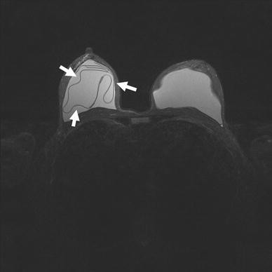
Magnetic resonance imaging (MRI) signs: the “linguine sign.” The T2-weighted STIR MRI sequence on the axial plane shows that the wall of the prosthesis is broken, whereas the fibrous capsule is intact. Thus, the wall is collapsed in its content of silicone on the right prosthetic implant (arrows). This sign is commonly known as the “linguine sign.”
-
Undercapsular streaks (“pull-away sign”). These hypointense lines form when a certain amount of gel, large or small (gel bleeding), comes out and causes a small displacement of the capsule. These lines have a characteristic trend parallel to the capsule and are of great help in differential diagnosis with radial folds [26]. This sign also is known as the “open loop sign.”
-
Teardrop sign or salad oil sign. This sign comprises actual water drops of extracapsular origin that penetrate through the semipermeable prosthetic shell. When more droplets merge to form larger droplets, the sign known as the “salad oil sign” appears (Fig. 8). The penetration of an abundant quantity of liquid at the intracapsular level is indicative of implant rupture [27].
Fig. 8 
Magnetic resonance imaging (MRI) signs: the “salad oil sign.” The turbo inversion recovery (TIRM) axial plane MRI sequence shows an intracapsular rupture of the left prosthetic implant (double lumen) with admixture of the contents (saline and silicone). This sign is commonly known as the “salad oil sign” (arrows)
-
C-sign. This sign originates from a variant of the previous sign when a real patch of silicone forms on the back of the capsule. When capsular collapse occurs, this patch takes precisely the form of a “C,” hence its name of “C-sign” [28].
-
Bulging. This phenomenon occurs when the fibrous capsule meets minor resistance (Fig. 9). It occurs with intact implants but can seriously lead to a possible rupture [29].
Fig. 9 
Magnetic resonance imaging (MRI) signs: herniation of the prosthetic implant: the “bulging.” The T2-weighted axial plane MRI sequence with suppression of silicone (silicone saturation) shows an initial bulging of the right prosthetic implant on the ipsilateral axillary extension, which was clinically determined only as a small asymmetry of the breasts (arrows)


On the basis of the findings encountered in diagnostics, it is plausible to suspect rupture in double-chamber, hydrosaline solution implants and silicone gel implants by following the guidelines provided by the assessment scheme [30] reported in Table 1. This scheme is based on identifying the presence of silicone normally located in the inner and outer lumen, both in admixture with hydrosaline solution (Fig. 10) (i.e., with the other material contained in the implants) and alone.

Magnetic resonance imaging (MRI) signs: double-chamber implant intraprosthetic rupture. Admixture of silicone with hydrosaline solution occurred. The T2-weighted axial plane MRI sequence with suppressed water signal shows the rupture of two standard double-lumen implants. This rupture is significantly more pronounced in the left implant, in which the collapse of prosthetic limbs was observed. Moreover, the hydrosaline solution (arrows) still is present in the outer lumen but now is admixed with silicone gel deriving from the rupture of the inner lumen (arrowhead). The implant as a whole is intact
Regarding extraprosthetic ruptures, the pathognomonic sign is discontinuation of the fibrous capsule, resulting in the migration of silicone gel in soft tissue or resorption of the hydrosaline solution. In such cases, at a clinical level, there is deformation and volumetric reduction of the prosthetic implant (Fig. 11). This type of rupture leads to systematic accumulation of silicone in the periprosthetic tissues and sometimes in the axillary fold and the lymph nodes, a phenomenon also well evaluated with MRI.

Magnetic resonance imaging (MRI) signs: extraprosthetic rupture of double-chamber implants with leaking of silicone. The WAT-SAT STIR T2-weighted sagittal plane MRI sequence shows an initial extraprosthetic rupture of a double-chamber implant, with rupture of the lower capsular wall and leakage of silicone in breast tissue periprosthetically (arrows)
The leaking of silicone in the periprosthetic tissues may lead to the formation of siliconomas, which are well differentiated from seromas with the use of specific MRI sequences (Fig. 12) and from silicone cysts. Siliconomas, which give a high signal on T1 and T2 and a low signal on fat-suppression sequences, are frequently placed in differential diagnoses with seromas and require surgery.

Magnetic resonance imaging (MRI) signs: extraprosthetic rupture of double-chamber implants with leaking of silicone and siliconomas formation. The sagittal T-weighted spin-echo fat saturation sagittal plane MRI sequence shows the presence of a massive siliconoma that occurs in the form of a pseudonodular inhomogeneous hypointense area (arrows) as the result of an extraprosthetic rupture
Discussion
In recent years, the use of breast implants has considerably increased because of both the augmentation of diagnosed breast cancer, due to finer diagnostic facilities and technologies available, and the desire of many patients to have a more abundant or tonic breast. This proliferation in demand translates into the use of plastic surgery related to reconstructive surgery in the former case and to cosmetic surgery in the latter case.
With the increase in prosthetic breast implant operations, the need for diagnostic imaging has become more frequent. The diagnostic techniques are therefore required to be more advanced, accurate, and sophisticated to ensure an effective study of the implanted breasts so as to allow a definite diagnosis of any breast disease but also to establish without doubt the integrity or damage of the implants themselves. Diagnostic imaging should therefore go hand in hand with the new needs of patients, and the techniques that were sufficient to date may not be so anymore. Hence, more accurate and sophisticated instruments that allow early diagnosis of diseases are needed, especially for those cases in which the gathered evidence was clinically undetectable, not due to misinterpretations of the operator but to technical limitations of the diagnostic methods used.
Diagnostic imaging for the study of patients with suspected breast implant ruptures uses different techniques for radiologic investigation. The most common radiologic techniques to date have been mammography and ultrasonography. However, the current gold standard is MRI, which has a sensitivity close to 100% because this exam has an extraordinary ability to detect even the smallest specimen. However, its specificity depends on the experience of the radiologist interpreting the findings [13].
The indications leading to the decision to use MRI and the answers expected from this technique can be summarized as assessment of the prosthesis implant’s integrity, description of the possible presence of extracapsular silicone, help in prosthetic implant replacement surgery planning, and more accurate assessment of those patients with known or suspected malignancy in the presence of implants. For a better understanding of the many actual positives and the few limitations in the application of MRI for diagnosing breast implants ruptures, we also highlight the strengths and weaknesses of the other techniques commonly used for these purposes, namely, mammography and ultrasound. We then focus on evaluation of the main MRI signs. In fact, MRI is able to highlight these signs, the so-called “MRI signs,” that once identified can provide useful guidance for the formation of contracture or rupture of the prosthesis (i.e., phenomena not clinically detectable).
Use of MRI has made it possible to determine an ultimate diagnosis and proper presurgical planning for cases in which prosthetic rupture should be present. This is a non–operator-dependent technique that offers a comprehensive and multiplanar vision due to its wide panoramic view [14, 15]. We therefore believe that this technique provides plastic surgeons with the appropriate instrument to identify, with certainty, any potential alteration of the prosthetic implants otherwise not diagnosed with traditional techniques.
Moreover, as shown in Table 1, MRI signs can be grouped and considered as parameters on which to base the assessment of alterations that may be present in the prosthetic implants. In addition, we must stress the importance of teamwork. In fact, proper cooperation and coordination between radiologists and dedicated plastic surgeons is vital to the proper management of patients and the complications they may experience.
Finally, it should be noted that to date, no universally recognized guideline exists for the normal follow-up evaluation of women with breast implants. In our opinion, for women who undergo surgery both for breast cancer and for aesthetic reasons, MRI must be preceded by mammography and ultrasonography because these two techniques can detect some findings not visible by MRI, such as microcalcifications [31].
Abbreviations
- MRI:
-
Magnetic Resonance Imaging
- CM:
-
Contrast medium
- SE:
-
Spin echo
- FSE:
-
Fast SE
- GE:
-
Gradient echo
- STIR:
-
Short tau inversion recovery sequence
- WAT SAT-STIR:
-
Water saturation with STIR sequence
- WS STIR:
-
STIR sequence with water saturation
- TIRM:
-
Turbo inversion recovery sequence
References
Eklund GW, Busby RC, Miller SH, Job JS (1988) Improved imaging of the augmented breast. AJR Am J Roentgenol 151:469–473
Miglioretti DL, Rutter CM, Geller BM, Cutter G, Barlow WE, Rosenberg R, Weaver DL, Taplin SH, Ballard-Barbash R, Carney PA, Yankaskas BC, Kerlikowske K (2004) Effect of breast augmentation on the accuracy of mammography and cancer characteristics. JAMA 291:442–450
Ahn CY, DeBruhl ND, Gorczyca DP, Shaw WW, Bassett LW (1994) Comparative silicone breast implant evaluation using mammography, sonography, and magnetic resonance imaging: Experience with 59 implants. Plast Reconstr Surg 94:620–627
Samuels JB, Rohrich RJ, Weatherall PT, Ho AM, Goldberg KL (1995) Radiographic diagnosis of breast implant rupture: current status and comparison of techniques. Plast Reconstr Surg 96:865–877
Theophelis LG, Stevenson TR (1986) Radiographic evidence of breast implant rupture. Plast Reconstr Surg 78:673–675
Beekman WH, van Straalen WR, Hage JJ, Taets van Amerongen AH, Mulder JW (1998) Imaging signs and radiologists’ jargon of ruptured breast implants. Plast Reconstr Surg 102:1281–1289
Caskey CI, Berg WA, Hamper UM, Sheth S, Chang BW, Anderson ND (1999) Imaging spectrum of extracapsular silicone: correlation of US, MR imaging, mammographic, and histopathologic findings. Radiographics 19:S39–S51; quiz S261–S262
Scaranelo AM, Marques AF, Smialowski EB, Lederman HM (2004) Evaluation of the rupture of silicone breast implants by mammography, ultrasonography and magnetic resonance imaging in asymptomatic patients: correlation with surgical findings. Sao Paulo Med J 122:41–47
DeBruhl ND, Gorczyca DP, Ahn CY, Shaw WW, Bassett LW (1993) Silicone breast implants: US evaluation. Radiology 189:95–98
Hölmich LR, Fryzek JP, Kjøller K, Breiting VB, Jørgensen A, Krag C (2005) McLaughlin JK: The diagnosis of silicone breast-implant rupture: clinical findings compared with findings at magnetic resonance imaging. Ann Plast Surg 54:583–589
Levine RA, Collins TL (1991) Definitive diagnosis of breast implant rupture by ultrasonography. Plast Reconstr Surg 87:1126–1128
Cilotti A, Marini C, Iacconi C, Mazzotta D, Moretti M, Giaconi C, Bartolozzi C (2006) Ultrasonographic appearance of breast implant complications. Ann Plast Surg 56:243–247
Di Benedetto G, Cecchini S, Grassetti L, Baldassarre S, Valeri G, Leva L, Giuseppetti GM, Bertani A (2008) Comparative study of breast implant rupture using mammography, sonography, and magnetic resonance imaging: correlation with surgical findings. Breast J 14:532–537
Piccoli CW (1994) Invited discussion: Imaging modalities for breast implants. Ann Plast Surg 33:256–257
Reynolds HE, Buckwalter KA, Jackson VP, Siwy BK, Alexander SG (1994) Comparison of mammography, sonography, and magnetic resonance imaging in the detection of silicone-gel breast implant rupture. Ann Plast Surg 33:247–255 discussion 256–257
Huch RA, Kunzi W, Debatin JF, Wiesner W, Krestin GP (1998) MR imaging of the augmented breast. Eur Radiol 8:371–376
Pfleiderer B, Garrido L (1995) Migration and accumulation of silicone in the liver of women with silicone gel-filled breast implants. Magn Reson Med 33:8–17
Silver RM, Sahn EE, Allen JA et al (1993) Demonstration of silicone in sites of connective-tissue disease in patients with silicone-gel beast implants. Arch Dermatol 129:63–68
Hirmand H, Hoffman LA, Smith JP (1994) Silicone migration to the pleural space associated with silicone gel augmentation mammaplasty. Ann Plast Surg 32:645–647
Gorczyca DP, Sinha S, Ahn CY, DeBruhl ND, Hayes MK, Gausche VR, Shaw WW, Bassett LW (1992) Silicone breast implants in vivo: MR imaging. Radiology 185:407–410
Ahn CY, Shaw WW, Narayanan K, Gorczyca DP, Sinha S, Debruhl ND, Bassett LW (1993) Definitive diagnosis of breast implant rupture using magnetic resonance imaging. Plast Reconstr Surg 92:681–691
Soo MS, Kornguth PJ, Walsh R, Elenberger CD, Georgiade GS (1996) Complex radial folds versus subtle signs of intracapsular rupture of breast implants: MR findings with surgical correlation. AJR Am J Roentgenol 166:1421–1427
Berg WA, Anderson ND, Zerhouni EA, Chang BW, Kuhlman JE (1994) MR imaging of the breast in patients with silicone breast implants: normal postoperative variants and diagnostic pitfalls. AJR Am J Roentgenol 163:575–578
Dobke MK, Middleton MS (1994) Clinical impact of breast implant magnetic resonance imaging. Ann Plast Surg 33:241–246
Stroman PW, Rolland C, Dufour M, Grondin P, Guidoin RG (1996) Appearance of low signal intensity lines in MRI of silicone breast implants. Biomaterials 17:983–988
Quinn SF, Neubauer NM, Sheley RC, Demlow TA, Szumowski J (1996) MR imaging of silicone breast implants: evaluation of prospective and retrospective interpretations and interobserver agreement. J Magn Reson Imaging 6:213–218
Berg WA, Caskey CI, Hamper UM, Anderson ND, Chang BW, Sheth S, Zerhouni EA, Kuhlman JE (1993) Diagnosing breast implant rupture with MR imaging, US, and mammography. Radiographics 13:1323–1336
Gorczyca DP, DeBruhl ND, Mund DF, Bassett LW (1994) Linguine sign at MR imaging: does it represent the collapsed silicone implant shell? Radiology 191:576–577
Berg WA, Nguyen TK, Middleton MS, Soo MS, Pennello G, Brown SL (2002) MR imaging of extracapsular silicone from breast implants: diagnostic pitfalls. AJR Am J Roentgenol 178:465–472
Middleton MS, McNamara MP Jr, Edelman R, Hesselink J (2005) Clinical magnetic resonance imaging. Elsevier, pp 2473–2474
Bazzocchi M, Zuiani C, Panizza P, Del Frate C, Sardanelli F, Giuseppetti GM, Simonetti G, Lattanzio V, Del Maschio A (2006) Contrast-enhanced breast MRI in patients with suspicious microcalcifications on mammography: results of a multicenter trial. AJR Am J Roentgenol 186:1723–1732
Conflicts of interest
The authors declare that they have no conflicts of interest to declare.
Author information
Authors and Affiliations
Corresponding author
Rights and permissions
About this article
Cite this article
Colombo, G., Ruvolo, V., Stifanese, R. et al. Prosthetic Breast Implant Rupture: Imaging—Pictorial Essay. Aesth Plast Surg 35, 891–900 (2011). https://doi.org/10.1007/s00266-011-9694-z
Received:
Accepted:
Published:
Issue Date:
DOI: https://doi.org/10.1007/s00266-011-9694-z