Abstract
Objective
To retrospectively analyze magnetic resonance (MR) findings in patients with popliteal arterial entrapment syndrome.
Materials and methods
This study was a retrospective MRI and CT scan review of 12 patients with 23 limbs with popliteal artery entrapment syndrome (PAES) treated over a 10-yr period. All 12 patients (23 limbs) were evaluated with MR and CT scan (11 patients—bilateral sides; one patient—unilateral side). All cases were classified as to various types of anomalous relationships between the popliteal artery and the neighboring muscles. The PAES was classified to gastrocnemius medial head and lateral head anomaly. Gastrocnemius medial head anomaly was classified according to the classification made by Whelan and Rich, from type 1 to type 6 [12, 13]. Gastrocnemius lateral head anomaly was defined as popliteal artery entrapment due to medially inserted gastrocnemius lateral head or aberrant accessory head of gastrocnemius lateral head.
Results
The gastrocnemius medial head anomaly was found in 14 limbs (14/23). The classic type 1 was found in none, type 2 in five patients (six limbs), type 3 in four patients (five limbs), type 4 in none, type 5 in one patient (one limb) and type 6 in one patient (two limbs). The uncommon type, i.e. lateral head of gastrocnemius anomaly, was found in five patients (eight limbs).
Conclusion
The gastrocnemius medial head anomaly was the cause of PAES, and PAES was classified by medial head anomaly. However the gastrocnemius lateral head anomaly was also the cause of PAES, and most cases of gastrocnemius lateral head anomaliy showed aberrant accessory slip which entrapped the popliteal artery and vein.
Introduction
PAES is a congenital anomaly of muscle or tendon insertion in relation to the popliteal artery that causes functional occlusion of the artery.
Since the first report by Stuart in 1879 [1], many clinical and radiologic articles about popliteal artery entrapment syndrome (PAES) have been published, including digital subtraction arterography (DSA), CT arterography (CTA) and MR arterography of PAES [2–8]. However, there has been only limited published data of radiologic findings using MRI for bilateral limbs [2–10], and PAES caused by gastrocnemius lateral head anomaly has only rarely been reported [23–25].
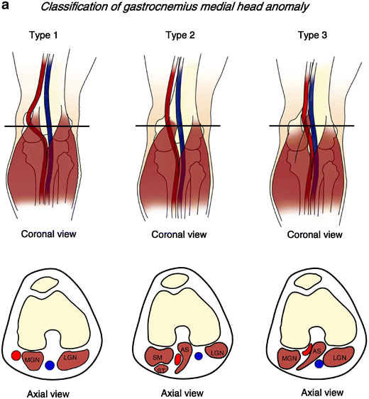
Due to the complexity of the embryologic development, various classifications of the anatomical abnormalities that can lead to PAES have been proposed [1, 5, 10–12]. The mostly widely accepted classification was made by Whelan and modified by Rich, and divides PAES into six types [12, 13]. According to this classification, type 1 is described as an aberrant medial arterial course around the normal medial head of the gastrocnemius muscle. Type 2 is the abnormal gastrocnemius medial head with lateral insertion on the distal femur and medial displacement of the popliteal artery. In type 3, an aberrant accessory slip from the medial head of the gastrocnemius muscle wraps around the normally positioned popliteal artery and entraps it. In type 4, the popliteal artery is located deep in the popliteus muscle or beneath fibrous bands in the popliteal fossa. Type 5 is any form of entrapment that involves the popliteal artery and vein [12–19]. Type 6, the functional type, has been described in symptomatic individuals with a normally positioned popliteal artery entrapped by normally positioned gastrocnemius with hypertrophy (Fig. 1) [20–22].
a,b Six subtypes of gastrocnemius medial head anatomical variations causing PAES. Type 1: an aberrant medial arterial course around normal medial head of gastrocnemius muscle. Type 2: abnormal head of the gastrocnemius muscle which is laterally inserted on the distal femur with medial displacement of popliteal artery. Type 3, an aberrant accessory slip from the medial head of the gastrocnemius muscle wraps around the normally positioned popliteal artery and entraps it. Type 4: the popliteal artery located deep in the popliteus muscle or beneath fibrous bands in the popliteal fossa. Type 5: any form of entrapment that involves the popliteal artery and vein. Type 6, functional type normally positioned popliteal artery which is entrapped by normally positioned gastrocnemius with hypertrophy
The rare types of PAES caused by anomalous lateral head of the gastrocnemius have not yet been classified, and there have been few case reports [23–25]. In this study, we described the MRI findings of popliteal artery entrapment syndrome, according to the most widely accepted classification of gastrocnemius medial head anomaly, and a more rare gastrocnemius lateral head anomaly. We also analyzed the correlation of the anatomic variations which were proven on axial study, with severity of symptom.
Materials and methods
Materials
Our study included 12 patients with 23 limbs who underwent MR (12 patients, 21 limbs) and CT (five patients, ten limbs) for calf claudication between January 1995 and September 2005. Both MR and CT were performed in five patients with eight limbs.
Four of the 12 patients (eight limbs) were evaluated with MR angiography, five patients (ten limbs) with CT angiography and five patients (ten limbs) with digital subtraction angiography, with provocative movement, plantar flexion and dorsi flexion of foot. The patients ranged in age from 18 to 54 years with a mean age of 25 years. All patients were physiologically active males. Seven patients (58%) had symptoms in both limbs. All patients were suspected of PAES before undergoing imaging studies, as a result of clinical findings, ABI (ankle brachial index) and screening continuous wave Doppler ultrasonography of the popliteal artery. All patients were treated by diverse treatments, including conservative treatment, myotomy and myotomy with angioplasty.
Methods
MR was performed with a 1.5 T Magnetom Vision (Siemens, Erlangen, Germany), 1.5 T Gyroscan (Philips, Eindhoven, Netherlands) and 3.0 T Gyroscan (Philips, Eindhoven, Netherlands) scanners. The knee was centered in a standard knee coil and immobilized with foam inserts. The MR sequence included spin echo T1-weighted axial, sagittal (TR/TE 400–500 ms/17–20 ms, 2 NEX), FLASH 2d axial (TR/TE=700/18, flip angle 20°, 2 NEX), turbo spin echo T2-weighted sagittal (TR/TE=3000–3500/90–100, 2 NEX), proton sagittal and coronal (TR/TE=2500–3700/20, 2 NEX). We applied more extended field of view (18 cm) than routine knee MRI (16cm) in the case of PAES.
MRA was performed with Gd-enhanced 3D MRA with 1.5 T Gyroscan (Philips, Eindhoven, Netherlands), and the patient was in supine and foot plantar flexion position. A 0.2 mmol/kg dose of melgumine gadoterate (20 ml; Dotarem; Guerbet, Aulnay-sous-Bois, France) was injected at 4 ml/sec. Single-station coronal Gd-enhanced 3D MRA using 3D-TFE(turbo field echo) (TR 5–6 msec/TE 1–3 msec, flip angle 40 degree, FOV 36–40 cm, matrix 368–512, slice thickness 0.75 mm) using elliptical centric k-space sampling was then preformed. We obtained arterial and venous phase and it took 5 minutes.
CT angiography was performed with a 16-detector row CT scanner (Sensation 16; Siemens, Forchheim, Germany). All patients were placed in the supine and neutral position of their feet. The scanning range included the aortoiliac and lower extremity arteries. Twenty seconds after the bolus injection of contrast agent, scanning was obtained. A nonionic iopromide (Ultravist 370; Schering, Berlin, Germany) was administered via an 18-gauge needle, in the antecubital vein, at a flow rate of 4 ml/sec, 150 ml. After reaching the preset contrast-enhancement level of 100 HU at descending aorta, data acquisition was performed with the patient in foot plantar flexion, in a craniocaudal direction with a section thickness of 0.75 mm and a 0.5-second gantry rotation time (pitch, 1.5). The X-ray tube voltage setting was 120 kV, and mean tube current was 210 mA. Transverse sections were reconstructed for both extremities on a work station, with a section thickness of 0.75 mm at an interval of 0.4 mm. For each patient, maximum intensity projection images were created.
DSA was the gold standard for diagnosis of PAES, but due to invasiveness it was replaced with CTA, after CTA was available.
All images were retrospectively reviewed by two experienced musculoskeletal radiologists. Differences of opinions were resolved by consensus. The reviewing radiologists were not masked to the clinical information.
Imaging findings were analyzed according to the following classification and criteria.
All patients were classified into gastrocnemius medial head anomaly and lateral head anomaly. The medial head anomaly was classified according to the most widely accepted classification made by Whelan and Rich, from type 1 to type 6 [12, 13]. In our study, these types were sub-classified according to the high insertion of the gastrocnemius medial head and the combined abnormal structure (bony tubercle, ganglion cyst, accessory slip not originated from femoral condyle). As there is no classification of gastrocnemius lateral head anomaly, in our study we classified the lateral head anomaly according to two types, i.e. the abnormal insertion type and the aberrant accessory slip.
In the abnormal insertion type, the lateral gastrocnemius muscle was medially inserted onto the distal femur. In the aberrant accessory slip type, an aberrant accessory slip which originates from the midline posterior cortex of the distal femoral metaphysis with entrapment of the popliteal vessel and joins the gastrocnemius lateral head in the caudal portion.
Two subtypes of gastrocnemius lateral head anatomical variations causing PAES. Abnormal insertion type: lateral gasrocnemius muscle medially inserted on the distal femur. Aberrant accessory slip type: an aberrant accessory slip from the lateral head of the gastrocnemius muscle wraps around the normally positioned popliteal artery and entraps it
Eleven patients (22 limbs) were evaluated for bilateral limbs, and we analyzed the relationships of bilaterality of radiologic anomaly and the presentation of symptomatic severity. Five patients (eight limbs) were evaluated with both CT and MRI, and the findings of CT and MRI were compared. MRA were performed on four patients (eight limbs), CTA on five patients (ten limbs), and DSA on five patients (ten limbs), and the findings of CTA, MRA and DSA were then compared.
Results
On screening continuous wave Doppler ultrasonography, all 12 patients showed normal flow or disturbed but sustained flow in neutral position and absence of arterial flow in plantar flexion and dorsiflexion.
The clinical profiles and imaging findings of popliteal fossa in the study patients with PAES are summarized in Table 1. Fourteen limbs in eight patients (14/23, 60%) had gastrocnemius medial head anomaly, and eight limbs in five patients (8/23, 34%) had gastrocnemius lateral head anomaly. One patient showed normal finding in one limb and gastrocnemius medial head anomaly in the other limb. The medial head anomaly was classified into six types. There were no examples of Types 1 or 4. Six limbs (26%) had type 2, laterally inserted MGN, (Fig. 3).
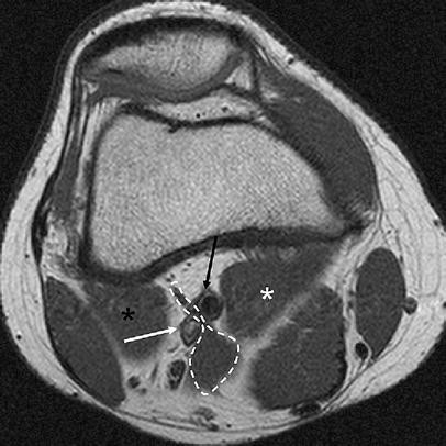
Five limbs (21%) had type 3, an aberrant accessory slip from MGN (Fig. 4). One limb (4%) had type 5, any form of entrapment that involves the popliteal artery and vein (Fig. 5). One patient had type 6, no anatomic variant, but bilateral gastrocnemius medial and lateral head hypertrophy in both limbs (8%) (Fig. 6). The gastrocnemius lateral head anomaly with abnormal insertion type, was present in one limb (1/23, 4%) (Fig. 7), while the aberrant accessory head type was found in seven limbs (7/23, 30%) ( Fig. 8) (Table 2). All gastrocnemius lateral head anomaly cases showed entrapment of both popliteal artery and vein.
Right knee of a 17-year-old male with PAES, type 3 MGN anomaly. T1-weighted axial image shows aberrant accessory muscle slip of medial head of right gastrocnemius muscle (outlined) and entrapped popliteal artery (black arrow). Medial head of right gastrocnemius muscle from medial femoral condyle (white star) and lateral head of right gastrocnemius muscle from lateral femoral condyle (black star), are normally positioned. Abnormal accessory muscle slip of medial head of right gastrocnemius muscle joins to normally positioned medial head of right gastrocnemius muscle in caudal portion (not illustrated). Popliteal vein; white arrow
Right knee of a 21-year-old male with PAES, type 5 MGN anomaly. T1-weighted axial image shows a laterally displaced medial head of right gastrocnemius muscle (outlined). Both popliteal artery (black arrowhead) and popliteal vein (white arrowhead) are displaced laterally. Lateral head of right gastrocnemius muscle; black star
Left knee of a 24-year-old male with PAES type 6 MGN anomaly. T1-weighted axial image shows hypertrophied but normally positioned medial head of left gastrocnemius muscle (white star) and normally positioned lateral head of left gastrocnemius muscle (black star) which wrap around normally positioned popliteal artery (black arrow head) and popliteal vein (white arrow head)
a Right knee of an 18-year-old male with PAES, LGN anomaly with abnormal insertion type. Fl2d-axial image shows medially positioned lateral head of right gastrocnemius muscle (outlined); popliteal artery and veins (arrowheads) are entrapped between lateral head of right gastrocnemius muscle and semimembranosus muscle (black star) b caudal portion shows gastrocnemius lateral head (black star) and gastrocnemius medial head (white star)
a,b Left knee of an 18-year-old male with PAES, LGN anomaly with aberrant accessory slip. Axial T1-weighted image shows aberrant accessory slip of lateral head of left gastrocnemius muscle (outlined). Popliteal artery and veins (arrowheads) are entrapped between aberrant accessory slip of lateral head of left gastrocnemius muscle (outlined) and semimembranosus muscle (black star). c Left knee of an 18-year-old male with PAES, LGN anomaly with aberrant accessory slip. Axial T1-weighted image at caudal level shows aberrant accessory slip of lateral head of left gastrocnemius muscle (outlined) passing parallel to normally positioned lateral head of gastrocnemius lateral head (two black stars). Medial head of gastrocnemius muscle (white star), semimembranosus muscle (black star), popliteal artery and veins (black arrowheads) are illustrated
One patient with type 2 gastrocnemius medial head anomaly also had aberrant accessory slip of the lateral head. This type was classified as gastrocnemius lateral head anomaly with variant (Fig. 9).
a and b Right knee of a 54-year-old male with LGN anomaly combined MGN type 2. Axial T1-weighted image shows aberrant accessory slip of lateral head of right gastrocnemius muscle (outlined; white line) and laterally displaced medial head of gastrocnemius muscle (outlined; black line). Popliteal artery and veins (arrowheads) are entrapped between two abnormal muscle slips
Eleven patients were evaluated for bilateral limbs and one patient was evaluated for a unilateral symptomatic site. Ten patients showed an anatomic anomaly in both limbs (10/11, 90%), of which six had symptoms of PAES in both limbs, and four patients had symptoms of PAES in one side. All six patients with gastrocnemius medial head anomaly with combined abnormal structure were symptomatic unilaterally, or had more severe symptoms in the unilateral side; these abnormal structures included ganglion cyst, bony tubercle, accessory slip with more prominent muscle belly than the less symptomatic site, and combination of gastrocnemius medial head anomaly and gastrocnemius lateral head anomaly (Table 3).
In 11 patients who were evaluated for bilateral limbs, four patients (eight limbs) showed the same type of classification and seven patients (14 limbs)- showed a mixture of different types of classification (Table 1).
Five patients (8 limbs) were evaluated with both CT and MRI, and there were no discrepancies in classifying the anatomic variants between the findings of CT and MRI.
Five patients (ten limbs) were evaluated using two methods: three patients (six limbs) with both MRA and DSA, and two patients (four limbs) with both CTA and DSA. Three patients (six limbs) showed similar results between CTA and DSA or MRA and DSA, but in two patients (four limbs), the arterial luminal narrowing was underestimated on MRA compared to DSA. These cases showed normal lumen (one limb), segmental narrowing (one limb), and eccentric narrowing (two limbs) on MRA , but all these cases showed total occlusion on DSA with foot dorsi flexion and plantar flexion views (Fig. 10).
Right knee of a 24-year-old male with LGN anomaly with aberrant accessory slip. a Axial T1-weighted image shows aberrant accessory slip (outlined) of lateral head of gastrocnemius muscle. Popliteal vessels (arrowheads) are entrapped between medial head of gastrocnemius (white star) and aberrant accessory slip. b MRA shows unremarkable popliteal artery in right knee. c Angiography with neutral position shows unremarkable right popliteal artery. d Angiography with foot plantar flexion shows luminal narrowing of right popliteal artery
Discussion
Popliteal artery entrapment is second only to atherosclerosis as the most common surgically correctible cause of leg claudication in young adults [11, 12, 14, 16, 17]. The true incidence of anatomical PAES is not known, but is likely to be more common than currently thought. Early reports cited a prevalence of 0.165% in young males entering military service, and a postmortem study found PAES in 3.5% of typically young athletic male patients (60% <30 years old, 15:1 male predilection) [16–19]. Our study showed similar results, in that all patients were men whose ages at presentation ranged from 18 to 54 years.
PAES is caused by an abnormal relationship between the popliteal artery and the gastrocnemius medial head. Rosset et al. reviewed 280 cases of popliteal entrapment, and using Whelan’s classification classified 19% as type 1, 25% as type 2, 30% as type 3, 8% as type 4, and 18% as other types [26, 27].
In our study, type 2 was also the most common, but we had no type 1 or type 4 cases.
In addition, in our study the gastrocnemius lateral head anomaly that was not described in the study by Rosset et al. was relevant. Most of the cases of gastrocnemius lateral head anomaly showed aberrant accessory slip of gastrocnemius lateral head, and entrapment of both popliteal artery and vein.
Type 6, functional PAES, a term first coined by Rignault et al., describes a situation where the popliteal artery, vein, and adjacent musculature are in normal anatomic positions but the muscles are hypertrophied [20–22]. Our case also showed hypertrophy of the gastrocnemius and entrapment between the hypertrophied gastrocnemius medial and lateral head. The gastrocnemius lateral head anomaly is known as a rare cause of PAES; in a review of the medical literature, we found only seven reported medical records and two radiologic case reports [23–25]. In our study, the gastrocnemius lateral head anomaly was relatively common (8/23, 34%) and most of these patients had aberrant accessory head.
In contrast, with a gastrocnemius medial head anomaly, only the type 5 MGN type showed involvement of both the popliteal artery and vein; in all gastrocnemius lateral head anomalies, both the artery and vein were involved in our study.
PAES has been found to occur bilaterally in approximately 25% of cases. However, in 1989 Collins et al. found bilateral popliteal entrapment in 8/12 patients for an incidence of 67%; however, only three of 12 patients (25%) had symptoms in both legs [28]. In our study, bilateral popliteal entrapment was observed in most patients (10/12, 81%), and the patients who had a symptomatic dominant side showed combined anomalies such as ganglion cyst, bony tubercle, accessory slip with more prominent muscle belly than less symptomatic site, and combination of gastrocnemius medial head anomaly and gastrocnemius lateral head .
In the classification of PAES, there was no discrepancy between CT and MRI in our study, but the advantages of MRI include its noninvasive nature, its lack of ionizing radiation and its ability to provide surgically relevant anatomic detail with higher soft-tissue contrast.
In PAES, angiographic evaluation alone might lead to overlooking the underlying cause of the thrombosis, which could lead to unsuccessful angioplasty. MR evaluation gives a detailed description of the anatomy and diverse findings of abnormalities, and these are more useful for deciding the appropriate surgical procedure rather than trying to fit every case into a reclassified subtype. In particular, variations in the insertion point of the muscle are so different in every case that this aspect must be evaluated separately in each patient [2, 4].
The limitation of MRA in contrast to DSA is the tendency of MRA to underestimate the degree of stenosis, especially stenosis less than 50%, in peripheral vessels. The underestimation is due to the decreased spatial resolution of MRA and related volume-averaging effects. Forster et al. suggested that as dynamic MRA underestimates the degree of stenosis relative to DSA, MR examinations demonstrating stenosis of 50% or less might be false-negative studies. They recommended that these patients undergo arteriography [4].
The differential diagnosis of PAES in young patients without generalized atherosclerosis includes trauma and cystic adventitial disease [29, 30]. In CAD, the cystic change of adventitia induces concentric or eccentric stenosis of the arterial lumen. The MR finding of CAD is cystic lesion with hyperintensity on T2-weighted MR images and variable signal intensity on T1-weighted images, due to the variable amount of mucoid material within the cyst and luminal compression on axial images [31, 32].
Our study has some limitations. As this was a retrospective study, many patients with popliteal entrapment syndrome were undoubtedly not included in the data analysis. This is unavoidable, however, as the study patients were selected on the basis of the clinical signs of popliteal artery entrapment. Other limitations of our study include the small sample size and the limited number of MRI and MRA scans. As for PAES provocative maneuvers in MRA or CTA, there is also the potential for lack of patient cooperation. Finally, the evaluation of the MR studies lacked blinding of clinical diagnosis.
In summary, we analyzed the MR findings in a subset of patients with clinical and surgical evidence of popliteal entrapment syndrome. PAES is classified by a medial head and also a lateral head anomaly. The bilaterality of anatomic anomaly was common in our study, and in the cases with combined abnormal structure (ganglion cyst, bony tubercle, accessory slip with prominent muscle belly, associated other type of gastrocnemius muscle anomaly) showed symptomatic unilaterality and more severe symptom in ipsilateral side.
MRI provides detailed information including classification of PAES, combined anomaly and the relationship of the artery to adjacent structures.
References
Stuart TP. Note on a variation in the course of the popliteal artery. J Anat Physiol 1879;13:162
Atilla S, Ilgit ET, Akpek S, Yucel C, Tali ET, Isik S. MR imaging and MR angiography in popliteal artery entrapment syndrome. Eur Radiol 1998;8:1025–1029
Takase K, Imakita S, Kuribayashi S, Onishi Y, Takamiya M. Popliteal artery entrapment syndrome: Aberrant origin of gastrocnemius muscle shown by 3D CT. J Comput Assist Tomogr 1997;21:523–528
Forster BB, Houston JG, Machan LS, Doyle L. Comparison of two-dimensional time-of-flight dynamic magnetic resonance angiography with digital subtraction angiography in popliteal artery entrapment syndrome. Canadian Assoc Radiol J 1997;48:11–18
McGuinness G, Durham JD, Rutherford RB, Thickman D, Kumpe DA. Popliteal artery entrapment: findings at MR imaging. J Vasc Interv Radiol 1991;2:241–245
Wehmann TW. Computed tomography in the diagnosis and management of popliteal artery entrapment syndrome. J Am Osteopath Assoc 1993;93:1039–1042, 1047–1050
Macedo TA, Johnson CM, Hallett JW, Breen JF. Poplieal artery entrapment syndrome; Role of imaging in the diagnosis. AJR Am J Roentgenol 2003;181:1259–1265
Ring DH Jr, Haines GA, Miller DL. Popliteal artery entrapment syndrome: arteriographic findings and thrombolytic therapy. J Vasc Interv Radiol 1999;10:713–721
Ohta M, Kusaba A, Shrestha D, et al. Popliteal artery entrapment syndrome. J Cardiovasc Surg 1991;32:697–701
Haidar S, Thomas K, Miller S. Popliteal artery entrapment syndrome in a young girl. Pediatr Radiol 2005;35:440–443
Baltopoulos P, Filippou DK, Sigala F. Popliteal entrapment syndrome: anatomic or functional syndrome? Clin J Sport Med 2004;14:8–12
Whelan TJ, Haimovici H (eds) Vascular surgery: principles and techniques, 2nd edn. McGraw-Hill, New York 1984;557–567
Rich NM, Collins GJ Jr, McDonal PT, Kozloff L, Clagett GP, Collins JT. Popliteal vascular entrapment: its increasing interest. Arch Surg 1979;114:1377–1384
Love JW, Whelan TJ. Popliteal artery entrapment syndrome. Am J Surg 1965;109:620–624
Fujiwara H, Sugano T, Fujii N. Popliteal artery entrapment syndrome: accurate morphological diagnosis utilizing MRI. J Cardiovasc Surg 1992;33:160–162
Gerkin TM, Beebe HG, Williams DM, Bloom JR, Wakefield TW. Popliteal vein entrapment presenting as deep venous thrombosis and chronic venous insufficiency. J Vasc Surg 1993;18:760–766
Fowl RJ, Kempczinski RF. Popliteal artery entrapment. In: Rutherford RB (eds) Vascular surgery, 5th edn. Saunders, Philadelphia, Pa 2000;1087–1093
Gibson MH, Mills JG, Johnson GE, Downs AR. Popliteal entrapment syndrome. Ann Surg 1977;185:341–348
Levien LJ, Veller MG. Popliteal artery entrapment syndrome: more common than previously recognized. J Vasc Surg 1999;30:587–598
Chernoff DM, Walker AT, Khorasani R, Polak JF, Jolesz FA. Asymptomatic functional popliteal artery entrapment: demonstration at MR imaging. Radiology 1995;195:176–180
Rignault DP, Pailler JL, Lunel F. The functional popliteal artery entrapment syndrome. Int Angiol 1985;4:341–343
de Almeida MJ, Bonetti Yoshida W, Habberman D, Medeiros EM, Giannini M, Ribeiro de Melo N. Extrinsic compression of popliteal artery in asymptomatic athlete and non-athlete individuals. A comparative study using duplex scan (color duplex sonography). Int Angiol 2004;23:218–229, Sep
Liu PT, Moyer AC, Huettl EA, Fowl RJ, Stone WM. Popliteal vascular entrapment syndrome caused by a rare anomalous slip of the lateral head of the gastrocnemius muscle. Skelet Radiol 2005;34:359–363
Turnipseed WD. Clinical review of patients treated for atypical claudication: a 28-year experience. J Vasc Surg 2004;40:79–85
Choghari C, Bosschaerts T, Locufier JL, Barthel J, Barroy JP. Variant form of popliteal artery entrapment syndrome. Acta Chir Belg 1993;93:34–37
Levien LJ. Popliteal artery entrapment syndrome. Semin Vasc Surg 2003;16(3):223–231
Rosset E, Hartung O, Burnet C, et al. Popliteal artery entrapment syndrome. Surg Radiol Anat 1995;17:161–169
Collins PS, McDonald PT, Lim RC. Popliteal artery entrapment: an evolving syndrome. J Vasc Surg 1989;10:484–490
Wright LB, Matchett WJ, Cruz CP, et al. Popliteal artery disease: diagnosis and treatment. Radiographics 2004;24:467–479
Rutherford RB, Baker JD, Ernst C, et al. Recommended standard for reports dealing with lower extremity ischemia: revised revision. J Vasc Surg 1997;26:517–538
Elias DA, White LM, Rubenstein JD, Christakis M, Merchant N. Clinical evaluation of MR imaging features of popliteal artery entrapment and cystic adventitial disease: pictorial essay. AJR Am J Roentgenol 2003;180:627–632
Jasinski RW, Masselink BA, Partridge RW, Deckinga BG, Bradford PF. Adventitial cystic disease of the popliteal artery. Radiology 1987;163:153–155
Author information
Authors and Affiliations
Corresponding author
Rights and permissions
About this article
Cite this article
Kim, H.K., Shin, M.J., Kim, S.M. et al. Popliteal artery entrapment syndrome: morphological classification utilizing MR imaging. Skeletal Radiol 35, 648–658 (2006). https://doi.org/10.1007/s00256-006-0158-5
Received:
Revised:
Accepted:
Published:
Issue Date:
DOI: https://doi.org/10.1007/s00256-006-0158-5


