
Abstract
Objective
To assess the clinical and radiological features of systemic sclerosis (SSc) joint involvement in a prospective cross-sectional study.
Design and patients
Seventy-six consecutive patients with SSc divided into clinical and serological subsets were investigated. Clinical and radiological assessments of the hands and feet were carried out. Three radiological patterns of inflammatory, degenerative and fibrotic changes were predefined. The Health Assessment Questionnaire (HAQ) disability index (DI) and individual components of the HAQ-DI were also evaluated.
Results
The highest impairments on the HAQ-DI (median 0.44; range 0–2.87) were detected in subdimensions such as hygiene, grip and activity components. Clinically articular involvement, arthralgia and finger contractures were seen more frequently than arthritis, and a significantly higher prevalence of finger flexion was found in patients with diffuse cutaneous SSc (P=0.03) compared with the other SSc subtypes. Radiologically, distal interphalangeal joint space narrowing and flexion deformity indicating periarticular fibrosis were frequently detected. Juxta-articular osteoporosis, joint space narrowing and flexion contractures of the fingers were seen significantly more frequently in the hands. A significantly higher frequency of fibrotic pattern were found in the hands whereas a degenerative pattern was more frequent in the feet (P<0.05). Finally, significant correlations were detected between flexion contractures and a radiological fibrotic pattern (P<0.001), and the severity scores of peripheral vascular impairment (P=0.026) and skin (P=0.007).
Conclusion
This cross-sectional prospective study confirms that an arthropathy is common in SSc patients and shows that it is a major determinant of disability. A classification of radiological alterations into three specific patterns is proposed.
Introduction
Systemic sclerosis (SSc) is a connective tissue disease characterized by deposition of collagen and other components of extracellular matrix in the skin and target internal organs [1]. During the disease course, many patients develop joint involvement which is manifest clinically as arthralgia and/or arthritis and/or flexion contractures, and radiologically as osteopenia, joint space narrowing, erosions and subluxation [2, 3]. Articular involvement in SSc could depend either on periarticular fibrosis or synovitis or even on an overlapping rheumatoid arthritis [4], the prevalence of each type of joint involvement not having been accurately defined. We have recently investigated articular involvement in a longitudinal retrospective study of 100 SSc patients [5]. We present the results of a cross-sectional prospective study on the clinical and radiological features of joint involvement in a cohort of consecutive SSc patients observed during 1 year.
Materials and methods
Seventy-six patients with SSc, all of whom fulfilled the ACR preliminary criteria for the classification of systemic sclerosis (formerly, ARA) [6], consecutively admitted to the Rheumatology Unit of the Second University of Naples from January 1 to December 31 2002 were invited to take part to study, and gave their informed consent.
The patients were classified into three clinical SSc subsets according to Giordano et al. [7], Ferri et al. [8] and Scussel-Lonzetti et al. [9](i.e., limited cutaneous systemic sclerosis (lcSSc), including SSc sine scleroderma; intermediate cutaneous systemic sclerosis (icSSc); and diffuse cutaneous systemic sclerosis (dcSSc)), and into three serological subsets (i.e., anticentromere antibody (ACA)-positive; anti-DNA topoisomerase I antibody (anti-Scl-70)-positive; and antinuclear antibody (ANA)-positive, with undetectable ACA and/or anti-Scl-70.
Clinical study
All the patients were investigated according to procedures and methods proposed to improve the comparability among different series [10, 11, 12, 13, 14, 15, 16, 17, 18, 19]. The disease duration was measured from the onset of the first symptom, usually Raynaud’s phenomenon. Table 1 lists the epidemiological and clinical features of the patients investigated.
Articular involvement was assessed by recording the minimal distance (mm) between the nailtip of the middle finger and the transverse palmar creases in both hands (normally no distance can be measured) [16] and the active extension of the hands was evaluated by measuring the distance (mm) between the nailtips of the thumb and little finger [20].
The Italian version of the Stanford Health Assessment Questionnaire (HAQ) disability index (DI) [21, 22] was used to evaluate functional disability. The results were expressed both as the highest score in each subdimension and as HAQ-DI.
ESR was measured according to the Westergren method; C-reactive protein (CRP) (cutoff level 0.5 mg/ml) and rheumatoid factor were tested by immunonephelometric assay (using a Behring nephelometer) (cutoff level 18 UI/ml). ANA and ACA were assessed by indirect immunofluorescence on HEp-2 cells as substrate (cutoff level 1:40), and anti-Scl-70 by ELISA (cutoff level 20 EU/ml).
Radiographic study
Radiographic evaluation included posteroanterior and oblique views of the hands and wrists, and posteroanterior and lateral views of the feet. All radiographs were evaluated without knowledge of the clinical or serological data of the patients examined.
Radiographic abnormalities of joints were differentiated according to our previous experience [5] into three predefined categories:
- Inflammatory joint pattern::
-
occurrence of juxta-articular osteoporosis, space narrowing of the proximal interphalangeal (PIP) and/or carpal joints, and/or erosive changes, either with or without digital flexion (Fig. 1).
Fig. 1 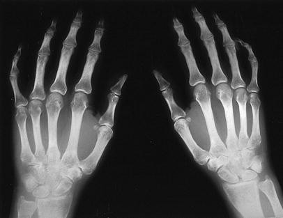
Juxta-articular osteoporosis, and joint space narrowing in the proximal interphalangeal and carpal joints. Associated marginal erosions demonstrate the inflammatory pattern
- Degenerative joint pattern::
-
occurrence of space narrowing of the proximal and or distal interphalangeal (DIP) joints, and/or tarsal joints, subchondral sclerosis and/or osteophytes (Fig. 2).
Fig. 2 
Degenerative abnormalities of distal and proximal interphalangeal joints in an SSc patient demonstrate the degenerative pattern. Changes in the ulnar styloid process on the left hand are concomitant
- Periarticular fibrotic pattern::
-
occurrence of digital flexion, space narrowing, particularly of the DIP joints, with or without subchondral sclerosis (Fig. 3).
Fig. 3 
Posteroanterior (A) and oblique views (B) of the hands show joint space narrowing and finger flexion contractures in an SSc patient and demonstrate the periarticular fibrotic pattern


Statistical analysis
The analysis of results was performed by the statistical Package for Social Sciences (SPSS) for Windows release 8.0. Data were expressed as the median and range or mean±standard deviation (SD) and 95% confidence interval (95% CI), when appropriate. Differences between groups were analyzed using nonparametric methods. Linear correlation between continuous variables was evaluated, using Spearman’s rho coefficient. The significance was set at a P value <0.05 using two-tailed tests.
Results
Clinical data
Table 2 shows the subdivision into clinical and serological subsets of the 76 SSc patients investigated. A significantly greater prevalence of ACA positivity was found in the lcSSc subset (P<0.001), and of anti-Scl-70 positivity in the dcSSc subset, respectively(P=0.007).
Table 3 shows the modified Rodnan skin score (mRSS) , HAQ-DI and articular features assessed in the whole series and in the patients subdivided into clinical subsets. The mRSS was higher in dcSSc patients; the median value in the whole series was 8.5 (P<0.001). HAQ-DI ranged from 0 to 2.87 (median 0.44). No difference was found among the subsets. The highest functional impairments were detected in the hygiene, grip and activity domains (data not shown). Arthralgias and finger contractures were identified more frequently than frank arthritis. A significantly higher prevalence of finger flexions was found in dcSSc patients (P=0.03).
As far as mRSS, HAQ-DI and articular abnormalities are concerned, no difference was found among the three serological subsets.
Finger-palm distance was found to be impaired in 22 patients in the right hand (median 25 mm, range 10–80 mm; 95% CI 21.6 to 36.5) (P>0.05), and in 25 patients in the left hand (median 30 mm, range 3–75 mm; 95% CI 21.5 to 35.6). Median active extension of the right hand was 173 mm, range 0–222 mm (95% CI 150 to170), that of the left hand 180 mm, range 0–227 mm (95% CI 158 to176) (P>0.05).
No significant correlations were found among the clinical features of joint involvement and disease duration.
A disease activity index ≥3 was found in 22 patients (51%). The median value was 2.5 (range 0–6.5; (Table 1). dcSSc patients showing more active disease (P=0.016).
The prevalence of tendon friction rubs was higher in dcSSc patients, but the difference did not reach statistical significance.
ESR ranged from 2 to 68 mm/h (median 16 mm/h). CRP ranged from 0.1 to 4.2 mg/dl (median 0.4 mg/dl). No differences were detected among subsets.
Radiological data
Table 4 summarizes the prevalences of each radiological abnormality in the hands and feet in the whole series. A high prevalences of DIP space narrowing and flexion deformity, both indicative of periarticular fibrosis, was found. Moreover, joint space narrowing, flexion contractures of the fingers and absorption of the distal phalanges were seen more frequently in the hands than in the feet (P<0.05).
Table 5 shows the prevalence of each radiological pattern in the hands and feet in patients subdivided into the clinical subsets. No differences emerged.
As far as the radiological patterns are concerned, 75 of the 76 SSc patients could be classified exclusively into one or other of the predefined patterns. One patient only showed two distinct coexisting patterns (i.e., inflammatory and degenerative) both in the hands and in the feet. A significantly higher frequency of the fibrotic pattern in the hands (P<0.001), and a significantly higher frequency of the degenerative pattern in the feet (P<0.001) was detected. As regards the inflammatory pattern, small and non-marginal erosions were found in only four patients (4 in hands, 1 in feet). Thus, the occurrence of an inflammatory pattern was essentially based on coexisting joint space narrowing and juxta-articular osteoporosis. No differences in the prevalence of any radiological pattern were found among the clinical or serological subsets.
Relationship among the clinical, radiological features of articular involvement and internal organ disease
Arthralgia/arthritis was associated with a fibrotic pattern in eight patients (8 in hands, 1 in feet), with a degenerative pattern in 14 (9 in hands, 11 in feet) and in three cases (3 in hands, 2 in feet) with an inflammatory pattern. No statistically significant correlation emerged among the clinical features of articular involvement and radiological patterns.
Significant correlations were found between flexion contractures in the hands and a radiological fibrotic pattern (r=0.54; P<0.001), and between these two aspects and the severity score of peripheral vascular involvement (r=0.25; P=0.025), and that of skin (r=0.30; P=0.007) as evaluated according to Medsger et al. [18]. No other correlation was detected.
Discussion
Clinical rheumatic complaints have long been recognized in SSc patients. Most patients show arthralgia, particularly of the hands and feet. Occasionally, joint swelling occurs at the onset or during the course of the disease, or as a manifestation of an overlapping rheumatoid arthritis, this last association being quite uncommon [4, 23, 24, 25, 26]. Articular and bone radiographic changes are also well described, with digital tuft resorption and soft tissue calcinosis being considered as the most typical abnormalities[2]. In our previous longitudinal retrospective study carried out on 100 SSc patients, we showed that hand involvement was more frequent than that of the feet, especially with regard to acro-osteolysis, calcinosis and erosions [5].
This is the first prospective study on clinical and radiological aspects of SSc arthropathy. The results confirm the data of other retrospective studies [5, 27, 28, 29].
As far as the single radiological abnormalities are concerned, our study confirms that some radiological changes in the hands and feet are frequently observed, i.e., DIP and/or PIP joint space narrowing, flexion deformities, soft tissue calcinosis and absorption of distal phalanges. Among the infrequent changes, we would underline the detection of a pencil-in-cup deformity in the hands in one case and in two cases in the feet. The occurrence of a pencil-in-cup deformity suggests that an enthesitis could play a role in SSc in determining the erosions that are usually minimal and not marginal [27]. This feature has been considered to be relatively specific for psoriatic arthropathy [30], but no psoriatic lesions were seen in our SSc patients. One case of pencil-in-cup deformity was concomitant with extensive calcinosis (Fig. 4). In this regard, it has been suggested that soft tissue calcification occurring at the insertions of ligaments into the phalanges could cause erosions of the contiguous bone when calcium deposits are observed [31]. There were no cases that satisfied the classification criteria of rheumatoid arthritis [32] and rheumatoid factor was present at a low titer only in three patients who did not show any erosions.

Joint space narrowing and a pencil-in-cup type erosion of the fourth distal interphalangeal joint in an SSc patient with massive soft tissue calcinosis
The predefined radiological patterns proved mutually exclusive, with the exception of one case. This may be due to the inclusion of digital flexion among the features of the inflammatory as well as the fibrotic periarticular pattern and this could be debatable, but we thought that it should not be considered peculiar to one pattern. In addition, the correlation detected between flexion contractures and the fibrotic pattern is worth noting. The greater prevalence of degenerative changes in the feet could represent weight-bearing as a risk factor.
The absence of significant differences in the prevalence of either individual abnormalities or radiological patterns in the clinical SSc subsets indicates that joint involvement is an integral feature of the disease.
Among the predefined radiological patterns we observed a greater prevalence of the fibrotic pattern in the hands and of the degenerative pattern in the feet. A significant correlation between the fibrotic pattern in the hands and mRSS suggests that cutaneous and subcutaneous sclerosis have a determinant role in conditioning the joint anatomy.
Nevertheless, it cannot be excluded that other factors, such as tenderness and/or swelling of joints, play a role. It has been demonstrated that a mild synovial inflammation and fibrosis similar to those found in the dermal lesions occur in SSc [33, 34]. Thus, arthralgia as well as joint tenderness and/or swelling could reflect inflammation, while deformities without radiological evidence of arthritis could be related to synovial fibrosis.
Since synovial fibrosis has been reported to be more frequent late in the course of the disease [33], and the median disease duration in our patients was 8 years, it is conceivable that some patients from an initial inflammatory phase progress to a fibrotic-non-inflammatory joint stage. The identification of three distinct patterns of radiological involvement in SSc must be considered tentative at present. The intra- and inter-observer variation of these three patterns has not been tested in this study and, therefore, the reproducibility of the patterns cannot be confirmed. The patterns described are based on the radiographic appearance and should be more specifically evaluated by a magnetic resonance imaging (MRI) study. MR examination could directly evaluate cartilage, synovium, joint fluid, ligaments, tendons and periarticular bone abnormalities associated with inflammation, and differentiate individual alterations. Moreover, it would be possible to monitor changes in those tissues over time [35].
In conclusion, this prospective study confirms that an arthropathy is common in SSc patients. Articular involvement has no relationship with either clinical or serological SSc subsets, but constitutes a major determinant of disability. The classification of the radiological aspects of SSc joint disease into three patterns needs to be confirmed by further studies.
References
Medsger TA Jr. Systemic sclerosis (scleroderma): clinical aspects. In: Koopman WJ, ed. Arthritis and allied conditions, 14th edn. Philadelphia: Lippincott Williams & and Wilkins, 2001:1590–1624.
Blocka K. Organ involvement: musculoskeletal. In: Clements PJ, Furst D, eds. Systemic sclerosis. Philadelphia: Williams & Wilkins, 1996:411–424.
Seibold JR. Scleroderma and mixed connective tissue diseases. In: Ruddy S, Harris ED, Sledge CB, eds. Kelley’s textbook of rheumatology, 6th edn. Philadelphia: WB Saunders, 2001:1211–1239.
Zimmerman C, Steiner G, Skriner K, Hassfeld W, Petera P, Smolen JS. The concurrence of rheumatoid arthritis and limited systemic sclerosis: clinical and serologic characteristics of an overlap syndrome. Arthritis Rheum 1998; 41:1938–1945.
La Montagna G, Baruffo A, Tirri R, Buono G, Valentini G. Foot involvement in systemic sclerosis. Semin Arthritis Rheum 2002; 31:248–255.
Subcommittee for Scleroderma Criteria of the American Rheumatism Association Diagnostic And Therapeutic Criteria Committee. Preliminary criteria for the classification of systemic sclerosis (scleroderma). Arthritis Rheum 1980; 23:581–590.
Giordano M, Valentini G, Migliaresi S, Picillo U, Vatti M. Different antibody patterns and different prognoses in patients with scleroderma with various extent of skin sclerosis. J Rheumatol 1986; 13:911–916.
Ferri C, Valentini G, Cozzi F, et al. Systemic Sclerosis Study Group of the Italian Society of Rheumatology (SIR-GSSSc). Systemic sclerosis: demographic, clinical, and serological features and survival in 1,012 Italian patients. Medicine (Baltimore) 2002; 81:139–153.
Scussel-Lonzetti L, Joyal F, Raynauld JP, et al. Predicting mortality in systemic sclerosis: analysis of a cohort of 309 French Canadian patients with emphasis on features at diagnosis as predictive factors for survival. Medicine (Baltimore) 2003; 81:154–167.
Akesson A, Fior G, Krieg T, van Den Hoogen FHJ, Seibold JR. Assessment of skin, joint, tendon muscle involvement. Clin Exp Rheumatol 2003; 21 (Suppl 29):S5―S8.
Kahaleh B, Meyer O, Scorza R. Assessment of vascular involvement. Clin Exp Rheumatol 2003; 21(Suppl 29):S9―S14.
Clements PJ, Becvar R, Drosos AA, Ghattas L, Gabrielli A. Assessment of gastrointestinal involvement. Clin Exp Rheumatol 2003; 21(Suppl 29):S15―S18.
Matucci Cerinic M, D’Angelo S, Denton C, Vlachoyiannopoulos P, Silver R. Assessment of lung involvement. Clin Exp Rheumatol 2003; 21 (Suppl 29):S19―S23.
Ferri C, Emdin M, Nielsen H, Bruhlmann P. Assessment of heart involvement: Clin Exp Rheumatol 2003; 21(Suppl 29):S24―S28.
Steen VD, Mayes MD, Merkel PA. Assessment of kidney involvement. Clin Exp Rheumatol 2003; 21(Suppl 29):S29―S31.
McHugh NJ, Distler O, Giacomelli R, Riemekasten G. Non organ based laboratory markers in systemic sclerosis. Clin Exp Rheumatol 2003; 21(Suppl 29):S32―S38.
Valentini G, Silman AJ, Veale D. Assessment of disease activity. Clin Exp Rheumatol 2003; 21(Suppl 29):S39―S41.
Medsger TA Jr, Bombardieri S, Czirijak I, Scorza R, Della Rossa A, Bencivelli W. Assessment of disease severity and prognosis. Clin Exp Rheumatol 2003; 21(Suppl 29):S42―S46.
Valentini G, Medsger TA Jr, Silman AJ, Bombardieri S. Conclusion and identification of the core set of variables to be used in clinical investigations. Clin Exp Rheumatol 2003; 21 (Suppl 29):S47–S48.
Clements PJ, Wong WK, Hurwitz EL, et al. Correlates of the disability index of the health assessment questionnaire. A measure of functional impairment in systemic sclerosis. Arthritis Rheum 1999; 42:2372–2380.
Ranza R, Marchesoni A, Calori G, et al. The Italian version of the functional disability index of the health assessment questionnaire. A reliable instrument for multicenter study on rheumatoid arthritis. Clin Exp Rheumatol 1993; 11:123–128.
Fries JF, Spitz P, Kraines G, Holman HR. Measurement of patients outcome in arthritis. Arthritis Rheum 1980; 23:137–145.
Baron M, Srolovitz H, Lander P, Kapusta M. The coexistence of rheumatoid arthritis and scleroderma: a case report and review of the literature. J Rheumatol 1982; 9:947–950.
Misra R, Darton K, Jewkes RF, Black CM, Maini RN. Arthritis in scleroderma. Br J Rheumatol 1995; 34:831–837.
Horiki T, Moriuchi J, Takaya M, et al. The coexistence of systemic sclerosis and rheumatoid arthritis in five patients. Clinical and immunogenetic features suggest a distinct entity. Arthritis Rheum 1996; 39:152–156.
Jinnin M, Ihn H, Yamane K, Asano Y, Yazawa N, Tamaki K. Clinical features of patients with systemic sclerosis accompanied by rheumatoid arthritis. Clin Exp Rheumatol 2003; 21:91–94.
Lovell CR, Jayson MIV. Joint involvement in systemic sclerosis. Scand J Rheumatol 1979; 8:154–160.
Bloka KLN, Bassett LW, Furst DE, Clements PJ, Paulus H. The arthropathy of advanced progressive systemic sclerosis. A radiographic survey. Arthritis Rheum 1981; 234:874–884.
Armstrong RD, Gibson T. Scleroderma and erosive polyarthritis: a disease entity ? Ann Rheum Dis 1982; 41:141–146.
Gladman DD, Rahman P. Psoriatic arthritis. In: Ruddy S, Harris ED, Sledge CB, eds. Kelley’s textbook of rheumatology, 6th edn. Philadelphia: WB Saunders, 2001:1071–1079.
Fischer F. Development of erosions in the finger phalanges during absorption of periosseous calcification. ROFO Fortschr Geb Rontgenstr Nuklearmed 1984; 141:87–91.
Arnett FC, Edworthy SM, Bloch DA, et al. The American Rheumatism Association 1987 revised criteria for the classification of rheumatoid arthritis. Arthritis Rheum 1988; 31:315–324.
Rodnan GP. The nature of joint involvement in progressive systemic sclerosis (diffuse scleroderma). Ann Intern Med 1962; 56:422–429.
Clarke MD, Winkelman RK, McDuffe FC, Ward CE. Synovial tissue changes and rheumatoid factor in scleroderma. Mayo Clin Proc 1971; 46:97–101.
Peterfy CG. MR imaging. In: Bird HA, Dougados M, eds. Imaging techniques. Part II: Modern methods. Baillieres Clin Rheumatol 1996; 10:645–678.
Author information
Authors and Affiliations
Corresponding author
Rights and permissions
About this article
Cite this article
La Montagna, G., Sodano, A., Capurro, V. et al. The arthropathy of systemic sclerosis: a 12 month prospective clinical and imaging study. Skeletal Radiol 34, 35–41 (2005). https://doi.org/10.1007/s00256-004-0830-6
Received:
Revised:
Accepted:
Published:
Issue Date:
DOI: https://doi.org/10.1007/s00256-004-0830-6