
Abstract
The SCSA® is one of the most widely utilized tests of sperm DNA damage. There are now a number of commercial kits available for testing of sperm DNA fragmentation in which great variations of clinical thresholds exist both within the same test and between tests. This presents a real problem for the clinics in providing a correct diagnosis and prognosis to patients. The greatest utility of the SCSA® has been to suggest when the %DFI is >25% to do changes in lifestyle and/or medical intervention to reduce this value. In addition, such couples should avoid spending time in unsuccessful IUI treatment but instead move on to IVF and preferably ICSI for the greatest success.
Access provided by Autonomous University of Puebla. Download chapter PDF
Keywords
The SCSA® is one of the most widely utilized tests of sperm DNA damage: as recently stated, “the SCSA® remains the most robust test, and the one for which most clinical data are available and, indeed, many of the current indications for sperm DNA fragmentation testing were derived from SCSA® testing – it is the only test of sperm DNA/chromatin for which validated clinical interpretation criteria exist, and these are based on many thousands of tests and hundreds of clinical treatment cycles” [1]. There are now a number of commercial kits available for testing sperm DNA fragmentation, in which great variations of clinical thresholds exist both within the same test and between tests. This presents a real problem for the clinics in providing a correct diagnosis and prognosis to patients.
The SCSA® sperm DNA fragmentation test was invented 30 years ago and has been tested over these years by measuring over 100,000 animal and human sperm samples derived from many etiologies. The SCSA® test was extensively tested for accuracy and precision over decades prior to offering it commercially for human clinical diagnosis and prognosis. In 2005, the SCSA® test was commercialized with a national reference lab, SCSA Diagnostics (http://www.SCSATest.com) and two SCSA licensed European labs: SPZ lab (http://www.spzlab.com) Copenhagen, and Biomnis (http://www.biomnis.com) Lyon, France.
Frozen clinical samples are sent to these centers via overnight courier for processing, and the electronic data are returned to the clinic within a few days following semen collection.
The SCSA® is technically much less demanding than any other DNA fragmentation test and can be conducted within minutes rather than hours. The SCSA® has two straightforward biochemical steps: (1) treat the raw semen dilution with a pH 1.20 buffer for 30 s and then stain with acridine orange (AO). Both the 30-s low-pH-induced opening of the DNA strands at the site of DNA breaks and the AO labeling are highly specific and repeatable in exacting patterns. No other DNA fragmentation test, whether classified artificially as direct or indirect, has this level of biochemical specificity for biochemical probe interaction with damaged chromatin/DNA.
The greatest utility of the SCSA® has been to suggest when the %DFI is >25% to do changes in life style and/or medical intervention to reduce this value. In addition, such couples should avoid spending time in unsuccessful IUI treatment but instead move on to IVF and preferably ICSI for the greatest success.
Pioneering the First Sperm DNA Fragmentation Test: SCSA®
Thirty years ago, this author conducted early studies on flow cytometry and acridine orange (AO) biochemistry in collaboration with laboratories that pioneered in the new field of flow cytometry [2, 3]. Following those efforts, we published [4] our pioneering study showing green (intact DNA) and red (damaged DNA) colored sperm in light microscopy, as in Fig. 9.1.

Fluorescence photomicrograph of sperm from a subfertile bull heated at 100°C for 5 min and stained with acridine orange (AO)
Of much greater significance, we obtained flow cytometry (FCM) data on the susceptibility of sperm obtained from subfertile/infertile men and bulls to heat-induced nuclear DNA denaturation [4]. This DNA denaturation was considered to occur at the sites of sperm double-stranded (ds) and/or single-stranded (ss) DNA breaks. The biochemical probe for detection of DNA strand breaks was AO (Fig. 9.2). AO is a flat planar molecular that intercalates into dsDNA and fluoresces green (F 515–530 nm) when exposed to 488 nm light, while it stacks on single-stranded nucleic acids (DNA and RNA) that then collapses into a crystal that produces a metachromatic shift to red fluorescence emission (F > 630 nm).

The acridine orange (AO) molecule. Molecular weight (MW) is 265 g/mol
Development of SCSA®
After numerous trials with buffers of varying pH, ionic strength, etc., we concluded that a 30-s pretreatment of sperm with pH 1.20 buffer opens up the DNA double strand at the sites of DNA strand breaks followed by staining with AO [5] and that measurement by flow cytometry was the most efficient and effective method to detect DNA strand breaks without known loss of sperm in the heated test tubes.
The last sentence of the Science article (4) stated: “We expect this assay to have application in many research areas, including animal husbandry, human infertility, and environmental and public health.” Thirty years later, it is very satisfying to confirm that this prediction has come true and beyond our initial expectations.
In short, with the SCSA®, raw semen aliquots can be flash-frozen, placed in a box with dry ice, and shipped through overnight courier to a SCSA® licensed lab. The samples arrive to the lab by early morning and can be prepared and analyzed in ∼10 min each and the results sent back to the doctor via Fax or Web. This method is much more efficient in both time and cost than a clinic sending a few samples to a core FCM facility with no SCSA® experience and poor quality control. All samples analyzed by a SCSA® licensed lab can be precisely referenced to the thousands of other samples sent to SCSA Diagnostics Inc., over the past 6 years.
Before we could claim that the SCSA® was a unique and clinically useful test, we had to show that this new SCSA® test achieved the following:
-
1.
Measured sperm cellular features related to infertility that were not duplicated by existing semen analysis measures.
-
2.
Provided measurements that were practically feasible.
-
3.
Repeat measurements of the same sample had a very low CV (1−2%) between measures.
-
4.
Results provided diagnosis/prognosis for clinic patients.
-
5.
Samples from infertility clinics could easily and quickly be prepared and measured on site, or packaged and sent to a diagnostic lab.
Power of the SCSA® Test: Six Important Parameters
-
1.
An aliquot of fresh, liquefied semen can be measured within a few minutes after collection. Thus, the newly collected sperm sample can be immediately analyzed by a SCSA® trained technician for DNA integrity that may direct a clinical decision regarding treatment.
-
2.
Flow cytometry provides for rapid measures of thousands of single cells resulting in very high statistical robustness, far beyond any light microscope evaluation.
-
3.
In contrast to human eye observations, flow cytometry provides high precision, machine set specifications that gives objective and precise measures (sensitivity = <5/1,024 increments of fluorescence intensity).
-
4.
SCSA® data are dual parameter measures of both green (native DNA) and red (broken DNA) fluorescence – thus providing scattergram patterns that give additional insight into sperm chromatin structure.
-
5.
Uniquely, biochemical interaction between AO and DNA/chromatin is precisely repeatable with any single sample. This is proven by comparing cytograms (X vs. Y scatter plots) of repeat measures of a single semen sample. The dot pattern from replicate measures is virtually identical. Thus, a specific cluster of <1% of the cell population identified in the first measurement will be located on the second measurement at virtually the same X and Y coordinates – this strongly argues against implications from some authors who state that “the acid treatment tends to denature the DNA,” as if the DNA denaturation was poorly specific. Both the 30-s low-pH-induced opening of the DNA strands at the site of DNA breaks and the AO labeling are highly specific and repeatable in exacting patterns. No other DNA fragmentation test, whether classified artificially as direct or indirect, has this level of biochemical specificity for biochemical probe interaction with damaged chromatin/DNA.
-
6.
Five populations of sperm are identified as having various classes of DNA integrity and chromatin structure, including the following:
-
a.
No measurable DNA fragmentation.
-
b.
Moderate level of DNA fragmentation.
-
c.
High level of DNA fragmentation.
-
d.
Total level (% moderate + % high) of DNA fragmentation (the %DFI threshold for reduced natural fecundity is currently set at `∼25% DFI).
-
e.
High DNA Staining (HDS) sperm due to abnormally retained histones. This population, identifiable only by the SCSA®, has a threshold of ∼15% HDS for increased probability for miscarriage or lack of fertilization; however, HDS data have been equivocal in various studies.
-
a.
SCSA® Method Overview
-
1.
After arrival of the samples on dry ice, they can be measured that day, or transferred to an ultracold freezer (<−70°C) or preferably a LN2 tank.
-
2.
In a SCSA® licensed flow cytometry laboratory, the samples are individually removed, thawed at 37°C for 30 s, and an aliquot transferred to TNE buffer to a final concentration ∼ 1–2 × 10⁶/ml.
-
3.
200 μl of this sperm suspension is mixed with 400 μl solution of 0.1% Triton X-100 at pH 1.2.
-
4.
After 30 s 1.20 ml of AO staining solution is added and the sample is placed in the flow cytometer sample chamber and flow is initiated to bring the sheath flow and sample flow to equilibrium.
-
5.
5,000 sperm are analyzed at an event rate of 100–200 cells/sec. If the event rate is above 250, a new sample must be prepared to ensure precise equilibrium between the AO dye and the sperm.
-
6.
The data are analyzed for the % of cells with (%DFI) measurable increased red fluorescence (sperm with fragmented DNA).
SCSA® Data
SCSA® Raw and Computer Reoriented Data
When the sperm are passing through the flow cytometer, small variations in the green and red emission light will occur due to the flattened shape of the sperm head [5]. This problem is overcome by use of the SCSAsoft® software where the DFI (red/red+green) signal is analyzed against the total fluorescence from the sperm (red plus green).
Typical examples of good sperm DNA integrity and poor DNA integrity are shown in Figs. 9.3 and 9.4, respectively. Two analyses are performed from each patient sample to ensure that no instrument or biochemical problems exist. Note the extremely high repeatability between the two replicates for each patient. This level of precision is not accomplished by any other measure in the andrology lab.

Left panel: Green vs. red scattergram (cytogram) showing 5,000 dots, each representing a single event with specific green (native DNA) and red (fragmented DNA) coordinates on a scale from 0 to 1024. The horizontal dashed line lays at the top of the highest green fluorescence values for normal sperm. Sperm above this line have “High DNA Stainabilty” (HDS) and are characterized by immature sperm lacking full protamination. Center panel: SCSAsoft® software (SCSA Diagnostics., Brookings, SD) converts the data in the left panel to total DNA stainability vs. the DNA Fragmentation Index (DFI). This reorients the data into a vertical/horizontal pattern of dots. Right panel: The data in the middle panel is converted to a frequency histogram of DFI which is divided into (a) nondetectable DNA fragmentation, (b) moderate level of DNA fragmentation, and (c) high level of DNA fragmentation. Total %DFI is Moderate + High level of DNA fragmentation, a parameter that is most frequently used in expressing the extent of sperm DNA fragmentation in a sample. This method, derived from SCSAsoft®, provides a much more accurate calculation of total %DFI due to the difficulties for a significant proportion of the samples to gate between the populations with no or moderate fragmentation in the left hand panel

SCSA® data from a sample with very poor DNA integrity, in this case, 64.9% of sperm demonstrate sperm DNA fragmentation. In this case, the two replicates provided exactly the same %DFI, resulting in a SD of 0%
It is very important for the SCSA® that the flow cytometer is set up according to a reference sperm sample each day and that repeated analyses of the reference sample is performed after measuring every 6–10 patient samples. When the software analysis is performed with SCSAsoft®, the gates are set according to the reference samples. Subsequently, all analyses of patient samples are done in a batch without changing the gates. This procedure ensures that no bias is introduced during the software analysis. Figure 9.5 shows an example of an analysis where 78% of the sperm displayed moderate DNA fragmentation. In this case, it was virtually impossible to correctly gate between the populations without fragmentation and the ones with moderate levels of DNA fragmentation in the dot plot from the FCM. However, the SCSAsoft® gating between these two populations was unproblematic [34].

SCSA® data from a sample with a high frequency of sperm with moderate DNA fragmentation. In this case, it is nearly impossible to gate between sperm with no or moderate DNA fragmentation in the FCM dot plot (left panel). With the SCSAsoft®, gating between the two populations is unproblematic (right panel, [34])
Characterization of Sperm Populations Identified in a SCSA® Analysis
We conducted an experiment [6] with sorted sperm to characterize more precisely the different sperm populations indentified in the SCSA® analysis. A SCSA® analysis was performed on a FACSort flow cytometer (BD Biosciences, San Jose, CA, USA), and each sperm population was sorted into a test tube and aliquots were cytocentrifuged onto glass microscope slides. One aliquot was Feulgen-stained for computer image analysis, while the second aliquot was prepared for comet assay. The image analysis photos are shown for each population in Fig. 9.6.

The figure shows computer gating around each population of SCSA® measured sperm including: (a) normal population (Norm), (b) HDS population (HDS) (c) sperm with moderate DNA fragmentation (Mod) (d) sperm with high DNA fragmentation (Hi). Examples of sperm morphology and Feulgen-stained sperm and comets are shown for each population
The Feulgen-stained slides were examined with a Nikon E800 fluorescence microscope fitted with a digital camera and computer image analysis system. Various sperm nuclear parameters were analyzed for 500 sperm, and the data for nuclear roundness and area are shown in Fig. 9.7. It was observed that the populations of normal and moderate DNA fragmentation essentially had the same morphology. However, the population of sperm with high DNA fragmentation had a smaller area. The HDS fraction, known to be immature sperm, had significantly more area and roundness as would be expected from immature sperm.

Bar graphs for 500 sperm per category stained with Feulgen and analyzed for various nuclear parameters with a Nikon E800 fluorescence microscope fitted with a digital camera and image analysis software. Roundness and area are shown for sperm populations without DNA fragmentation (Norm), with moderate (Mod) or high DNA fragmentation (Hi), as well as sperm with high DNA stainability (HDS)
The comet analysis showed that approximately 75% of the sperm with moderate and high DNA fragmentation also had positive comets (Fig. 9.8). The population without sperm DNA fragmentation (Norm) and the population of sperm with high DNA stainability (HDS) only showed a minor degree of background noise level of comets. The “noise” in the mechanical FACSort FCM system probably caused a less than unity between % comets and %DFI. Several conclusions can be drawn from this: (1) Sperm with fragmented DNA in a SCSA® analysis demonstrate true DNA strand breaks, (2) HDS sperm, lacking full protamination and having increased ratio of histones to protamines, do not have any significant amount of DNA strand breaks.

Percent positive comets in the SCSA® populations of sperm without DNA fragmentation (Normal), with moderate or high DNA fragmentation (DFI) or sperm with HDS
Other Probes that Shed Light on SCSA® Data
Disulfide Bonding of Chromatin
Mammalian sperm are unique cells that have highly condensed chromatin and other unique structures. Transmission electron microscope images of human sperm show a great variation of chromatin condensation [7]. Flow cytometry of sperm treated with dithiothreitol (DTT) and/or proteases shows great variation of decondensation [8]. It may be questioned whether such variations of chromatin packaging allow biochemical probes of chromatin structure to interact with the chromatin as equally as a nucleus with highly compacted chromatin. The highly condensed chromatin and/or intertwined mesh of fibers may inhibit access for large DNA probes (enzymes and tagged antibodies); furthermore, this same meshwork may inhibit the complete washing out of nonreacted labeled probes from this meshwork. A great advantage of the SCSA® test is that it requires no washing, fixing, and centrifugation or digestion steps. Following the highly repeatable opening of the DNA strands at the sites of damage, the very small AO molecules are kept in equilibrium (∼2 AO molecules DNA base pair) during the measuring time, making the entire procedure highly exacting and independent of agents such as enzymes and tagged antibodies.
A unique feature of mammalian sperm nuclei is the high level of disulfide bonding (S=S) between the cysteine residues of nuclear protamines, which provides high structural strength and protection to paternal genome DNA. A study was done [9] on stallion sperm to determine the relationship between the extent of free nuclear –SH groups and SCSA® data. Semen samples from 30 stallions were sonicated to liberate sperm nuclei, purified through a 60% sucrose gradient, stained with an –SH-specific fluorochrome (CPM (7-diethylamino-3-(4’-maleimidylphenyl)-4-methyl-coumarin)) and the blue fluorescence of 5,000 sperm per sample was measured by flow cytometry. If S=S bonds stabilized chromatin, and thus, inhibited the low-pH-induced DNA strand separation, low blue intensity would correlate with low DFI values. However, this study showed no significant correlation (Fig. 9.9, r = −0.199, P = 0.31). Another study [10] claimed a correlation between these two parameters; however, this study, was done on whole sperm which included the measurement of a high level of –SH groups on the sperm tails.

A plot of the coordinates for each stallion semen sample related to %DFI vs. relative fluorescence intensity of CPM labeled nuclear –SH groups [8]
Chromomycin A3 (CMA3) Staining of HDS Sperm
The HDS sperm have an increased histone to protamine ratio [11]. Chromomycin A3 (CMA) staining is thought to reflect underprotamination of sperm DNA, a phenomenon that could result in incomplete condensation. To further examine this relationship, semen samples from 182 men (aged 18–40) were analyzed by SCSA® and CMA staining [12]. The %DFI and %HDS were not significantly correlated (r = 0.038, P = 0.61), showing that they measure independent features of sperm nuclei. %HDS, on the other hand, was significantly correlated with %CMA+ sperm (r = 0.610, P < 0.0001 [9]. This correlation suggests that these two assays measure a common feature of sperm nuclei. As has been reported previously, %DFI correlated with neither sperm morphology nor sperm concentration. By contrast, both %HDS and %CMA+ were significantly correlated with both of these routine measures. Together, these observations provide insights into the interpretation of sperm nuclear integrity assays. As has been shown in infertility patients, DNA fragmentation may be present in the absence of other semen abnormalities; therefore, %DFI can be considered a relatively independent predictor of infertility or abnormal pregnancy outcomes. On the other hand, %HDS and %CMA appear to be less independent of routine semen measures such as sperm concentration and morphology.
Comparison Between SCSA® and TUNEL
In some TUNEL assays [13–15], sperm are first washed with phosphate-buffered saline (PBS), resuspended in paraformaldehyde, and fixed for approximately one hour. The sperm are then washed again to remove the paraformaldehyde, resuspended in ETOH, and stored. The sperm are then washed twice to remove the ETOH and the final sperm pellet is resuspended in a staining solution containing TdT enzyme/reaction buffer and FITC-tagged dUTP for an hour. The resulting batch of specimens are then washed again in rinse buffer, resuspended in a propidium iodide/RNAse solution, incubated for 30 min, and then measured by flow cytometry. A potential concern for worldwide utility of the TUNEL assay is that TdT enzyme kits may vary in activity not only between batches from commercial firms but also between products. Data in Fig. 9.10 show a comparison of TUNEL and SCSA® data. Although there was a statistical significant relationship between the two tests, correlations were relatively moderate and varied from 0.56 to 0.78, with the lowest correlation observed for human semen and the highest for bull semen. In conclusion, the measures obtained by TUNEL and SCSA® should not be regarded as identical.

Regression analysis showing the relationship between percentage of sperm with fragmented DNA after SCSA® analysis (x-axis) and TUNEL (y-axis, 12). (a) Human sperm (n = 25, r = 0.56, P = 0.004). (b) Ram sperm (n = 29, r = 0.84, P = 0.002). (c) Bull sperm (n = 36, r = 0.78, P < 0.001). (d) Stallion sperm (n = 36, r = 0.65, P < 0.001)
Validation of Flow Cytometry and AO Biochemistry on Sperm DNA Integrity
Requirements for Validating a New DNA Fragmentation Test
The requirements are as follows:
-
1.
Precision of interaction between the detector probe and the damaged DNA.
-
2.
Repeatability of different sources and lots of kits used for DNA damage detection (SCSA® is the only assay not susceptible to commercial kit variation).
-
3.
High repeatability (low CV) between repeat measures, both within a diagnostic lab, and importantly, between labs.
-
4.
Meaningful detection of DNA damage with a variety of etiologies including toxicology, disease, and environmental-induced damage.
The development and validation of the SCSA® has extensively gone through all the above required steps over the past 20 years with well over a hundred thousand SCSA® measures of sperm obtained from animals and humans of known fertility and those being exposed to a variety of reproductive toxicants.
Examples of Repeatable High-Quality SCSA® Data
Genotoxicant Exposure
An excellent means to determine the precision and utility of the SCSA® was to conduct studies on sperm from animals exposed in a time–dosage fashion to genotoxicants. In addition, repeatability studies were done between samples measured as freshly collected sperm and frozen aliquots accumulated over time and then measured at a single time period. What makes the male particularly susceptible to toxin-induced damage is that the testis is characterized by a very high rate of cell proliferation with millions of sperm produced daily. Furthermore, the precursor stem cells undergo highly complex cell differentiation with specific steps known to be highly susceptible to certain types of chemical exposures.
Mouse
-
1.
Genotoxic actions of triethylenemelamine (TEM) on mouse sperm DNA integrity was studied [16] by examining effects of TEM for 44 weeks after exposure. Fresh epididymal sperm were assayed by SCSA® each week over 44 weeks and the data were compared to samples frozen each week and then measured at one time period. As shown in Fig. 9.11, freezing had little to no effect on SCSA® data. Correlation of %DFI between fresh and frozen sperm for 1.0 mg/kg treated mice (n = 55) collected over 44 weeks (no controls included) was 0.93 (P < 0.001). This evidence also shows that instrument settings over the 11 months study period can be adjusted to provide highly repeatable measurements.
Fig. 9.11 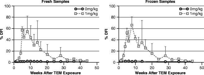
Effects of 1.0 mg/kg (daily ×5) TEM on %DFI in epididymal sperm during a 44-week period. Left: %DFI on fresh samples. Right: Aliquots of the same samples frozen and measured later at a single time period
-
2.
X-radiation. The scrotal region of male mice was exposed to X-rays ranging from 0 to 400 rads [17]. Forty days after exposure, the mice were killed and the caudal epididymal sperm were removed. The SCSA® detected increased DNA fragmentation after 12.5 rads of X-ray exposure, with significant increases following 25 rads. These data not only show that the SCSA® is a very sensitive method of detecting X-ray damage to sperm DNA but also show the very high repeatability of the measurements (Fig. 9.12.).
Fig. 9.12 
SCSA® data on epididymal sperm from scrotum-exposed mice to 0–400 rads X-ray. Epididymal sperm were surgically removed from mice at 40 days post exposure. n = 3 for each point
-
3.
Dominant lethal agents. The effects of 150 mg/ml methyl methane sulfate on mouse epididymal sperm DNA fragmentation can be seen in Fig. 9.13 [18]. By day 3 post exposure, about 85% of the sperm have extensive DNA damage; however, mating of these exposed mice to nonexposed females did not result in embryo death until 5 days post conception [18a]. Thus, the molecular events leading to embryo death can be derived from SCSA® data. Of interest, glutathione depletion potentiates ethyl methanesulfonate induced susceptibility of rat sperm DNA fragmentation [19].
Fig. 9.13 
Effect of methyl methanesulfonate on mouse sperm chromatin structure and subsequent embryo death


Human
-
1.
Pesticides: Men exposed to various insecticides and pesticides showed significantly increased levels of sperm DNA fragmentation. A dramatic effect of exposure to organophosphorous pesticides showed that 3/4 pesticide operators, not using protective gear, had DFI values above 30%, whereas those not exposed showed an average of 9.9% DFI [20] Fig. 9.14 shows SCSA® cytograms from a nonexposed and an exposed worker.
Fig. 9.14 
SCSA® cytograms from a nonexposed (left) and an exposed (right) worker
-
2.
Air Pollution: For the first time, SCSA® data showed a dose–response relationship for men exposed to winter time air pollution [21]. Residents of Teplice, Czech Republic, a town with heavy winter-time air pollution, generated by burning soft brown coal, experienced a higher than normal rate of infertility and spontaneous miscarriages. Czech army conscripts, 18–20 years of age, provided semen samples in a 2-year longitudinal study that went through periods of clean summer air and polluted winter air. Sperm DNA fragmentation measured by the SCSA® was the only semen quality measure to detect a statistically significant correlation between air pollution levels and semen quality in these young men. One fourth of these young men had %DFI above 30, placing them in a statistical group known to be at an increased risk for infertility.

Potential RNA Staining Artifacts for SCSA®
Since AO stains both single-stranded DNA and RNA in the fluorescent color red, it was very important to know if cytoplasmic or nuclear RNA contributed to the red fluorescence that might be erroneously attributed to denatured DNA.
First, any small amount of nuclear mRNA should be of small consequence to the total red fluorescence and, furthermore, should be a constant amount making only a constant background. RNAse treatment of mouse sperm did not reduce the red fluorescence caused by genotoxicant treatment [5]. Also of question was whether any residual cytoplasmic RNA contributed to ssDNA values. We addressed this question [22] by sonicating whole bull, mouse, stallion, and human sperm, purifying each sample of nuclei through a sucrose gradient and measuring both the sonicated and nonsonicated sperm by SCSA®. Somatic cells are fully destroyed and removed from the purified nuclei fraction. As illustrated in Fig. 9.15, the unsonicated and sonicated sperm produced cytograms that were practically identical. In the upper panel, note that the small percent of sperm with increased red fluorescence is in the same location (just to right of lower edge of main sperm population) after as before sonication. Of importance, note in the bottom panel that sperm with a high level of red fluorescence also produced essentially the same cytogram pattern. Given the rigor of sonication that destroys somatic cells and frees sperm nuclei, these data may suggest that sperm with a high %DFI are not fragmented nuclei that would be ripped apart by sonication. Since histone complexed chromatin is likely broken apart by sonication, it is hypothesized that the 15% histone complex in human sperm nuclei is not at the nuclear periphery where it might be highly susceptible to being removed by the sonication process.

Each human semen sample was diluted to a final volume of 0.5 ml with TNE buffer to obtain a count of approximately 2 × 10⁶ sperm/ml. The samples in the right column were sonicated for 30 s with a Branson 450 Sonifier operating at a power setting of 3 and utilizing 70% of 1-s pulses
Repeatability of SCSA® Data Over Time for Men
Forty-five men provided a semen sample once per month for nine months [22]. While the CV for the classic sperm parameters varied considerably, the SCSA® data showed that, as discussed above, the AO/DNA biochemistry as well as the flow cytometry measurements were highly repeatable with great precision. The sample to sample variation was only 3.4%, indicating that %DFI is a much more stable parameter than the classic sperm parameters. Of significant interest in the cytograms seen in Fig. 9.16 is the repeatability of the pattern of the scattergrams within a man, and also, the repeatability of a very small percent of the populations appearing exactly with the same green and red values – it is speculated from these data that a fraction of a percent of germ cells have a mutation such that the altered chromatin has a highly distinct pattern of DNA damage.

Green vs. Red fluorescence cytograms from monthly semen samples provided by three donors. Examples are selected from the 45-men illustration of different types of cytogram patterns by Evenson et al. [22]
Repeatability of %DFI Values of Human Sperm Samples from Two Commercial SCSA® Laboratories
While repeatability of SCSA® values, as well as other sperm DNA fragmentation tests, may be repeatable within a laboratory, it is important for any used test to be highly repeatable between laboratories that may have different types of flow cytometers and different technicians. SCSA Diagnostics has a licensed agreement with SPZ Lab in Copenhagen, Denmark). As part of this agreement, all SCSA® samples must be done according to strict protocols to ensure that any patient gets a result that is repeatable. Figure 9.17 shows the correlation of %DFI obtained from aliquots of the same sample in the SCSA Diagnostics Inc. lab in Brookings, SD and SPZ Lab (Copenhagen, Denmark). The data show a R 2 = 0.98 solidifying the high repeatability of the SCSA® between two SCSA® certified laboratories.

SCSA® %DFI values obtained from SCSA Diagnostics Inc. (Brookings, SD, x-axis) and SPZ Lab (Copenhagen, Denmark, y-axis) Two aliquots were made for each human semen sample, which were frozen in LN2. One aliquot was measured in each laboratory and the results are mean values of two replicates per aliquot
Animal Fertility
Given the great complexity of human fertility, we considered it important to conduct mammalian animal fertility trials for validation of the SCSA® prior to doing human clinical studies.
Bulls
Semen from individual bulls is often used for hundreds to thousands of cow inseminations. Thus, fertility rankings can be made between bulls in a stud service. Following the preliminary study of bull fertility as reported in the Science paper [4], the relationship between nuclear chromatin structure and fertility was evaluated in two groups of Holstein bulls: Group 1, 49 mature bulls, and Group 2, 18 young bulls [23]. Fertility ratings had been estimated for Group 1 and nonreturn rates were known for group 2. Intraclass correlations of the SCSA® values were high (>0.70), based on four collections obtained over several years from Group 1 bulls. Negative correlations were seen between fertility ratings and both SD DFI (−0.58, P < 0.01) and %DFI (−0.40, P < 0.01) in Group 1, and between nonreturn rates and both SD DFI (0.65, P < 0.01) and %DFI in Group 2 (−0.53, P < 0.05). These data showed that the SCSA® is a useful tool for identification of low fertility bulls and poor quality semen samples (Fig. 9.18).

Relationship of the competitive fertility index for bulls with (a) Standard deviation of DFI (SD DFI) and (b) %DFI
Inherent in studies mentioned above, and much more so with human studies, are the variables in the females and a host of other factors such as experience of the artificial insemination team. To get around this problem, animal studies can use what is known as heterospermic insemination protocols in which equal numbers of motile sperm from two or more phenotypically different bulls are mixed prior to insemination. The parentage of calves resulting from these matings is determined, and based on the number of calves sired with each phenotype, a competitive fertility index is derived for each bull [24]. Correlations of SD DFI and %DFI with competitive index were −0.94 (P < 0.01) and −0.74 (P < 0.05), respectively.
Boars
The advantage of investigating the relationship between SCSA® data and boar fertility is that pigs are multiparous, thus allowing a determination of both fertility rate and number of piglets per litter. The SCSA® was used [25] retrospectively to characterize sperm from 18 sexually mature boars having fertility information. Boar fertility was defined by farrow rate (FR) and average total number of pigs born (ANB) per litter of gilts and sows mated to individual boars. Fertility data were compiled for 1,867 matings across the 18 boars. In contrast to humans and other mammals studied, where the threshold for reduced fertility is an approximate 25–30% DFI, the threshold for boars is about 6% DFI. The %DFI and SD DFI showed the following significant negative correlations with FR and ANB; %DFI vs. FR, r = −0.55, P < 0.01; SD DFI vs FR, r = −0.67; %DFI vs. ANB, r = −0.54, P < 0.01 and SD DFI vs. ANB, r = −0.54, P < 0.02. The present data suggest that boar sperm possessing fragmented DNA can affect embryonic development corroborating earlier studies in mice showing that fertilization occurs whether the sperm has damaged DNA or not [26] but may cause embryonic death. In a recent study by Boe-Hansen et al. [27], fertility has been studied for 155 boars with 2,593 experimental litters. Using a threshold of 3% for DFI, it was found that the number of piglets born decreased from 14.94 piglets per litter (below threshold to 13.90 piglets per litter (P < 0.01).
Human Fertility
As stated above, the SCSA® or any other sperm DNA fragmentation test cannot predict fertility for a couple. Good fertility for the couple also depends on many female factors, and a low DFI value for a couple attending a fertility clinic may, therefore, imply that another cause of the infertility exists. However, the SCSA® can be predictive of male subfertility or infertility. Other chapters in this book provide more details than that outlined here.
Natural Conception
The SCSA® was the first flow-cytometric test to suggest that abnormal sperm chromatin structure was predictive of failed natural conception [4]. Following the pioneering study described in Science, the Georgetown fertility study [28] suggested an odds ratio of approximately 8 if the %DFI was above 30%. In this study [28], 200 couples with no known infertility factors were enrolled in a natural conception male factor infertility study. Monthly semen samples were obtained for the first 3 months or up to the time of biochemical or clinical pregnancy. Pregnancies were recorded over the first 12 months. The results showed that the men who had a <15% DFI had the shortest time to establish a pregnancy. Men with DFI between 15 and 30% had the next longest time period, while men with DFI above 30% had the longest time to pregnancy or no pregnancy. This latter group also had the highest level of miscarriages.
The “first pregnancy planner” study by Spano et al [29] also suggested for natural conception an odds ratio of 8–10 when the DFI was between 30 and 40%. A lower level of %DFI (20%) as a significant clinical threshold has been very recently reported by Giwercman et al. [30]. A value of 20–25% DFI appears to be a clinically significant threshold for natural conception.
SCSA® Test and ART Clinics
The first studies relating %DFI with IVF pregnancies consisted of 26 patients [31], IUI and IVF patients [28], and 89 IVF patients [31, 33] for a total of 148 patients with no pregnancies when DFI was above 27%. This led to the early concept that pregnancies were difficult to obtain when %DFI was above 27–30%. Boe-Hansen et al. [33] used SCSA in a clinical study for IUI, IVF, and ICSI treatments with reproductive outcomes of biochemical pregnancy (BP), clinical pregnancy (CP) and implantation ratio (IR). 385 semen samples from 234 couples were frozen for SCSA, and smears were prepared for morphology: 48 IUI, 139 IVF, and 47 ICSI. The results showed no significant difference in the fertility variables BP, CP, and IR when <27% DFI was used between the IVF and ICSI groups. A low number of patients received IUI with low success rate, and statistical analysis was therefore not performed. Ongoing pregnancy was achieved for both IVF and ICSI couples with DFI levels >27%, and six couples in ICSI treatment achieved CP full-term. DFI >27% had a high prognostic power for predicting no CP for IVF patients, with a specificity of 97%. Similar results were obtained from a study of 249 couples undergoing their first IVF and/or ICSI cycle conducted in the Markham clinic [35]. However, later studies showed that SCSA® values above 30% DFI could result in pregnancies after ART treatment.
While the TUNEL test has shown a wide variation of thresholds for clinical pregnancy outcomes ranging from about 4 to 36%. By contrast, the threshold for human semen with the SCSA® appears to be close to 30% and has changed only slightly downward (25%) since it was estimated many years ago. The SCSA® is now implemented routinely for all couples considered for IUI in the Southern Sweden hospital region, and a threshold of 25% was selected as a compromise. Bungum et al. [36] observed that the success for IUI started to decrease at a DFI value of 20% and approached zero when the DFI was 30%. A recent study by Giwercman et al. [31] also included information regarding sperm morphology in the assessments and suggested that the SCSA® %DFI threshold for reduced fecundity appears to be at 20%.
The greatest utility of the SCSA®, as shown by Bungum et al. [36] is that couples with a DFI above 25% should move on to IVF and preferably ICSI for the greatest success. IUI for these couples may not be cost-effective.
One hypothesis as to why ICSI can achieve a pregnancy when the %DFI >25%, is that the ICSI technician will pick up sperm with the best morphology and the greatest motility. Also, ICSI fertilization avoids potential additional DNA damage from oxidative stress either in the female reproductive tract or during in IVF. Finally, one to several of the best-grade embryos will be transferred to the female.
TESA for Failed ICSI Cycles with High %DFI
The %DFI thresholds for ICSI are likely to be higher than for IUI or natural conception since ICSI is the best method for avoiding potential additional DNA damage to the sperm prior to fertilization. However, a precise threshold for ICSI is difficult to establish, since only 3–5% of fertility patients have a %DFI above 50.
Previous and new data show that the use of testicular sperm in combination with ICSI provides an efficient treatment option for couples who fail multiple IVF cycles due to high levels of sperm DNA fragmentation. Initially, Greco et al. [37] found that for couples with failed ICSI cycles and the man had a high TUNEL defined %DFI, pregnancy success was dramatically increased with the use of testicular sperm (TESA). The overall incidence of DNA fragmentation in the testicular sperm samples was 4.8 + 3.6%, which was significantly lower (P < 0.001) compared with the ejaculated sperm samples from the same individuals (23.6 + 5.1%). (Note: DFI levels reported here cannot be compared directly DFI levels reported for the SCSA®). Greco et al. [37] did not observe differences in fertilization and cleavage rates and in embryo morphological grade found between the ICSI attempts performed with ejaculated and with testicular spermatozoa. However, eight ongoing clinical pregnancies (four singletons and four twins) were achieved by ICSI with testicular spermatozoa (44.4% pregnancy rate; 20.7% implantation rate), whereas ICSI with ejaculated spermatozoa led to only one pregnancy that was spontaneously aborted.
A recent study [38] has included couples who had undergone between one and seven prior ICSI attempts with a mean of three failed cycles. A pregnancy rate of 62.5% was achieved when testicular sperm were used. An 83% pregnancy rate was achieved when the SCSA® defined DFI was >65%. A 75% pregnancy rate was achieved in couples who underwent four or more prior failed IVF cycles. Likewise, among the thousands of measurements done at our SCSA Diagnostics lab, we have numerous ad hoc cases where several to a dozen unsuccessful ICSI cases have failed when the %DFI is above 50–60%. Thus, there is utility for the SCSA® for those patients that have had several ICSI failures. As noted by Carrell et al. [39], those patients that had two or more failed ICSI cycles, the %DFI by TUNEL was about fourfold higher than that found in sperm donors.
SCSA® Defined Etiologies of Increased DNA Fragmentation
The most likely common factor in causing sperm DNA fragmentation is oxidative stress [40] in response to reactive oxygen species (ROS). Simply stated, we need oxygen to live, but excess ROS activity is a negative consequence of this fact. Many of the environmental factors discussed here are related to increased oxidative stress. Thus, many physicians and patients are well aware of the need to have a diet rich in antioxidants.
Age
While it has become socially acceptable to father children at an older age, this increased age of fatherhood has been correlated with an increased time to establish a pregnancy or no pregnancy. Since 1980, US birth rates have increased up to 40% for men aged 35–49 years and have decreased up to 20% for men under 30 years of age.
The first study on the relationship between age of nonsmoking, healthy men and sperm DNA integrity [41] showed that among all the sperm genomic end points measured, age had the strongest effects on sperm DNA integrity. A healthy 20 year old man typically has about 5% DFI. A gradual upward trend in the average frequency of sperm with increased %DFI was observed, beginning in the early reproductive years as seen in Fig. 9.19.

Age of men vs. %DFI. The horizontal line is placed at 30% DFI, the approximate clinical threshold for risk of reduced natural fertility potential
In this age study, men in their 50s ranged from excellent %DFIs (5%) to very poor levels (73%). Even men in their 20s and 30s had abnormal DFI values, suggesting they too might experience diminished fertility and/or abnormal pregnancy outcomes. This factor is likely related to the other factors as discussed below.
The statistical odds in this study to reach the 30% DFI threshold for negative natural pregnancy outcome was age 48 as seen in Fig. 9.20, even though these men may have fathered children in their 20s. Thus, the reproductive biological clock also ticks for men, but the time window is not as narrow as for women.

Statistical probability of a man reaching a 30% DFI by age alone
Genetics
Although the evidence is very limited, it would be fully expected that genetics plays an important role in susceptibility to sperm DNA fragmentation. One example is from a study [42] on a group of men who were participants in the Teplice, Czech Republic study described above [21]. The hypothesis was as follows: men who are homozygous null for glutathione-S-transferase M1 (GSTM1-) are less able to detoxify reactive metabolites of carcinogenic polycyclic aromatic hydrocarbons (c-PAHs) found in air pollution. Consequently, they are more susceptible to the effects of air pollution on sperm chromatin. Using a longitudinal study design in which men provided semen samples during periods of both low (baseline) and episodically high air pollution, this study revealed a statistically significant association between GSTM1 null genotype and increased SCSA®-defined %DFI (beta = 0.309; 95% CI: 0.129, 0.489). Furthermore, GSTM1 null men also showed higher %DFI in response to exposure to intermittent air pollution (beta = 0.487; 95% CI: 0.243, 0.731). This study, thus, provides novel evidence for a gene–environment interaction between GSTM1 and air pollution (presumably c-PAHs).
Varicocele
Varicoceles are found in approximately 15% and 19–41% of the general and infertile populations, respectively, and have long been recognized as a common cause of infertility.
The exact pathways of damage by varicocele are difficult to explain and may be due to apoptotic events, oxidative stress, or heat [40, 43]. Zini et al. found that sperm DNA fragmentation was significantly increased in infertility patients with varicocele in comparison with patients with normal results on genital examination [44]. Furthermore, it has been shown that sperm DNA fragmentation decreases after varicocele repair [45]. Recently, Werthman et al. [46] have found a 31% increase in pregnancy rate after varicocelectomy, whereas no pregnancy occurred before surgery. In this study, %DFI values were assessed by SCSA® before and after varicocelectomy (Fig. 9.21). Although this study was small, 10 of the 11 patients with varicocele showed a significant decrease in sperm DNA fragmentation after varicocele repair.

%DFI values obtained from a man with a varicocele prior to surgical repair and at later time points. Note that a return to the lowest level %DFI shown occurred at 5 months post surgery
Cancer
Not unexpectedly, the majority of young patients with newly diagnosed testicular cancer is concerned about future fertility and wants to be informed about the different treatment modalities’ influence on spermatogenesis. In the first study of effects of cancer on sperm DNA fragmentation, 14 patients with testicular cancer, assessed after orchiectomy but before further treatment [47], displayed considerable variability in the SCSA® results, most often revealing an increased percentage of sperm cells with abnormal chromatin structure.
As a follow-up to this initial study [48], semen samples from 39 patients with testicular cancer were analyzed by the SCSA® after orchiectomy but before further treatment, and in 28 patients the SCSA® was repeated 12–26 months after orchiectomy. Figure 9.22 shows the pretreatment %DFI for the patients compared to %DFI for 18 healthy semen donors.

Distribution of individual %DFI values. Black bars, 18 samples from healthy semen donors; gray bars, 39 samples from patients with testicular cancer after unilateral orchiectomy and before further treatment
The results from 19 patients undergoing cytotoxic treatment (radiotherapy, 13 chemotherapy, 6) indicate that posttreatment recovery of spermatogenesis (recovery in 4 of 5 patients) is observed more often in patients with a normal pretreatment chromatin structure than in those with abnormal SCSA® values before treatment. This study suggested that pretreatment SCSA® results may help clinicians to identify those testicular cancer patients with a high risk of long-lasting posttreatment disturbance of spermatogenesis.
It is not known whether childhood cancer and its treatment are associated with sperm DNA damage, which subsequently affects fertility and might be transmitted to the offspring. In 99 children cancer survivors (CCS) and 193 age-matched healthy controls, %DFI was assessed using the SCSA® [49]. In the whole group of CCS, %DFI was increased compared with the controls, with borderline statistical significance. Those treated with radiotherapy only or surgery only had statistically significantly higher %DFI than the controls. The odds ratio (OR) for having DFI >20%, which is associated with reduced fertility, was significantly increased in CCS compared with the control group. (OR, 2.2) For the radiotherapy-only group, the OR was even higher (OR, 4.9). %DFI was not associated with dose of scattered testicular irradiation or type of chemotherapy given. It was concluded that %DFI was increased in CCS, with those treated with chemotherapy being the only exception. This sperm DNA impairment may be associated with the disease per se, rather than due to the treatment, and may have negative consequences in terms of fertility and risk of transmission to the offspring.
Environmental Heat
The purpose of a scrotum is to keep sperm function and maturation at an approximate 2°C lower than body temperature. Mammalian sperm, including maturing epididymal sperm, are very sensitive to excess heat. Studies on bulls that had a wool sock placed over the scrotum for 48 h showed significant damage to sperm DNA [50]. Three samples were collected for 3 time periods and the %DFI measured. For day 0 = 4%, days 3–9 = 11%, and days 12–21 = 22% DFI. These data clearly show environmentally induced sperm DNA damage.
In another experiment [51], mice were anesthetized and the scrota exposed on the underside of a Styrofoam raft floating in a high precision water bath at 2° and 4° degrees above body temperature for 60 min. The higher temperature caused a significant amount of SCSA® defined sperm DNA damage. Figure 9.23 shows significantly increased epididymal sperm DNA damage after 3 days post exposure. Caudal epididymal sperm at this time point would have been traversing the caput and corpus epididymides during exposure to the elevated temperatures. Sperm at this stage of maturation would be undergoing further condensation including intra- and intermolecular S–S bonding between protamine cysteine–SH residues. The 38°C mice exhibited SCSA® values close to controls for most days. The SD DFI values showed the largest difference between controls and 40°C treated mice with a significant increase in value by day 11 (P < 0.001) and a return to control values by day 35, or about one spermatogenic cycle.

SCSA® data on epididymal sperm obtained from scrotal heated mice. The scrotal regions of anesthetized mice were placed on the underside of a Styrofoam raft floating on a water bath (38° or 40°C) for 60 min. Three mice were used for each time point studied for each temperature
Fever
High fever has long been known to be a negative factor for pregnancy. A man who had a 104°F fever for 1 day showed [52] a dramatic increase to 36% DFI 18d post fever (dpf). The %DFI then decreased, while the %HDS increased to 49% at 33 dpf (Fig. 9.24).

Native DNA stainability vs. fragmented DNA for 66 days post fever
Sperm nuclear proteins were isolated from this 33 dpf sample; amino acid sequencing of the first 8 N-terminal residues identified this unique protein as the precursor to protamine 2. Flow-cytometric measurements of nuclear –SH groups revealed the greatest reduction in free nuclear thiols at 33 dpf, and then returned to normal by 45 dpf. Increased DNA staining is likely due to the increased histone/protamine ratio. By 60 days the sperm chromatin structure was back to normal – an approximate waiting time that physicians should suggest to such patients until trying to achieve conception.
Medications
Given the myriad of prescription and over-the-counter medications, it would not be surprising that some single agents or unstudied combination of agents will cause sperm DNA damage. Publications are sparse in this area.
Recently, several manuscripts have been published on the effects of SSRI’s on sperm DNA fragmentation. Tanrikut et al. [53] showed that the mean sperm DNA fragmentation index (TUNEL assay) was significantly higher for men while on paroxetine (30.3%) vs. baseline (13.8%). Before paroxetine, 9.7% of patients had a TUNEL score ≥ 30% compared with 50% at week 4 of treatment. The odds ratio (OR) of having abnormal DNA fragmentation while taking paroxetine was 9.33 (95% confidence interval, 2.3–37.9). Multivariate logistic regression correcting for age and body mass index confirmed this correlation (OR, 11.12). Of interest, standard semen parameters were not significantly altered during paroxetine treatment; however, the fertility potential of a substantial number of men on paroxetine may be adversely affected by these changes in sperm DNA integrity.
Diabetes and Insulin Resistance
Agbaje et al. [54] studied a cohort of 27 diabetic and 29 nondiabetic men. The level of sperm DNA fragmentation was significantly different between the two groups. Pittleloud et al. [55] reported that insulin resistance leads to a decrease in testosterone secretion at the testicular level (Leydig cell). Stigsby (personal communication, 2010) have also observed a link between insulin resistance (as measured by blood c-peptide level) and DFI value in a group of 10 men. When these men were consuming a diet with low glycemic index (GI) for a period of 4 months, both the c-peptide as well as the DFI values deceased. Although this study was very small, it appears that reduction of dietary intake of carbohydrates with a high GI may be advisable. It is recommended that identification of such individuals is based on blood levels of c-peptide (normal reference 200–700 pm/l). C-peptide is the “connecting peptide” that is cleaved from proinsulin when this is activated to insulin. This is a more stable parameter than traditional measurements of blood sugar or insulin. High insulin seems to increase the level of tumor necrosis factor alfa (TNF-alfa). TNF-alfa has a negative effect on sperm motility [56] and induces DNA fragmentation [57].
Conclusions
Thirty years ago, human infertility was considered to be a female problem if the man’s semen analysis was within a reasonable range of normal. Today, couple infertility is almost equally shared between the man and woman. The routine semen analysis may in some cases identify subfertility or infertility when sperm motility is very poor or sperm concentration is very low. However, in many cases the cause of the decreased or absent fertility remains undetected unless sperm DNA fragmentation is considered. According to our experience and the data from Bungum et al. [36], sperm DNA fragmentation is the cause for every fourth couple attending the infertility clinic. In many cases, this problem is overlooked because other problems coexist, e.g., PCOS. In such cases, detection of sperm DNA fragmentation is essential for successful treatment of the couple.
The SCSA® is technically the easiest sperm DNA fragmentation test, and the repeatability of the assay is high within and between certified SCSA® laboratories. This is in contrast to many laboratories performing TUNEL where threshold ranging from 4 to 36% in DFI has been reported.
Clinical Utility of the SCSA®
The SCSA® has currently established a 20–30% DFI threshold for reduced pregnancy via natural or IUI. When %DFI reaches 20%, fertility starts to decline, and at 30% it reaches a very low level.
It appears that most unsuccessful IUI treatments can be avoided if couples with a DFI above 25% go on to IVF, or even better, ICSI treatment. However, if DFI is below 20–25% and no other causes of subfertility or infertility are detected for the couple, IUI treatment is likely to be successful.
A %DFI close to or above 50 is found in 3–5% of the couples with failed ART cycles. Currently, no threshold for %DFI is detected for ICSI treatment, but when %DFI is above 50, standard IVF is likely to be unsuccessful. Couples with >50% DFI might consider combination of TESA with ICSI, although there are few data to support this practice.
It is recommended that possible causes and lifestyle factors producing a high DFI be ruled out early in the treatment process. Repair of varicocele and corrections of other factors are likely to reduce the DFI level and will maximize the chances of a successful fertility treatment.
References
Bjorndahl L, Mortimer D, Barratt C, et al. A practical guide to basic laboratory andrology. Cambridge: Cambridge University Press; 2010.
Kametsky LA, Melamed MR. Spectrophotometer: spectrophotometer cell sorter. Science. 1967;156:1364–5.
Darzynkiewicz Z, Traganos F, Sharpless T, et al. Thermal denaturation of DNA in situ as studied by acridine orange staining and automated cytofluorometry. Exp Cell Res. 1975;90:411.
Evenson DP, Darzynkiewicz Z, Melamed MR. Relation of mammalian sperm chromatin heterogeneity to fertility. Science. 1980;240:131–1133.
Evenson DP, Higgins PH, Grueneberg D, et al. Flow cytometric analysis of mouse spermatogenic function following exposure to ethylnitrosourea. Cytometry. 1985;6:238–53.
Evenson DP, Tritle D. Platform Presentation Abstract: “Characterization of SCSA Resolved Sperm Populations by Comet Assay and Image Analysis”. IFFS 8th World Congress on Fertility and Sterility, Palais des congres de Montreal, Montreal, Quebec Canada. 2004; May 23/28.
Evenson D, Witkin S, de Harven E, et al. Ultrastructure of partially decondensed human spermatozoal chromatin. Ultrastructure.1978;63:178–87.
Evenson D, Darzynkiewicz Z, Melamed M. Comparison of human and mouse chromatin structure by flow cytometry. Chromosoma. 1980;78:225–38.
Evenson DP, Jost LK, Varner DD. Stallion sperm nuclear protamine -SH status and susceptibility to DNA denaturation are not strongly correlated. J Reprod Fertility Suppl. 2000;56:401–6.
Love CC, Kenny RM. Scrotal Heat stress induces altered sperm chromatin structure associated with a decrease in protamine disulfide bonding in the stallion. Biol Reprod. 1999;60:615–20.
Evenson DP, Jost LK, Corzett M, et al. Characteristics of human sperm chromatin structure following an episode of influenza and high fever: a case study. J Androl. 2001;21:739–46.
Jeffay SC, Strader LF, Buus RM, et al. Relationships among semen endpoints used as indicators of sperm nuclear integrity. Am Soc Androl. Abstract. 2006.
Gorczyca W, Gong J, Darzynkiewicz Z. Detection of DNA strand breaks in individual apoptotic cells by the in situ terminal deoxynucleotidyl transferase and nick translational assays. Cancer Res. 1993;53:1945–51.
Sharma RK, Sabenegh E, Mahfouz R, et al. TUNEL as a test for sperm DNA damage in the evaluation of male infertility. Urology. 2010;76:1380–86.
Sailer BL, Jost LK, Evenson DP. Mammalian sperm DNA susceptibility to in situ denaturation associated with the presence of DNA strand breaks as measured by the terminal deoxynucleotidyl transferase assay. J Andrology. 1995;16:80–7.
Evenson DP, Baer RK, Jost LK. Long term effects of triethylenemelamine exposure on mouse testis cells and sperm chromatin structure assayed by flowcytometry. Environ Mol Mutagen. 1989;14:79–89.
Sailer BL, Jost LK, Erickson KR, et al. Effects of X-ray irradiation on mouse testicular cells and sperm chromatin structure. Environ Mol Mutagen. 1995;25:23–30.
Evenson DP, Jost L, Baer R. Effect of methyl methanesulfonate on mouse sperm chromatin structure and testicular cell kinetics. Environ Mol Mutagen. 1993;21:144–53.
Sega GA, Owens JG. Methylation of DNA and protamine by methyl methane sulfonate in the germ cells of male mice. Mutat Res. 1983;111:227–44.
Evenson DP, Jost LK, Gandy JG. Glutathione depletion potentiates ethyl methanesulfonate-induced susceptibility of rat sperm DNA denaturation in situ. Reprod Toxicol. 1993;7:297–304.
Sanchez-Pena LC, Reyes BE, Lopez-Carrillo L, et al. Organophosphorous pesticide exposure alters sperm chromatin structure in Mexican agricultural workers. Toxicol Appl Pharmacol. 2004;196:108–13.
Rubes J, Selevan SG, Evenson DP, et al. Episodic air pollution is associated with increased DNA fragmentation in human sperm without other changes in semen quality. Hum Reprod. 2005;20:2776–83.
Evenson DP, Jost L, Baer R, et al. Individuality of DNA denaturation patterns in human sperm as measured by the sperm chromatin structure assay. Reprod Toxicol. 1991;5:115–25.
Ballachey BE, Hohenboken WD, Evenson DP. Heterogeneity of sperm nuclear chromatin structure and its relationship to fertility of bulls. Biol Reprod. 1987;36:915–25.
Ballachey BE, Saacke RG, Evenson DP. The sperm chromatin structure assay: relationship with alternate tests of sperm quality and heterospermic performance of bulls. J Androl. 1988;9:l09–115.
Didion B, Kasperson K, Wixon R, et al. Boar fertility and sperm chromatin structure status: a retrospective report. J Androl. 2009;30:655–60.
Ahmadi A. Ng S-C Fertilizing ability of DNA-damaged spermatozoa. J Exp Zool. 1999;284:696–704.
Boe-Hansen GB, Christensen P, Vibjerg D, et al. Sperm chromatin structure integrity in liquid stored boar semen and its relationships with field fertility. Theriogenology. 2008;69:728–36.
Evenson DP, Jost LK, Zinaman MJ, et al. Utility of the sperm chromatin structure assay (SCSA) as a diagnostic and prognostic tool in the human fertility clinic. Hum Reprod. 1999;14(4):1039–49.
Spano M, Bonde J, Hjollund HI, et al. Sperm chromatin damage impairs human fertility. Fertil Steril. 2000;73:43–50.
Giwercman A, Lindstedt L, Larsson M, et al. Sperm chromatin structure assay as an independent predictor of fertility in vivo: a case-control study. Int J Androl. 2010;33:221–7.
Larson KL, DeJonge CJ, Barnes AM, et al. Sperm chromatin structure assay parameters as predictors of failed pregnancy following assisted reproductive techniques. Hum Reprod. 2000;15(8):1717–22.
Larson-Cook K, Brannian JD, Hansen KA, et al. Relationship between assisted reproductive techniques (ART) outcomes and DNA fragmentation (DFI) as measured by the sperm chromatin structure assay (SCSA). Fertil Steril. 2003;80:895–902.
Boe-Hansen GB, Ersboll AK, Greve T, Christensen P. Increasing storage time of extended boar semen reduces sperm DNA integrity. Theriogenology. 2005;26(3):360–8.
Boe-Hansen GB, Fedder J, Ersboll AK, et al. The sperm chromatin structure assay as a diagnostic tool in the human fertility clinic. Hum Reprod. 2006;21(6):1576–82.
Virro MR, Larson-Cook KL, Evenson DP. Sperm chromatin structure assay (SCSA®) related to blastocyst rate, pregnancy rate and spontaneous abortion in IVF and ICSI cycles. Fertil Steril. 2004;81:1289–95.
Bungum M, Humaidan P, Axmon A, et al. Sperm DNA integrity assessment in prediction of assisted reproduction technology outcome. Hum Reprod. 2007;22:174–9.
Greco E, Scarselli F, Iacobelli M, et al. Efficient treatment of infertility due to sperm DNA damage by ICSI with testicular spermatozoa. Hum Reprod. 2005;20:226–30.
Werthman P, Boostanfar R, Chang W. Use of testicular sperm/intracytoplasmic sperm injection yields high pregnancy rates in couples who failed multiple in vitro fertilization cycles owing to high levels of sperm DNA Fragmentation. 2010 Pacific Coast Reproductive Society Abstract.
Carrell DT, Liu L, Peterson CM, et al. Sperm DNA fragmentation is increased in couples with unexplained recurrent pregnancy loss. Arch Androl. 2003;49:49–55.
Saleh RA, Agarwal A, Nada EA, et al. Negative effects of increased sperm DNA damage in relation to seminal oxidative stress in men with idiopathic and malefactor infertility. Fertil Steril. 2003;79: 1597–605.
Wyrobek AJ, Eskenazi B, Young S, et al. Advancing age has differential effects on DNA damage, chromatin integrity, gene mutations, and aneuploidies in sperm. Proc Natl Acad Sci USA. 2006;103:9601–6.
Rubes J, Selevan SG, Sram RJ, et al. GSTM1 genotype influences the susceptibility of men to sperm DNA damage associated with exposure to air pollution. Mutat Res. 2007;625:20–8.
Chen SS, Huang WJ, Chang LS, et al. 8-hydroxy-20-deoxyguanosine in leukocyte DNA of spermatic vein as a biomarker of oxidative stress in patients with varicocele. J Urol. 2004;172:1239–40.
Zini A, Blumenfeld A, Libman J. et al; Beneficial effect of microsurgical varicocelectomy on human sperm DNA integrity. Hum Reprod. 2005;20:1018–21.
Yamamoto M, Hibi H, Tsuji Y, et al. The effect of varicocele ligation on oocyte fertilization and pregnancy after failure of fertilization in in vitro fertilization–embryo transfer. 1994;40:683–7.
Werthman P, Wixon R, Kasperson K, et al. Significant decreases in sperm deoxyribonucleic acid fragmentation after varicocelectomy. Fertil Steril. 2008;90: 1880–4.
Evenson DP, Klein FA, Whitmore WF, et al. Flow cytometric evaluation of sperm from patients with testicular carcinoma. J Urol. 1984;132:1220–25.
Fossa SD, De Angelis P, Kraggerud SM. Predication of post treatment spermatogenesis in patients with testicular cancer by flow cytometric sperm chromatin structure assay. Cytometry (Communications in Clinical Cytometry). 1997;30:192–6.
Romerius P, Stahl O, Moell C, et al. Sperm DNA integrity in men treated for childhood cancer. Clin Cancer Res. 2010;16:3843–7.
Karabinus DS, Vogler CJ, Saacke RG, et al. Chromatin structural changes in sperm after scrotal insulation of holstein bulls. J Androl. 1997;18:549–55.
Sailer B, Sarkar LJ, Bjordahl JA, et al. Effects of heat stress on mouse testicular cells and sperm chromatin structure. J Androl. 1997;18:294–301.
Evenson DP, Jost LK, Corzett M, Balhorn R. Characteristics of human sperm chromatin structure following an episode of influenza and high fever: a case study. J Androl. 2000;21:739–46.
Tanrikut C, Feldman AS, Altemus M, et al. Adverse effect of paroxetine on sperm. Fertil Steril. 2010;94:1021–6.
Agbaje IM, Rogers DA, McVicar CM, McClure N, Atkinson AB, Mallidis C, et al. Insulin dependant diabetes mellitus: implications for male reproductive function. Hum Reprod. 2007;22:1–7.
Pitteloud N, Hardin M, Dwyer AA, et al. Increasing insulin resistance is associated with decrease in Leydig cell testosterone secretion in men. J Clin Endrocrinol Metab. 2005;90:2636–41.
Koçak I et al. Relationship between seminal plasma interleukin-6 and tumor necrosis factor alpha levels with semen parameters in fertile and infertile men. Urol Res. 2002;30:263–7.
Perdichizzi A et al. Effects of tumour necrosis factor-alpha on human sperm motility and apoptosis. J Clin Immunol. 2007;27(2):152–62.
Author information
Authors and Affiliations
Corresponding author
Editor information
Editors and Affiliations
Rights and permissions
Copyright information
© 2011 Springer Science+Business Media, LLC
About this chapter
Cite this chapter
Evenson, D.P. (2011). Sperm Chromatin Structure Assay (SCSA®): 30 Years of Experience with the SCSA® . In: Zini, A., Agarwal, A. (eds) Sperm Chromatin. Springer, New York, NY. https://doi.org/10.1007/978-1-4419-6857-9_9
Download citation
DOI: https://doi.org/10.1007/978-1-4419-6857-9_9
Published:
Publisher Name: Springer, New York, NY
Print ISBN: 978-1-4419-1781-2
Online ISBN: 978-1-4419-6857-9
eBook Packages: MedicineMedicine (R0)