
Abstract
The ease and simplicity of lipofilling techniques combined with the broadened indications for lipofilling is attracting the interest of many surgeons who want to improve aesthetic results after breast cancer treatment. We are also looking forward to having the maximum stable volume without reabsorption and the least number of complications and to proving the oncological safety of lipofilling. Especially in the tissue engineering era, adipose tissue is being experimented upon and used by many scientists and companies worldwide. Some novel products and machines may need approval and well-performed clinical studies before being accepted in surgery on a daily basis.
Access provided by Autonomous University of Puebla. Download chapter PDF
Keywords
1 Introduction
Lipofilling is an autologous technique used in breast reconstruction. It is also known as “fat transfer,”, “lipotransfer,” “fat injection,” or “fat transplantation” as well as many other terms. The procedure consists of two major steps: these are liposuction and lipoinjection of the patient’s own fat tissue and other tissue elements, either with or without specific preparation processes before lipoinjection. It is considered to be a minimally invasive procedure which can be effectively performed with the patient under local or general anesthesia.
This technique was initially introduced for aesthetic and scar correction purposes especially for the face and hands [1–6]. Recently, it has also been widely applied for breast indications including micromastia, postaugmentation deformity, tuberous breast, Poland’s syndrome, postlumpectomy deformity, postmastectomy deformity, deficits caused by conservative treatment or reconstruction with implants and/or flaps, tissue damaged by radiotherapy, and nipple reconstruction augmentation [7].
Despite various indications related to breast reconstruction after breast cancer treatment, there are different strategies for performing lipofilling procedures in different countries, without international consensus [8]. Up to now, the literature has provided evidence of only expert experience and clinical series trying to demonstrate the oncological safety and efficacy of lipofilling for the breast cancer patient. Nonetheless, there are fundamental and clinical researchers at the European Institute of Oncology who are dedicated to the oncological safety and technical application of lipofilling [9–12].
2 Biology of the Lipoaspirated Specimen
The fat specimen when injected into the breast is not just a physical filler or framework, but contains a significant number of cells which can survive and function. Viable and dead adipocytes, adipose-derived stromal cells, vascular endothelial cells, fibroblasts, hematopoietic cells, blood cells, and other cells can be found in the lipoaspirated specimen [13, 14]. Laboratory research from European Institue of Oncology also found that adipose tissue is a very rich reservoir of vascular progenitor cells. The current literature provides data on the endocrine, paracrine, and autocrine activities of the transplanted fat tissues. It is also interesting that in the future of medical bioengineering, stem cell culture and expansion may alter the composition and biology of the fat injection specimen.
3 Lipofilling and Oncological Concerns
When lipofilling was introduced for scar correction and aesthetic indications, there was rarely a question of cancer risk or cancer incidence. On the other hand, the concern for oncological safety obviously important becomes when performing lipofilling for the breast cancer patient. Theoretically, the “tumor–stroma interaction” can potentially induce cancer reappearance by “fueling” dormant breast cancer cells in the tumor bed. Our experimental findings also suggest that purified progenitor cells from liposuction specimens can stimulate angiogenesis, cell growth, and metastasis in animal models. No study on the effects of lipotransfer on human cancer breast cells in vivo is available [10].
In our clinical experience [8, 9, 11, 12], we have demonstrated that there is no increased risk of local recurrence in the invasive breast cancer patient who is treated with lipofilling. However, we recommend close oncological follow-up in this particular group, especially in the carcinoma in situ patient. If abnormal clinical or radiological signs are detected during follow-up, prompt pathological examination is highly recommended. We propose surgeons who perform lipofilling do a complete preoperative oncological examination and create a database of fat grafting patients.
4 Surgical Technique
4.1 Donor Site
The harvesting procedure can be performed with the patient under local or general anesthesia, depending on the patient’s clinical condition and risks. Local anesthesia is our preference, whereas general anesthesia is recommended in the case of harvesting a large amount of fat tissue or combined multiple procedures. The preferred donor sites are the abdomen and flank areas, outer thighs, buttocks, inner thighs, and knees. The donor site selection is based on excess fat tissue in the area, and then the amount of fat that can be removed without aesthetic damage of the donor site. The selected donor site is infiltrated with Klein solution, which consists of 1 ml of epinephrine diluted in 500 ml of 0.001 % lactate Ringer solution. Mepivacaine (2 %) is added in the solution if lipofilling with the patient under local anesthesia is indicated (Fig. 37.1).

Infiltrating the donor site with Klein solution
The amount of solution injected is double the volume of the preestimated fat tissue requirement. The whole procedure of fat harvesting and “lipofilling” is performed according to Coleman’s technique [15]. After the injection of the diluted solution, a two-hole, 3-mm-diameter Coleman cannula with a blunt tip attached to a 10-ml Luer-Lok syringe is inserted through the small incision. A combination of slight negative pressure and the curetting action of the cannula through the tissues allows fat harvesting [2] (Fig. 37.2). The method of liposuction with different machine models or a manual syringe and different sizes and numbers of cannula holes has been not proven to affect fat cell survival. However, the “nontraumatic” blunt cannula technique is preferred rather than a sharp cannula technique [16–19]. Other harvesting techniques such as water-assisted liposuction or the Body-Jet system [20], Cytori Therapeutics’s Celution system [21], and Adivive’s Lipokit system are also available. There is a debate between open-system and closed-system techniques but there is no definite conclusion regarding the difference in fat cell survival and clinical results in each group. At the end of the lipoaspiration procedure, the access site of the cannula is sutured with fine absorbable material and a pressure dressing is applied.

Harvesting the fat tissue with a Coleman cannula
There are different methods to process and purify the fat before grafting. The choice depends on several factors, such as the surgeon’s preference, costs, higher concentration of adipose-derived stem cells, volume requirement, and injection. Different techniques can be used:
-
1.
No preparation. This no-touch technique allows surgeons to inject the lipoaspirated fat into the recipient site without any preparation [22]. The advantages are that the specimen remains in a closed system and that it allows a shorter operating time compared with other techniques. However, it is suitable only when performing lipoinjection with small volume requirements, e.g., a few cubic centimeters to make a reconstructed nipple projection or a small linear scar correction. The disadvantage is the increase risk of calcification and cyst formation, because the oil is not eliminated.
-
2.
Mechanical preparation (centrifugation, decantation, or washing technique). The purpose of this technique is to remove cell debris, serum, tumescence solution, and the oily component from the adipocytes and the derivative cells. Centrifugation is the technique currently used by the authors [11, 12]. In our setting, the fat is centrifuged at 3,000 rpm for 3 min until the oily part and fluid are separated from adipose tissue (Fig. 37.3). The speed and duration of the centrifugation have no effect on adipocyte survival, but greater force seems to be better in removing oil and cell debris than lower centrifugal forces as was demonstrated by some authors [23]. Other authors prefer lower speeds and a shorter duration to avoid adipocyte damage [24] (Figs. 37.4 and 37.5). After the top (oily) layer and the bottom (fluid) layer have been removed, the middle (cellular) layer, which contains the adipocytes, endothelial cells, and mesenchymal stem cells, is immediately transferred to a 1-ml Luer-Lok syringe and prepared for injection [11, 12, 25].
Fig. 37.3 
Medical device for fat centrifugation
Fig. 37.4 
Specimen before centrifugation
Fig. 37.5 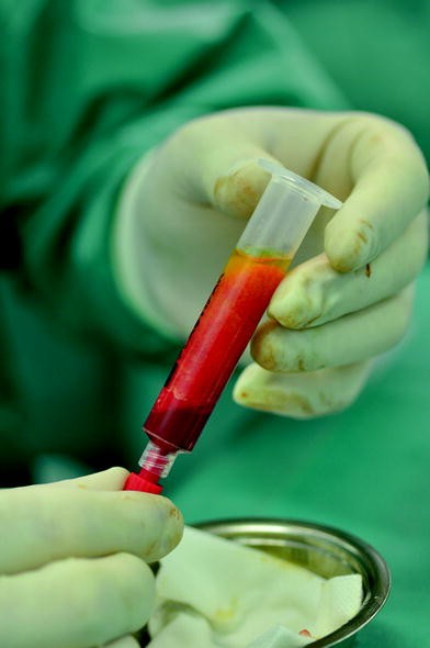
Specimen after centrifugation
-
3.
Other methods of preparation (enzymatic and biological preparation). Some scientists try to enhance fat graft survival with fibroblast growth factor β [26]. Some surgeons divide the lipoaspirate specimen in half and prepare each half separately before putting them together and performing lipoinjection. An example of this technique is called cell-assisted lipotransfer. This process increases the number of adipose-derived stromal cells before fat injection [27, 28]. Cytori Therapeutics’s Celution system also prepares the fat by separation of two equal parts of a lipoaspirate specimen before mixing them together [21].


4.2 Recipient Site
The recipient site is prepared by preoperative marking and estimating the area which is required for lipoinjection. A suitable local anesthetic agent is injected around the defect prior to the injection of purified fat if the procedure is done with the patient under local anesthesia. Prepared cellular component is then injected into the defect area through a blunt Coleman cannula. Retrograde injection with a thin-layer, multiple-tunnel and fan or cylindrical shape technique is performed (Fig. 37.6). We avoid placing the fat as an excessive deposit, which may result in liponecrosis and graft loss. We judge the amount of fat needed to be grafted in each individual case on the bass of the tissue quality and the shape and size of the defect. If the anatomical site allows, we try to avoid intraparenchymal injection. In the case of tight fibrosis from a surgical scar or irradiated tissue, a sharp needle is inserted to break up the fibrotic scar and create a space for lipoinjection (Fig. 37.7). In general, we overcorrect the volume deficit by approximately 30–40 % depending on the reconstructive indication and recipient site tissue quality. After finishing the injection, we suture the entrance site of the injection cannula in a conventional fashion.

Fat injection in the recipient site

A sharp needle is used to release fibrotic scars
Some authors have proposed the use of an external suction machine on the donor site to produce subcutaneous tissue expansion and allow a larger volume of fat to be harvested and injected. This machine (Brava system) is not comfortable for patients and needs to be used during the night 1 month before and after the procedure [29].
5 Indications
5.1 Breast Conservative Surgery Defect Correction
A patient with breast conservative treatment usually receives conventional radiotherapy and this therefore leads to difficulty in selecting a reconstructive procedure. However, lipofilling offers a simple and reliable method which does not increase the complication rates in the breast conservative treatment patient.
-
Immediate reconstruction after breast conservative treatment. Lipofilling can be used for reshaping of the breast immediately after conservative surgery as a sole procedure or in combination with other oncoplastic procedures. A good indication would be in the case of a small breast and an upper quadrant tumor. A quadrantectomy can be performed and the defect can be closed with glandular sutures. The defect created by these sutures can be repaired by fat grafting in the subcutaneous space. Circumcavity injection is recommended and intracavity injection should be avoided.
-
Delayed reconstruction after breast conservative treatment. This is one of the major indications for lipofilling performed by the authors. It is possible to correct the defects and also increase the skin quality after radiotherapy damage. Depending on the dimensions of the defect, the correction can be done in one or more sessions. The procedure can be performed with the patient under local anesthesia in the case of a monolateral procedure and with the patient under general anesthesia in cases that need a contralateral procedure, such as a reduction mammaplasty (Figs. 37.8 and 37.9).
Fig. 37.8 
Patient with huge asymmetry after right breast conservative surgery and radiotherapy
Fig. 37.9 
Postoperative view after 250 cm3 of fat grafting in the right side and left reduction mammaplasty


5.2 Defects After Mastectomy and Breast Reconstruction
Lipofilling is a main technique in breast reconstruction after mastectomy and is indicated in the following situations:
-
Immediate breast reconstruction, Lipofilling in immediate reconstruction is very difficult owing to the lack of a surgical plane for fat implantation. In special cases with a small breast and huge flank lipodystrophy, an implant can be positioned at the same time as the mastectomy. After complete expansion, the reconstructive steps start with deflation of the expander and fat grafting to twice the volume deflated. After two or three fat grafting sessions, the expander can be removed and the nipple and areola can be reconstructed to achieve the final result without an implant (Figs. 37.10, 37.11, and 37.12),
Fig. 37.10 
Preoperative view before mastectomy and immediate breast reconstruction with a tissue expander
Fig. 37.11 
Preoperative view before 280 cm3 of fat grafting and deflating the expander
Fig. 37.12 
Postoperative view after expander removal and a second session of 250 cm3 of fat grafting without any implant
-
Secondary total breast reconstruction using lipofilling as the primary reconstructive procedure. This is still an early procedure done in a few surgical centers and is usually performed with pre-expansion or vacuum systems [30–33 ]. It allows delayed total breast reconstruction with autologous fat tissue; however, the procedure can rarely be completed in a single stage. It is also difficult to obtain a good skin envelope, good definition of the inframammary fold, and a good breast mound.
-
Secondary defect corrections after breast reconstruction with implants or autologous flaps. Lipofilling can be used to correct upper breast fullness in the case of an anatomical implant defect, or also to correct the lower pole fullness (Figs. 37.13 and 37.14). It can also be used for secondary defects of reconstructions done with autologous flap procedures [34 ]. When an autologous flap reconstruction develops an early or delayed complication such as partial flap necrosis or delayed flap atrophy, especially for extended latissimus dorsi flap reconstructions, lipofilling can replace volume deficit without requiring flap or microvascular procedures (Figs. 37.15, 37.16, 37.17, and 37.18).
Fig. 37.13 
Upper breast fullness after immediate left breast reconstruction with an anatomical implant
Fig. 37.14 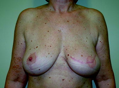
Postoperative view after 80 cm3 of fat grafting in the upper pole of the left breast
Fig. 37.15 
Upper outer defect after delayed reconstruction with a monopedicled transverse rectus abdominis myocutaneous flap
Fig. 37.16 
Cosmetic results 6 months after lipofilling
Fig. 37.17 
Cosmetic results 6 months after immediate right breast reconstruction with a latissimus dorsi flap plus an implant and prophylactic mastectomy of the left breast and immediate breast reconstruction with a definitive implant. A bilateral upper outer lipofilling was performed with the inner thighs as the donor site
Fig. 37.18 
Final cosmetic results after 6 months


5.3 Unusual Indications
-
Rippling correction To correct visible rippling after implant-based reconstruction (Figs. 37.19 and 37.20).
Fig. 37.19 
Upper pole rippling after immediate left breast reconstruction with ab implant
Fig. 37.20 
Cosmetic results at 6 months after injection of 50 cm3 of fat in the upper pole of the left breast
-
Capsular contracture Fat grafting around the implant and especially around the capsule can correct a visible rippling appearance by increasing the thickness of the capsular wall. Moreover, the effect of adipose-derived stromal cells in the cellular component of the lipoinjection may cause biological tissue remodeling of the cellular structure in the contracted capsule (Figs. 37.21 and 37.22).
Fig. 37.21 
Patient with Baker IV capsula contracture after mastectomy and immediate breast reconstruction with a definitive implant
Fig. 37.22 
Cosmetic result after four sessions of left breast lipofilling
-
Nonspecific pain therapy. There is still no clear explanation for this mechanism of action. Adipose tissue is a rich source of various types of progenitor, endothelial, and mesenchymal stem cells. Some of them have angiogenic potential which may resolve the nonspecific pain.
-
Improvement of irradiated local tissue damage (including postradiotherapy ulcer). The cellular component in the lipofilling specimen has angiogenic potential and is able to generate new stromal and cellular matrix, which benefits the chronic wound-healing process and irradiated tissue.
-
Contralateral symmetrical procedure. Lipofilling can also be used on the contralateral side to produce symmetry either immediately with the oncological procedure or later after reconstruction.


6 Complications and Sequelae
-
Immediate complications include seroma, hematoma, cellulitis, abscess, and liponecrosis. In published data from the European Institute of Oncology, we reported a rate of complications ranging from 2.8 to 3.6 % [11, 12]. The type of oncological resection, the type of reconstruction, and the type of radiation do not affect the occurrence of immediate complications.
-
Late complications include fat reabsorption, scar retraction, and donor site deformity. Donor site deformity can be avoided by the selection of the appropriate donor sites, obtaining the optimum volume of lipoaspiration, and avoiding superficial planes of lipoaspiration. Fat reabsorption is an expected sequela after lipoinjection and is estimated at 30–60 % in the first year [35]. However, a stable result may start to be observed at 6 months. The reabsorption also depends on the injection technique, recipient tissue quality, volume of injection, and methods of preparation. We prefer to perform more than one session of lipofilling in the case of large-volume defects.
7 Future Trends
The ease and simplicity of lipofilling techniques combined with the broadened indications for lipofilling is attracting the interest of many surgeons who want to improve aesthetic results after breast cancer treatment. We are also looking forward to having the maximum stable volume without reabsorption and the least number of complications and to proving the oncological safety of lipofilling. Especially in the tissue engineering era, adipose tissue is being experimented upon and used by many scientists and companies worldwide. Some novel products and machines may need approval and well-performed clinical studies before being accepted in surgery on a daily basis [36].
References
Coleman SR (1998) Structural fat grafting. Aesthet Surg J 18(5):386, 388
Coleman SR (2002) Hand rejuvenation with structural fat grafting. Plast Reconstr Surg 110(7):1731–1744; discussion 45–47
Coleman SR (2006) Facial augmentation with structural fat grafting. Clin Plast Surg 33(4):567–577
von Heimburg D, Pallua N (2001) Two-year histological outcome of facial lipofilling. Ann Plast Surg 46(6):644–646
Andre P (2002) Facial lipoatrophy secondary to a new synthetic filler device (Profill) treated by lipofilling. J Cosmet Dermatol 1(2):59–61
Bertossi D, Zancanaro C, Trevisiol L, Albanese M, Ferrari F, Nocini PF (2003) Lipofilling of the lips: ultrastructural evaluation by transmission electron microscopy of injected adipose tissue. Arch Facial Plast Surg 5(5):392–398
Gutowski KA (2009) Current applications and safety of autologous fat grafts: a report of the ASPS fat graft task force. Plast Reconstr Surg 124(1):272–280
Petit JY, Clough K, Sarfati I, Lohsiriwat V, de Lorenzi F, Rietjens M (2010) Lipofilling in breast cancer patients: from surgical technique to oncologic point of view. Plast Reconstr Surg 126(5):262e–263e
Petit JY, Botteri E, Lohsiriwat V, Rietjens M, De Lorenzi F, Garusi C et al (2012) Locoregional recurrence risk after lipofilling in breast cancer patients. Ann Oncol 23(3):582–588
Lohsiriwat V, Curigliano G, Rietjens M, Goldhirsch A, Petit JY (2011) Autologous fat transplantation in patients with breast cancer: “silencing” or “fueling” cancer recurrence? Breast 20(4):351–357
Petit JY, Lohsiriwat V, Clough KB, Sarfati I, Ihrai T, Rietjens M et al (2011) The oncologic outcome and immediate surgical complications of lipofilling in breast cancer patients: a multicenter study—Milan-Paris-Lyon experience of 646 lipofilling procedures. Plast Reconstr Surg 128(2):341–346
Rietjens M, De Lorenzi F, Rossetto F, Brenelli F, Manconi A, Martella S et al (2011) Safety of fat grafting in secondary breast reconstruction after cancer. J Plast Reconstr Aesthet Surg 64(4):477–483
Eto H, Suga H, Matsumoto D, Inoue K, Aoi N, Kato H et al (2009) Characterization of structure and cellular components of aspirated and excised adipose tissue. Plast Reconstr Surg 124(4):1087–1097
Suga H, Matsumoto D, Inoue K, Shigeura T, Eto H, Aoi N et al (2008) Numerical measurement of viable and nonviable adipocytes and other cellular components in aspirated fat tissue. Plast Reconstr Surg 122(1):103–114
Coleman SR (1995) Long-term survival of fat transplants: controlled demonstrations. Aesthetic Plast Surg 19(5):421–425
Sommer B, Sattler G (2000) Current concepts of fat graft survival: histology of aspirated adipose tissue and review of the literature. Dermatol Surg 26(12):1159–1166
Kaufman MR, Miller TA, Huang C, Roostaeian J, Wasson KL, Ashley RK et al (2007) Autologous fat transfer for facial recontouring: is there science behind the art? Plast Reconstr Surg 119(7):2287–2296
Gonzalez AM, Lobocki C, Kelly CP, Jackson IT (2007) An alternative method for harvest and processing fat grafts: an in vitro study of cell viability and survival. Plast Reconstr Surg 120(1):285–294
Erdim M, Tezel E, Numanoglu A, Sav A (2009) The effects of the size of liposuction cannula on adipocyte survival and the optimum temperature for fat graft storage: an experimental study. J Plast Reconstr Aesthet Surg 62(9):1210–1214
Sasaki GH (2011) Water-assisted liposuction for body contouring and lipoharvesting: safety and efficacy in 41 consecutive patients. Aesthet Surg J 31(1):76–88
Fraser JK, Zhu M, Wulur I, Alfonso Z (2008) Adipose-derived stem cells. Methods Mol Biol 449:59–67
Karacalar A, Orak I, Kaplan S, Yildirim S (2004) No-touch technique for autologous fat harvesting. Aesthetic Plast Surg 28(3):158–164
Pulsfort AK, Wolter TP, Pallua N (2011) The effect of centrifugal forces on viability of adipocytes in centrifuged lipoaspirates. Ann Plast Surg 66(3):292–295
Kim IH, Yang JD, Lee DG, Chung HY, Cho BC (2009) Evaluation of centrifugation technique and effect of epinephrine on fat cell viability in autologous fat injection. Aesthet Surg J 29(1):35–39
Conde-Green A, Baptista LS, de Amorin NF, de Oliveira ED, da Silva KR, Pedrosa Cda S et al (2010) Effects of centrifugation on cell composition and viability of aspirated adipose tissue processed for transplantation. Aesthet Surg J 30(2):249–255
Hong SJ, Lee JH, Hong SM, Park CH (2010) Enhancing the viability of fat grafts using new transfer medium containing insulin and beta-fibroblast growth factor in autologous fat transplantation. J Plast Reconstr Aesthet Surg 63(7):1202–1208
Yoshimura K, Sato K, Aoi N, Kurita M, Hirohi T, Harii K (2008) Cell-assisted lipotransfer for cosmetic breast augmentation: supportive use of adipose-derived stem/stromal cells. Aesthetic Plast Surg 32(1):48–55; discussion 6–7
Matsumoto D, Sato K, Gonda K, Takaki Y, Shigeura T, Sato T et al (2006) Cell-assisted lipotransfer: supportive use of human adipose-derived cells for soft tissue augmentation with lipoinjection. Tissue Eng 12(12):3375–3382
Smith CJ, Khouri RK, Baker TJ (2002) Initial experience with the Brava nonsurgical system of breast enhancement. Plast Reconstr Surg 110(6):1593–1595; author reply 5–8
Babovic S (2010) Complete breast reconstruction with autologous fat graft—a case report. J Plast Reconstr Aesthet Surg 63(7):e561–e563
Alexander Del Vecchio D, Bucky LP (2010) Breast augmentation using pre-expansion and autologous fat transplantation—a clinical radiological study. Plast Reconstr Surg 126:68–69
Del Vecchio D (2009) Breast reconstruction for breast asymmetry using recipient site pre-expansion and autologous fat grafting: a case report. Ann Plast Surg 62(5):523–527
Khouri R, Del Vecchio D (2009) Breast reconstruction and augmentation using pre-expansion and autologous fat transplantation. Clin Plast Surg 36(2):269–280, viii
Hamdi M, Andrades P, Thiessen F, Stillaert F, Roche N, Van Landuyt K et al (2010) Is a second free flap still an option in a failed free flap breast reconstruction? Plast Reconstr Surg 126(2):375–384
Zocchi ML, Zuliani F (2008) Bicompartmental breast lipostructuring. Aesthetic Plast Surg 32(2):313–328
Martin-Padura I, Gregato G, Marighetti P, Mancuso P, Calleri A, Corsini C et al (2012) The white adipose tissue used in lipotransfer procedures is a rich reservoir of CD34+ progenitors able to promote cancer progression. Cancer Res 72:325–334
Author information
Authors and Affiliations
Corresponding author
Editor information
Editors and Affiliations
Rights and permissions
Copyright information
© 2013 Springer-Verlag Italia
About this chapter
Cite this chapter
Rietjens, M., Lohsiriwat, V., Manconi, A., Urban, C. (2013). Fat Grafting in Breast Reconstruction . In: Urban, C., Rietjens, M. (eds) Oncoplastic and Reconstructive Breast Surgery. Springer, Milano. https://doi.org/10.1007/978-88-470-2652-0_37
Download citation
DOI: https://doi.org/10.1007/978-88-470-2652-0_37
Published:
Publisher Name: Springer, Milano
Print ISBN: 978-88-470-2651-3
Online ISBN: 978-88-470-2652-0
eBook Packages: MedicineMedicine (R0)