
Abstract
Invasive fungal sinusitis can be diagnosed early only if the primary clinician treating the patient, whether an ENT surgeon, a chemotherapist, an oncologist or a paediatrician, has a high index of clinical suspicion. In addition, there should be both microbiological as well as histopathological evidence for further definitive management. An analysis of tissue invasion and host immunological response is an important step in the evaluation of the patient.
Access provided by Autonomous University of Puebla. Download chapter PDF
Keywords
These keywords were added by machine and not by the authors. This process is experimental and the keywords may be updated as the learning algorithm improves.
Invasive fungal sinusitis can be diagnosed early only if the primary clinician treating the patient, whether an ENT surgeon, a chemotherapist, an oncologist or a paediatrician, has a high index of clinical suspicion. In addition, there should be both microbiological as well as histopathological evidence for further definitive management. An analysis of tissue invasion and host immunological response is an important step in the evaluation of the patient.
The clinical features of the disease vary depending upon the acuity of the fungal infection. While acute invasive fungal sinusitis is a rapidly progressing infection, chronic invasive and granulomatous are indolent and progress insidiously over several months to years.
Acute Invasive Fungal Sinusitis
It is the most lethal form of fungal sinusitis, and the incidence of reported mortality is 50–80 % [1]. It is usually seen in immunocompromised individuals but is also seen occasionally in immunocompetent persons. It is also postulated that the nasal cavity is the primary site of infection, with the middle turbinate being affected in two-thirds of the biopsy positive cases [2].
Initially patient may complain of nasal block with bloodstained or serosanguineous nasal discharge. There may be painless, necrotic nasal septal ulcer or eschar. There is rapid progression over a few days with angioinvasion and haematogenous dissemination with fungi invading the mucosa, submucosa, blood vessels and bony walls of the nose and paranasal sinuses. Hyperglycemia and acidosis provide ideal conditions for fungal growth and tissue invasion. Also ketoacidosis has been shown to adversely affect phagocytic activity [3]. There may be intracranial spread either through the cribriform plate or via the orbital apex or via septic emboli. Once the ophthalmic and other orbital arteries are involved, infection can further reach the cavernous sinus and carotid artery. Acute subdural hematoma, cavernous sinus thrombosis and internal carotid artery thrombosis may occur in rhinocerebral mucormycosis. Invasion of the carotid arteries can rapidly lead to cerebral ischemia and death [4, 5].
Depending upon the progression of the infection, Spellberg et al. [6] classified it as:
-
Stage 1: Rhinomaxillary (Fig. 1; Figs. 1, 2, 3 in Chapter “Radiology in Invasive Fungal Sinusitis”)
Fig. 1 
Stage 1: Early rhino-maxillary fungal sinusitis
-
Stage 2: Rhino-orbital (Fig 2; Figs. 4, 5, 6 in Chapter “Radiology in Invasive Fungal Sinusitis”)
Fig. 2 
Stage 2: Rhino-sino-orbital fungal sinusitis
-
Stage 3: Rhino-orbito-cerebral (Fig. 3; Fig. 7 in Chapter “Radiology in Invasive Fungal Sinusitis”)
Fig. 3 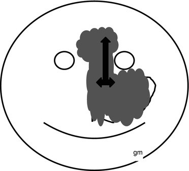
Stage 3: Rhino-sino-orbito-cerebral invasive fungal sinusitis


Symptoms
The following symptoms, especially in an immunocompromised patient, should arouse the suspicion of fungal disease:
-
Fever with spikes, not responding to antibiotics. It is the most common presenting feature [7].
-
Persistent nasal blockage with bloodstained serosanguineous nasal discharge with cough.
-
With progression of the disease, there could be facial or periorbital swelling (Fig. 4), facial pain, numbness and headache.
Fig. 4 
Facial and periorbital (arrow) swelling
-
Orbital symptoms: Spread of disease to orbit may cause chemosis, proptosis, ptosis, blurring of vision, loss of vision and ophthalmoplegia.
-
Cranial nerve palsies especially 2nd, 3rd, 4th, 5th, 6th and 7th nerve due to cavernous sinus thrombosis or temporal lobe mycotic infarcts.
-
CNS symptoms such as altered consciousness, delirium, convulsions, hemiparesis, hemiplegia or coma may be seen in patients with intracranial involvement.

Examination
The patient is usually under treatment with a physician for a metabolic disorder or oncologist for chemotherapy with neutropenia and then referred to either an ENT surgeon or an ophthalmologist.
ENT Examination
Initial or a cursory nasal examination may not reveal anything significant. Therefore, it is always advisable to decongest the nose and perform nasal endoscopy. A discolouration of the nasal mucosa and serosanguineous discharge should be looked for. Crusting, whitish discolouration (due to tissue ischemia) (Fig. 5) or black discolouration with eschar formation (Fig. 6) (due to tissue necrosis) may be present [8]. There could also be granulation or ulceration of the nasal mucosa. These changes have been most commonly found to occur on the middle turbinate, followed by the septum, palate and inferior turbinate [9]. There may be reduced nasal mucosal bleeding on account of tissue ischemia or infarction. The sentinel signs and symptoms are dark-coloured nasal septal (Fig. 6) or palatal ulcers or eschars (Figs. 7 and 8), fever, headache, nasal crusting, epistaxis, cough and mental changes [10]. There could also be skip lesions due to spread of the infection along the intima of blood vessels (Fig. 9 showing a tongue lesion in a patient of ALL with rhino-orbital mucormycosis).

Endoscopic appearance of discoloured nasal mucosa in a case of mucormycosis

Progressive nasal septal and lateral nasal wall infarction (arrows) in a case of acute fulminant mucormycosis

Discolouration of palate (black arrow) in early palatal involvement in a diabetic patient with rhino-orbital mucormycosis

Palatal infarction (white arrow) in progressive mucormycosis

Skip mucor lesion on tongue (arrow) in patient with ALL and rhino-orbital mucormycosis
Tissue samples should ideally be taken from discoloured areas of the middle and inferior turbinate or septum under vision during a diagnostic nasal endoscopy.
Teaching point: Samples taken from the nasal vestibule or swabs taken blindly do not help in the diagnosis and instead delay the diagnosis. The samples should be sent for both microbiological (KOH and fungal culture) and histopathological examination.
-
Decreased facial or nasal mucosal sensations may be present even in early stages of the disease before the development of other signs and symptoms.
-
Gingival or palatal eschars or ulceration may be found (Figs. 7 and 8).
Ascioglu et al. [11] have suggested major and minor criteria for clinical diagnosis.
Minor criteria | Major criteria |
|---|---|
Nasal discharge/stuffiness | Radiologic e/o sinus invasion, i.e. erosion of sinus walls, extension of infection to neighbouring structures, extensive skull base destruction |
Nasal ulceration/eschar/epistaxis | |
Periorbital swelling | |
Maxillary tenderness | |
Black necrotic lesions/perforation of palate |
Ophthalmic Examination
An ophthalmologist should assess the vision, field of vision and retinal pathology and look for ophthalmoplegia or chorioretinitis through the entire course of the patient’s treatment.
It has been recommended that nasal endoscopy and imaging studies are warranted if there is persistence of fever of unknown origin for more than 48 h despite appropriate antibiotic therapy and in the presence of localized sino-nasal symptoms in an immunocompromised patient [9, 12, 13].
Chronic Invasive Fungal Sinusitis
This usually develops in immunocompetent individuals but is also seen in diabetics and individuals with low level of immunocompromise [14]. There may be a history of chronic rhinosinusitis, and symptoms may consist of serosanguineous nasal discharge, epistaxis, nasal polyposis, fever or a persistent oroantral fistula (Fig. 10 and Fig. 8 in Chapter “Radiology in Invasive Fungal Sinusitis”). There could be development of sequestrum in the nose after several years (Figs. 11 and 12) or palatal ulcer (Fig. 13). The symptoms are persistent and recurrent and may take months and years to develop.

Oroantral fistula (black arrow) in a diabetic patient with chronic invasive mucormycosis

Nasal sequestrum (arrow) in a juvenile diabetic patient with chronic mucormycosis

Nasal sequestrum after removal from same patient as Fig. 11 in Chapter “Epidemiology, Pathogenesis, and Risk Factors”

Palatal ulcer (arrow) in diabetic patient with chronic mucormycosis
Chronic Invasive Granulomatous
This is usually seen in immunocompetent individuals and is usually caused by Aspergillus flavus. The presenting complaint is diplopia, and there may be proptosis in majority of the patients. There may be history of chronic rhinosinusitis.
An algorithm for diagnosis of invasive fungal sinusitis and for diagnostic criteria is shown in Figs. 14 and Table 1, respectively.

An algorithm for diagnosis
References
Waitzman AA, Birt BD. Fungal sinusitis. J Otolaryngol. 1994;23(4):244–9.
Gillespie MB, O’Malley Jr BW, Francis HW. An approach to fulminant invasive fungal rhinosinusitis in immunocompromised host. Arch Otolaryngol Head Neck Surg. 1998;124(5):520–6.
Abramson E, Wilson D, Arky RA. Rhinocerebral phycomycosis in association with diabetic ketoacidosis. Ann Intern Med. 1967;66:735–42.
Epstein VA, Kern RC. Invasive fungal sinusitis and complications of rhinosinusitis. Otolaryngol Clin North Am. 2008;41:497–524.
Lehrer RI, Howard DH, Sypherd PS, et al. Mucormycosis. Ann Intern Med. 1980;93:93–108.
Spellberg B, Edwards Jr J, Ibrahim A. Novel perspectives on mucormycosis: pathophysiology, presentation and management. Clin Microbiol Rev. 2005;18:556–69.
Talbot GH, Huang A, Provendar M. Invasive aspergillus rhinosinusitis in patients with acute leukemia. Rev Infect Dis. 1991;13:219–32.
Idris N, Lim LH. Nasal eschar: a warning sign of potentially fatal invasive fungal sinusitis in immunocompromised children. J Pediatr Hematol Oncol. 2012;34(4):e134–6.
Gillespie MB, O’Malley BW. An algorithmic approach to the diagnosis and management of invasive fungal rhinosinusitis in the immunocompromised patient. Otolaryngol Clin North Am. 2000;33(2):323–34.
DeShazo RD. Syndromes of invasive fungal sinusitis. Med Mycol. 2009;47(Suppl I):S 309–14.
Ascioglu S, Rex JH, de Pauw B, et al. Clinical infectious diseases. Clin Infect Dis. 2002;34(1):7–14. Epub 2001 Nov 26.
Ferguson BJ. Definitions of fungal rhinosinusitis. Otolaryngol Clin North Am. 2000;33(2):227–35.
Park AH, Muntz HR, Smith ME, et al. Pediatric invasive fungal rhinosinusitis in immunocompromised children with cancer. Otolaryngol Head Neck Surg. 2005;133(3):411–6.
Aribandi M, McCoy VA, Bazan C. Imaging features of invasive and non-invasive fungal sinusitis: a review. Radiographics. 2007;27:1283–96.
Author information
Authors and Affiliations
Corresponding author
Editor information
Editors and Affiliations
Rights and permissions
Copyright information
© 2014 Springer India
About this chapter
Cite this chapter
Mankekar, G., Chavan, K. (2014). Clinical Features and Diagnosis. In: Mankekar, G. (eds) Invasive Fungal Rhinosinusitis. Springer, New Delhi. https://doi.org/10.1007/978-81-322-1530-1_4
Download citation
DOI: https://doi.org/10.1007/978-81-322-1530-1_4
Published:
Publisher Name: Springer, New Delhi
Print ISBN: 978-81-322-1529-5
Online ISBN: 978-81-322-1530-1
eBook Packages: MedicineMedicine (R0)