
Abstract
Complex hand injuries are not well defined in the literature but generally refer to severe injuries of multiple important structures that may endanger the survival or severely impair functions of the hand. The quality of the treatment may determine the further usability of the entire upper limb in daily life and work, and thus the ability to work and quality of life of the affected individual. The management of such a complex hand injury requires:
Access provided by Autonomous University of Puebla. Download chapter PDF
Keywords
These keywords were added by machine and not by the authors. This process is experimental and the keywords may be updated as the learning algorithm improves.
14.1 Complex Hand Injuries
14.1.1 Introduction
Complex hand injuries are not well defined in the literature but generally refer to severe injuries of multiple important structures that may endanger the survival or severely impair functions of the hand. The quality of the treatment may determine the further usability of the entire upper limb in daily life and work, and thus the ability to work and quality of life of the affected individual. The management of such a complex hand injury requires:
-
Thorough anatomical understanding of the hand and carpus
-
Specific surgical planning
-
Surgical experience in bone surgery, microsurgery, and soft tissue reconstruction with local, pedicled, distant flap, and microsurgical flaps.
14.1.2 Treatment Principles
Complex hand injuries often result from high-energy trauma, such as car accidents and crush or blast injuries. Management includes ruling out injuries to other organ systems, which may be life-threatening, and adhering to the following priorities:
-
1.
Survival of the patient
-
2.
Maximum tissue preservation of the injured limb
-
3.
Preservation or restoration of function
-
4.
Aesthetic restoration
14.1.3 Evaluation of Injury
Accurate evaluation begins with a detailed report from the patient or other observers of how the injury occurred. The possibility of any coexisting injuries to other areas must be considered. The mechanism of injury gives clues to the degree of crushing, contamination, and inappropriate first aid. Complex injuries are also frequently combined injuries (e.g., circular saws accidents), which may affect soft tissue (vessels, nerves, muscle-tendon units), bones, and joints and involve different types of injury, including
-
Lacerations (with injury of structures) (Fig. 14.1)
Fig. 14.1 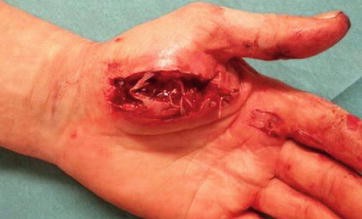
Deep laceration wound of the hand with injury of thenar muscles
-
Severe bruising (tissue contusion)
-
Avulsions (with traction injury) (Fig. 14.2)
Fig. 14.2 
Degloving injury of the hand
-
Thermal destruction (Fig. 14.3)
Fig. 14.3 
Entrance site of a low-voltage burn (a), after 5 days before debridement (b)


There are many classic injuries associated with certain types of accidents. Falling on the outstretched palm with hyperextension of the wrist is often the mechanism for fracturing the scaphoid.
14.1.4 Planning
Treatment planning is based on careful analysis of the initial injury and the resulting defect after surgical primary care, the lost functions, and the necessary reconstruction, including soft tissue coverage.
Surgical treatment is directed at achieving the following goals:
-
1.
Complete removal of infected or avital tissue
-
2.
Restoration of tissue perfusion
-
3.
Bony stabilization with minimal additional soft tissue trauma
-
4.
Stable, well-perfused, and aesthetically adequate soft tissue coverage
-
5.
Early mobilization of the limb in order to minimize post-traumatic scarring and restore function and motion of the hand
Many factors enter into the treatment plan for each individual patient, including age, education, vocation, hand dominance, expectations, and even hobbies.
14.1.5 Surgical Procedure
The surgical procedure may be divided into several steps:
14.1.5.1 Acute Care/Emergency Room Management
-
Review of the injury and possible additional injuries (ABC rule, exclusion of danger to life and involvement of sensory organs, especially eyes and ears)
-
Hemostasis of acute bleeding (by tourniquet, usually no clamping of vessels)
-
Reduction of bony deformities
-
Control of tetanus protection, if necessary booster or vaccination, antibiotic administration
-
Cooling of devascularized tissue (leaving intact skin bridges)
Polytraumatized patients, that is, patients with life-threatening injury to one or more organ systems, must always be judged in terms of the leading injury (“life before limb”). Treatment of hand injuries is limited to temporary stabilization of bone injuries and bleeding. For clinically unstable patients, amputations may be more useful than lengthy reconstructions.
After determining the leading injury, triage is performed and hemostasis and prevention of life-threatening blood loss is the first goal in the emergency room. Removal of a compression bandage often significantly improves the perfusion situation and enables further evaluation, including pulse status and sensibility testing.
The exact medical history includes factors such as age, occupation, recreational interests, additional illnesses, medications, allergies, and other factors, such as nicotine consumption. It is always important to document the exact circumstances of the accident and mechanism, as this can significantly influence further treatment and prognosis. Radiographs of the affected limb in two planes (also amputate) should be performed (Fig. 14.4).

Radiograph of amputate and hand
14.1.5.2 Anesthesia and Use of Tourniquet
Anesthetic requirements for hand surgery are few and simple. The patient should be free of all pain and lie quietly throughout the operation, including application of the dressing. The vast majority of patients are healthy young people for whom a wide variety of general or regional nerve block techniques are equally satisfactory.
In case of complex hand injuries or multiple amputations of finger with expected long operation times, the operation should be performed under general anesthesia, if necessary, with brachial plexus. Postoperative monitoring in the ICU should be considered.
Regular use of a pneumatic tourniquet to maintain a bloodless operation has been a major contribution to reparative hand surgery and is essential both for the identification of injured structures and to protect uninjured ones.
In most cases, one and a half hours ischemia time is sufficient for identification of injured parts, debridement, and dissection. Most of the repairs can be performed following the release of the tourniquet if necessary.
The pressure of the pneumatic tourniquet should not be above 300 mmHg on the adult upper extremity and proportionally less for children.
14.1.5.3 Surgical Debridement
Debridement of complex hand injuries follows several important rules
-
Wound excision and aggressive debridement of necrotic and ischemic tissue, especially muscle
-
Preservation of critical structures (nerves, tendons, and arteries)
-
Marking of important structures such as nerves, blood vessels and tendons for any secondary reconstructions (Fig. 14.5).
Fig. 14.5 
Preoperative view of severe self-wrist cutting injury (a); View after debridement: complete transection of tendon, nerve, and vessel with defect (b)

In amputation injuries, during initial debridement, well-preserved or vascularized tissue components (spare parts, such as bones, tendons, nerves, or blood vessels) should be preserved for use in reconstruction or soft tissue coverage (fillet flap, skin grafts).
The extent of debridement in “crush injuries” is often difficult to assess. Wound assessment can be complicated, especially with closed injuries, because a significant soft tissue injury case is sometimes detected preoperatively but underestimated in many cases.
14.2 Bone and Joint Reconstruction
Fractures of the hand are the most common of all fractures. Radiographs are required in at least two planes to confirm clinical impressions and to demonstrate the extent of injury. CT scan may be necessary with fracture of the hook of the hamate or of the scaphoid.
Stabilization of bone injuries is usually performed by simple, fast, and less traumatic methods, for example, K-wires or external fixation; it is less often performed with plating.
Important strategies in the care of bony injuries are:
-
Fracture visualization by minimal wound enlarging, without destruction of periosteum
-
Maximum length preservation; bone reduction only if for the primary soft tissue closure or for bone and nerve reconstruction necessary
-
Accurate anatomical fracture reduction with special consideration of joint surfaces
-
Bridging of defects with external fixation or plate (Fig. 14.6)
Fig. 14.6 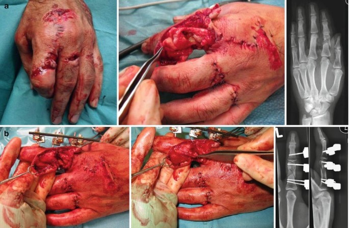
Open fracture with segmental bone loss and severe soft tissue trauma (a). The mini Hoffmann external fixation device applied for a complex proximal phalanx fracture; due to external fixation, a primary tendon repair can be achieved (b)
-
Complex bony reconstruction, for example, iliac crest bone graft is usually performed secondarily

Bony stabilization is recommended to enable early movement therapy. Examples include
-
Radius/ulna – plating (Fig. 14.7)
Fig. 14.7 
Fractured radius and ulna treated with plating of both bones
-
Wrist – compression screw/K-wires, reconstruction of injured ligamentous structures, stabilization with K-wires
-
Metacarpals – mini plate fixation for early mobilization (Fig. 14.8)
Fig. 14.8 
Circular saw injury of the hand with open metacarpal fractures, segmental bone loss, extensor tendon injury and dorsal soft tissue injury (a); metacarpal shaft fractures 3–5, mini plate fixation with some shortening of the metacarpal (b); repair of extensor tendon (c); radiograph preoperatively and after open reduction and internal fixation with 2.3 mm dorsal plate (d)


To exclude additional injuries of the wrist or fingers, they are moved under the imager to detect, for example, overlooked ligament injuries or dislocations.
In a perilunate dislocation, open reduction and suture of carpal ligaments (especially the SL ligament) should be performed (Fig. 14.9). In joint destruction a primary fusion (arthrodesis) in appropriate function may be advisable.

Preoperative radiograph showing dorsal perilunate dislocation in which the lunate dislocates from its fossa and is rotated more than 90° (a); intraoperative image showing the dislocated lunate (b); after open reduction and K-wire fixation (c); after 12 weeks and K-wire removal (d)
14.3 Digital Joint Injuries
Dislocations of the metacarpophalangeal (MP) finger joints are rare. They usually result from falling on the outstretched hand or another mechanism that forces the MP joint into severe hyperextension. They are limited to the index and the small fingers.
These are classified as simple when reduction is easily accomplished and complex when soft tissue elements preclude closed manipulative reduction. After reduction, protection against recurrent injury is provided initially by splinting and then by taping the involved finger to a normal adjacent digit for 2 or 3 weeks. If closed reduction is unsuccessful, then the injury becomes a complex type of dislocation that requires an open reduction.
Stability and pain-free motion of proximal interphalangeal (PIP) joints is extremely important to hand function. Therefore, injury to these joints warrants high priority in treatment. PIP joint dislocations are usually dorsal and result from force on the end of digit with hyperextension (Fig. 14.10).


Open PIP joint dislocation of the middle finger, palmar plate rupture of the ring- and small finger (a); preoperative radiograph (b); radiograph after treatment with protected motion (Extension Block Splint) (c); 12 month follow-up, the patient had no pain and achieved functional range of motion (d)
PIP joint injuries that are stable with active motion can be treated by immobilization with a dorsal splint, usually in 20–30° of flexion for comfort and to rest the soft tissues. The period of immobilization ranges from as little as 3–5 days for mild hyperextension injuries to 7–14 days for dislocations and stable fracture dislocations. Duration of immobilization is individualized, depending on the extent of the injury and resultant amount of soft tissue swelling.
14.4 Tendon Suture and Reconstruction
The prognosis for tendon repairs is determined primarily by what tissues lay in contact with the repair of the tendon.
Extensor tendon injuries of the hand represent common injuries that are underestimated in many cases. If there is a delay in diagnosis and the primary injury is overlooked, deformities (e.g., buttonhole or boutonniere deformity, swan neck deformity) may have already been made. Secondary reconstruction after extensor tendon injuries is more difficult and yields worse results than primary care.
14.4.1 Zones of Extensor Tendon Injury
The dorsum of the hand, wrist, and forearm are divided into eight anatomic zones to facilitate classification and treatment of extensor tendon injuries. The most widely used classification system is that described by Verdan.
Zones 1, 3, 5, and 7 lie over the distal interphalangeal joint (DIPJ), proximal interphalangeal joint (PIPJ), metacarpophalangeal joint (MCPJ), and wrist joint, respectively.
The even numbers are allocated to the intervening zones. The zones of the thumb differ from the finger as there are only two phalanges. They are numbered 1–5, with zones 1, 3 and 5 overlying the interphalangeal joint (IPJ), MCPJ, and wrist joint, respectively. The even numbers, 2 and 4, apply to the intervening zones.
-
Zone 1 (distal interphalangeal [DIP] joint)
-
Zone 2 (middle phalanx)
-
Zone 3 (proximal interphalangeal [PIP] joint)
-
Zone 4 (proximal phalanx)
-
Zone 5 (metacarpophalangeal [MCP] joint)
-
Zone 6 (dorsum of hand)
-
Zone 7 (wrist)
-
Zone 8 (dorsal forearm)
Extensor tendon injuries may require operative intervention, depending on the complexity of the injury and the zone of the hand involved.
-
Zone 1 injuries, otherwise known as mallet injuries, are often closed and treated with immobilization and conservative management where possible. Zone 2 injuries are conservatively managed with splinting. Closed Zone 3, or “boutonniere,” injuries are managed conservatively unless there is evidence of displaced avulsion fractures at the base of the middle phalanx, axial and lateral instability of the PIPJ associated with loss of active or passive extension of the joint, or failed nonoperative treatment. Open zone 3 injuries are often treated surgically unless splinting enables the tendons to come together. Zone 5 injuries are often caused by human bites and, require primary tendon repair after irrigation. Zone 6 injuries are close to the thin paratendon and thin subcutaneous tissue and require strong core-type sutures and then splinting; they should be placed in extension for 4–6 weeks. Complete lacerations to zone 4 and 6 involve surgical primary repair followed by 6 weeks of splinting in extension (Fig. 14.11).
Fig. 14.11 
No active extension of the small finger due to extensor tendon injury (a); intraoperative image showing tendon injury in zone 6 (b)
-
Zone 8 requires multiple figure-of-eight sutures to repair the muscle bellies and static immobilization of the wrist in 45° of extension.

14.4.2 Zones of Flexor Tendon Injury
The five flexor tendon zones are modifications of Verdan’s original work, which based zone boundaries from distal to proximal on anatomic factors that influenced prognosis following flexor tendon repair. The five zones discussed below apply only to the index through small fingers; separate zone boundaries exist for the thumb flexor tendon.
-
Zone 1 extends from just distal to the insertion of the superficialis tendon to the site of insertion of the flexor digitorum profundus tendon (FDP).
-
Zone 2 is often referred to as “Bunnell’s no man’s land,” indicating the frequent occurrence of restrictive adhesion bands around lacerations in this area. Proximal to zone 2, the flexor digitorum superficialis (FDS) tendons lie superficial to the FDP tendons. Within zone 2 and at the level of the proximal third of the proximal phalanx, the FDS tendons split into two slips, collectively known as Camper chiasma. These slips then divide around the FDP tendon and reunite on the dorsal aspect of the FDP, inserting into the distal end of the middle phalanx.
-
Zone 3 comprises the area of the lumbrical muscles origin between the distal margin of the transverse carpal ligament and the beginning of the critical area of pulleys or first annulus. The distal palmar crease superficially marks the termination of zone III and the beginning of zone II.
-
Zone 4 is the zone covered by the transverse carpal ligament, including the carpal tunnel and its contents (the nine digital flexors and the median nerve).
-
Zone 5 extends from the origin of the flexor tendons at their respective muscle bellies to the proximal edge of the carpal tunnel.
14.4.3 Tendon Healing
The following factors predict tendon healing: age, overall health condition, scar formation disposition, motivation, injury risk based on Verdan’s zones, injury type, synovial containment, and surgical technique. Three phases of tendon healing are defined. First, a migration of peripheral cells and invasion of blood vessels occurs and, second, the tendon and surrounding tissues heal. Remodeling happens in the third phase of healing as a result of movement and function of the tendon. This is the ratio of the widely recommended early passive movement therapy, which leads to better supply and strength of the tendon.
The tendon gains its daily life loading capacity after 12 weeks of healing, and sporting activities are allowed no sooner than 4 months after injury. The remodeling process can last up to 12 months.
Regarding restoration of tendon function, the following guidelines apply:
-
Excision of bruised hand intrinsic muscles (especially Mm. interossei and lumbricales) to prevent ischemia and subsequent contracture
-
Reconstruction and seam of the A2 and A4-ring belts to prevent bowstring phenomenon
-
If necessary, primary tenodesis or tendon transposition surgery
-
Late reconstruction using tenolysis, tendon transposition, transplantation, or functional muscle transplantation
Tendon sutures, if possible; the superficial digital flexor tendon can be used as a donor in a primary operation. In a secondary reconstruction, long segment splintering, usually with silastic rod insert and subsequent tendon transplant, is performed (e.g., the PL tendon). The injured structures must be identified during the initial stage and marked.
14.5 Treatment of Vascular Injuries
Important considerations in the reconstruction of vascular lesions, which generally take place only after the treatment of bone and tendon injuries, are:
-
Creation of temporary shunts in critical ischemia
-
Preparation of vessels with microsurgical instruments under direct vision with a tourniquet
-
Use of a Fogarty catheter with proximal and distal vascular injuries
-
Return reduction of vessels until healthy, uninjured tissue is reached (Caution: intima damage in avulsion injuries)
-
Regular flushing of the vessels with heparin solution (10 U/ml)
-
Vascular sutures under the microscope, outside the zone of injury
-
Length gain by ligation or removal of side branches, diffraction from adjacent joints of interposition vein grafts (in reverse flow direction)
-
Vein grafts are introduced outside the zone of injury and previously dilated (Fig. 14.12)
Fig. 14.12 
Traumatic partial amputation of ring and small finger (a); radiograph showing severe bone crush of metacarpals (b); revascularization with vein grafts (arrow) (c)
-
Reconstruction of both vascular trunks of the radial artery and the ulnar artery

In avascular limbs, especially macro-replantations, revascularization is the first priority, before tendon and nerve sutures. Bruised or avulsed vessels must be cut behind the zone of contusion. For vein harvesting for long defects, the greater saphenous vein or veins of the forearm are suitable as interposition grafts.
For revascularization proximal to the hand, a fasciotomy is usually required to prevent compartment syndrome as a result of reperfusion. If possible and appropriate, an attempt should always be made at revascularization. If this is not successful, an amputation can still be performed. The main indicative factor is always the expected functional result in the context of age, profession, and the favorite leisure activities of the patient.
In complex amputation injuries, for example, with severance of multiple fingers and the thumb, an extraanatomical reconstruction by heterotopic replantations is an option to achieve basic grip function.
14.6 Nerve Reconstruction
Nerve reconstruction surgery is normally performed as a last step before soft tissue coverage:
-
Shortening of the nerves until healthy fascicles become visible
-
Epineural nerve suture under a microscope to restore the fascicular structure
-
Nerve suture, always without tension (Fig. 14.13).
Fig. 14.13 
Injury of the proper digital nerve (PDN) (a); primary tensionless repair (b)

Nerve stump approximation is possible by flexion of adjacent joints. In replacement of nerves (e.g., ventralization of the ulnar nerve), for smaller defects (<2 cm) artificial nerve conduits can be used; otherwise, nerve grafting (sural nerve, sensory nerve forearm) is necessary. If a primary nerve suture without tension is not possible, nerve stumps are marked and nerve reconstruction is performed secondary to consolidation of wound conditions.
14.7 Soft Tissue Coverage
Final soft tissue coverage should never be enforced. If necessary, a temporary wound closure can be achieved by a synthetic skin substitute (Epigard®) or a vvacuum-assisted closure (V.A.C.®).
-
Definitive soft tissue wound closure is performed in a stable situation, possibly after repeated debridement (often after 5–10 days).
-
Temporary wound conditioning should be performed, possibly with (V.A.C.®) (contraindicated in infection or bleeding persists).
-
Movable joints and tendons should be covered with well-vascularized, thin, and movable tissues (flaps).
-
Exposed nerves or vessels must be primarily covered by local flaps, pedicled distant flaps, or microsurgical tissue transplantation
-
All “white structures” (tendons, bones, ligaments, joints) should be covered with a flap.
-
Violation of the fingertips without exposed bone often heals by secondary intention (OPSITE™ FLEXIGRID™ film dressing) (Fig. 14.14)
Fig. 14.14 
Horizontal amputation of the distal soft tissue of the index finger with exposure of the distal tuft (a); 6 weeks after secondary healing with dressing changes (b)

Time-proven and reliable pedicled flaps in finger, hand, and wrist wounds are the Moberg flap, cross finger flap, intrinsic flaps (e.g., Foucher or DMCA flap), groin flap, distally pedicled posterior interosseous artery flap (Fig. 14.15), and the radial forearm flap.

Massive hand infection due to a primary overlooked joint injury (a); defect with exposed tendon after debridement (b); Coverage of the defect with a distally pedicled, fasciocutaneous posterior interosseous artery flap (c)
In the groin flap, the sensory lateral femoral cutaneous nerve can be included and connected using microsurgery. Usually the pedicle is separated off after about 2 weeks; the final flap inset is performed after 3 weeks. Disadvantages include limited mobility of the patient with increased thrombotic risk, which makes prophylaxis necessary to prevent potentially life-threatening complications, such as pulmonary embolism. Thinning operations of the flap are usually necessary, in addition to long-term physiotherapy to prevent stiffness (Fig. 14.16).

Degloving injury of the hand (a); advantages of the groin flap are that it is rapidly and easily harvested (b); coverage of the hand with pedicled groin flap and skin graft for closure of the donor site (c); result after 3 month and two operation for separating fingers (d)
Defects in the region of the distal forearm are commonly covered using the gracilis, latissimus, lateral arm, or anterior lateral thigh (ALT) flaps (Fig. 14.17).

Deep self-inflicted wrist laceration as shown in Fig. 14.5 after tendon and nerve repair (a); free anterior lateral thigh flap (ALT) (b); Coverage of the forearm defect with the free flap (c)
Note that, when planning microsurgical flaps after avulsion injuries, occult vascular injuries with intima damages may complicate early tissue transplantations.
14.8 Amputation Injuries of the Hand
The establishment of microsurgery has enabled the restoration of the hand after traumatic amputation injuries, even in severe injuries; valuable basic function can often be reconstructed. Survival rates of replanted extremity parts have reached more than 80%. However, today, criteria for success includes not only survival but the functional value of the reconstructed hand, including their sensibility. Results even with modest functional recovery can justify such operations. This chapter presents indications, preoperative management, surgical technique, and postoperative treatment of amputation of the hand.
14.8.1 Definition
Amputation is defined as complete detachment of a body part, whereas subtotal amputation is an interruption of blood supply, which, by definition, without immediate restoration would lead to necrosis of the affected limb (no vessel and not more than 25 % of soft tissues intact). Consequently, replantation means restoring a completely separate body part, while revascularization is the restoration of the blood flow in a subtotal amputation.
14.8.2 Indications for Replantation
Absolute indications are:
-
Amputation of the thumb
-
Amputation of several fingers
-
Amputation with concomitant serious injury of several fingers
-
Amputation of the metacarpal
-
Amputation of wrist/forearm/elbow or upper arm (in sharp amputations and intact amputates)
-
Amputation injury in children
Relative indications include:
-
Isolated finger in intact neighboring fingers (exception: special function needed, e.g., for a profession)
-
Single distal phalanx amputation or destroyed MCP or PIP joint
No indication/contraindications are present in case of:
-
Life-threatening additional injury
-
Improper handling or destruction of amputates (e.g., freezing)
-
Amputation of the DIP joint/nail root
-
Incompliance of patient
-
Multilevel injuries
The indication in favor or against a replantation is often difficult and depends on various factors.
14.8.3 Assessment of the Patient
The decision is based on the history and mechanism of the accident, occupation, age, and smoking-related co-morbidities, and absence of associated injuries. The patient must be informed of opportunities and chances of success of the operation and included as far as possible in the decision-making process for or against replantation. The first requirement is that the patient is able to tolerate surgery over several hours and is not compromised by preexisting conditions or associated injuries. In children, replantation almost always should be attempted. Although the growth of the replanted body may be reduced, the results regarding motion and sensibility are usually much better than in adults. On the other hand, advanced age alone is not a contraindication for replantation; a mentally and physically active patient beyond the age of retirement can equally benefit from a microsurgical reconstruction. It must be remembered, however, that for prolonged and intense physical therapy and occupational therapy, the patient’s cooperation and ability to understand is required. The microsurgical reconstruction makes sense if the function is expected to be at least as good as or better than a prosthetic.
14.8.4 Assessment of the Amputate
The suitability of the amputate depends on:
-
Ischemia time
-
Amputation level
-
Amputation type (Guillotine type vs. avulsion or severe contusion)
Clear guillotine-like amputations without joint involvement have a better prognosis for replantation than avulsion or crush mechanisms (e.g., table saw injuries), which complicate the surgical management and reduce the chance of success (Fig. 14.18).

Amputate of the index finger by a table saw, hand and radiograph showing a guillotine-like amputation (a); assessment of the amputate reveals a severe crush with no opportunity for replantation (b)
14.8.5 Evaluation of Overall Injury
Due to its functional significance, replantation of the thumb is always desirable in order to achieve maximum length preservation. In case of injury to the palmar neurovascular bundle, the thumb can be revascularized with a long vein graft from the radial artery or the princeps pollicis artery. In multiple finger amputations, a heterotopic replantation of the least injured parts in the best functional position is advisable (e.g., to put the index finger on the thumb stump). Successful replantations of amputations at the metacarpal, wrist, or forearm level are usually functionally better than prosthesis.
If sensibility is sufficient for a protective function, the extrinsic forearm muscles lead to a satisfactory hand function, even if the intrinsic hand function is usually poor. Due to the slow nerve regeneration of about 1 mm per day, and a distance of 40–80 cm, functional return of the intrinsic hand muscles is unrealistic.
In macro-replantations, age of the patient, ischemia time (about 4–8 h, depending on cooling) and the suitability of the amputate for replantation represent the main determinants.
A special entity of amputations is ring avulsions of the fingers, which are classified using different systems, most commonly according to Urbaniak.
Classification of ring avulsion injuries according to Urbaniak:
-
I.
Perfusion of the finger distal to the injury intact, simple soft tissue injury or fracture, simple osteosynthesis sufficient.
-
II.
Perfusion disturbed and reconstruction of either arteries or veins required, or both, but no complete amputation
-
III.
Complete avulsion with amputation and fracture in middle and distal phalanx, with avulsion of functional structures (Fig. 14.19).
Fig. 14.19 
Ring avulsion of the index finger

Third-degree avulsions have the worst prognosis, especially proximal to the PIP joint.
If the amputation is distal to the insertion of the FDS tendon and the PIP joint is intact without further destruction of the proximal phalanx, the replantation may have a more favorable prognosis. Vessels should always be shortened sufficiently and restored by vein grafts.
Ischemia tolerance (cooled at about 4 °C) depends on the soft tissue components:
-
Without muscles (e.g., finger) 8–12 h up to 24 h
-
With muscles (e.g., forearm) 4–5 h up to 8 h
14.8.6 Technical Requirements
Instrumental and technical requirements for microsurgical replantation and revascularization include:
-
Magnifying loupes (3.5–4.5-fold magnification, working distance of 35–45 cm) for the surgeon and assistant
-
Surgical microscope (at least 20-fold magnification), best with two binocular eyepieces, for the surgeon and assistant
-
Precision microinstruments with forceps (thickness 0.2–0.3 mm), jeweler forceps and tweezers with coated tips, fine needle holders, microirrigation, and possibly a microdilatator
-
Suture material for vessels and nerves (fingers at 10-0, 10-0, and 9-0 in the metacarpal and proximal to the wrist, 8-0), nonresorbable (Ethilon, Prolene, or similar)
Although many plastic surgeons have received microsurgical training, replantation should take place in centers where microsurgical training options exist and such operations are performed regularly.
14.8.7 Transport of Amputates
The appropriate transportation and storage are critical to the success of replantation. Amputates should be recovered at the accident scene and, after careful cleaning, be cooled and transported in a wet compress; they should never be stored in direct contact with ice. Special bags should be used with an inner pouch in which the dry amputated can swim in an outer pouch filled with ice-water. Maceration of the amputate will induce swelling of the vessels and render replantation considerably more difficult or impossible.
14.8.8 The Surgical Management
After an initial X-ray examination of the injured limb and amputate, two members of the replantation team start to clean and prepare the amputate. In amputated fingers, medio-lateral incisions are used to identify and dissect the digital nerves and the accompanying palmar vessels; then the flexor and extensor tendons and dorsal veins. Nerves, arteries, and vessels are marked with micro clips or 9–0 sutures after arteries and veins were rinsed by Liquemin. The veins may be hard to find initially, a spilling of the arteries may be helpful. If the veins cannot be detected, wait until the first arterial anastomosis is accomplished and venous outflow can be seen. Marking of vessels and nerves in the still bloodless surgical field proves helpful and saves time, especially in multiple finger replantations and when, in the advanced stage of the operation, efficiency and patience of the surgeon may be reduced.
After the tendon is identified, the bone is shortened with an oscillating saw. If the PIP joint or DIP joint is destroyed, arthrodesis is performed.
The bony fixation should be performed quickly and without additional injury of the dorsal structures, preferably using K-wire (1.0 or 1.2 mm) and transosseous suture.
A perioperatively plexus catheter for pain and sympathetic management is important.
When the patient reaches the operating room, the second team starts to prepare the proximal stump under loupe magnification and tourniquet, also using medio-lateral incisions to identify and mark the important structures corresponding to the amputate. Finding veins in the fingers requires a subtle preparation, but it is important because venous outflow is critical for success.
Basically, the same procedures apply for micro- as for macro-replantations; although, in the latter case, because of the lower tolerance of ischemic time, restoring perfusion is considered the first priority.
The order of procedures in a replantation is usually:
-
1.
Cleaning and debridement of the soft tissues at stump and amputate
-
2.
Marking tendons, vessels, and nerves
-
3.
Debridement and possible shortening of the bone, joint arthrodesis in functional position if necessary
-
4.
Osteosynthesis (mostly by K-wires)
-
5.
Suture of flexor and extensor tendons
-
6.
Suture of palmar arteries
-
7.
Suture of nerves
-
8.
Suture of dorsal veins
-
9.
Soft tissue closure, possibly only temporarily (e.g., in severe swelling)
14.8.9 Dressing
The dressing is made with loosely laid gauzes and compresses. Constriction by a circular bandage, for example, by drying blood, should never occur. The hand should be well padded and kept warm by generous cotton wrapping. A volar splint in intrinsic plus position may be applied together with a plexus catheter and reduced active mobilization can be started.
14.8.10 Postoperative Management
Important points of the postoperative care are:
-
Plexus catheter to control of the sympathetic vasoconstriction and improve circulation for 4–7 days
-
Elevation of the arm on a cushion; in case of venous congestion the arm can also be suspended
-
If venous drainage if venous drainage is a problem, use of leeches for 3–4 days
-
For arterial inflow problems, the hand must be kept slightly lowered
Skin color, finger temperature, skin turgor and capillary refill time (preferably 1–2 s) are useful in assessing the perfusion situation. The perfusion status of the replanted finger or the hand can change suddenly. In order to react quickly, during the first 3–4 days a reliable and regular monitoring must be ensured
14.8.11 Evaluation of Results
The Chen classification evaluates the functional results after replantation in the upper limb:
-
I.
Able to resume previously held employment; range of motion (ROM) exceeds 60 % of normal; complete or nearly complete recovery of sensibility; muscle power of grades 4 and 5
-
II.
Able to resume professional activities; ROM exceeds 40 % of normal; nearly complete sensibility; muscle power of grades 3 and 4
-
III.
Able to lead normal daily life; ROM exceeds 30 % of normal; partial recovery of sensibility; muscle power of grade 3
-
IV.
Almost no useable function in survived limb
14.8.12 Complications
Typical risks after revascularization and replantation include
-
Reamputation in case of ischemia or functional disability
-
Motion deficits and disorders
-
Cold sensitivity, primarily dependent on the type of injury and also possibly in amputation stumps
-
Nail growth disorders
-
Stump neuroma
14.8.13 Postoperative Treatment, Rehabilitation
Surgical treatment usually includes immobilization with a splint, mostly in neutral position or extension of the wrist, flexion of the MCP joints, extension of the IP joints (intrinsic plus – positioned to minimize contractures). Early intensive physiotherapy is useful to optimize the sliding of the tissue layers. To prevent edema (with the risk of fibrosis caused by lymphatic congestion) limb elevation, drainage, intensive physiotherapy, and occupational therapy are recommended (Fig. 14.20).

Additionally, psychological and social support should be offered after a complex hand injury.
14.8.14 Secondary Operations
Secondary operations include operations requiring postoperative immobilization:
-
Bone grafts (Fig. 14.21)
Fig. 14.21 
Postoperative radiograph with external fixation after revascularization of ring and small finger as shown in Fig. 14.12 (a); change to internal fixation with 2.3 mm dorsal plate and bone graft after 12 weeks (b)
-
Corrective/joint reconstructions
-
Nerve transplants
-
Sensitive reconstructions
-
Muscle transpositions / functional muscle transplants
-
Soft-tissue reconstruction
-
Toe transplants

At a later stage, procedures requiring early mobilization are performed:
-
Tenolysis
-
Capsulotomies
-
Contracture releases
References
Biemer E, Duspiva W (1980) Rekonstruktive mikrogefäßchiurgie. Springer, Heidelberg
Büchler U (1990) Traumatic soft-tissue defects of the extremities. Implications and treatment guidelines. Arch Orthop Trauma Surg 109:321
Buncke HJ Jr (2000) Microvascular hand surgery—transplants and replants—over the past 25 years. J Hand Surg 25A:415
Chen ZW, Yu HL (1987) Current procedures in China on replantation of severed limbs and digits. Clin Orthop 215:15–23
Chuang DC, Lai JB, Cheng SL, Jain V, Lin CH, Chen HC (2001) Traction avulsion amputation of the major upper limb: a proposed new classification, guidelines for acute management, and strategies for secondary reconstruction. Plast Reconstr Surg 108(6):1624–1638
Dorf E, Blue C, Smith BP, Koman LA (2010) Therapy after injury to the hand. J Am Acad Orthop Surg 18(8):464–473
Gan AW, Neo PY, He M, Yam AK, Chong AK, Tay SC (2012) A biomechanical comparison of 3 loop suture materials in a 6-strand flexor tendon repair technique. J Hand Surg Am 37(9):1830–1834
Germann G, Levin S (1997) Intrinsic flaps in the hand: new concepts in skin coverage. Tech Hand Up Extrem Surg 1:48
Germann G, Sherman R, Levin S (1999) Decision making in reconstructive surgery. Springer, Berlin/Heidelberg/New York
Germann G, Karle B, Brüner S, Menke H (2000) Treatment strategies for complex hand injuries. Orthop 103:342–347
Griffin M, Hindocha S, Jordan D, Saleh M, Khan W (2012) An overview of the management of flexor tendon injuries. Open Orthop J 6:28–35
Kim JYS, Brown RJ, Jones NF (2005) Pediatric upper extremity replantation. Clin Plast Surg 32:1
Kleinert HE, Jablon M, Tsai TM (1980) An overview of replantation and results of 347 replants in 245 patients. J Trauma 29:390
Lee BI, Chung HY, Kim WK et al (2000) The effects of the number and ratio of repaired arteries and veins on the survival in digital replantation. Ann Plast Surg 44:288
McCabe SJ, Breidenbach WC (1999) The role of emergency free flaps for hand trauma. Hand Clin 15:275–288
Pederson WC (2001) Replantation. Plast Reconstr Surg 107:823
Schöffl V, Heid A, Küpper T (2012) Tendon injuries of the hand. World J Orthop 3(6):62–69
Tamai S, Michon J, Tupper J et al (1983) Report of the subcommittee on replantation. J Hand Surg 8:730
Verdan CE (1975) Primary and secondary repair of flexor and extensor tendon injuries. In: Flynn JE (ed) Hand surgery, 2nd edn. Williams & Wilkins, Baltimore
Weinzweig N, Sharzer LA, Startker I (1996) Replantation and revascularization at the transmetacarpal level: long-term functional results. J Hand Surg Am 21:877
Werdin F, Schaller HE (2008) Combined flexor tendon and nerve injury of the hand. Orthopade 37(12):1202–1209
Author information
Authors and Affiliations
Corresponding author
Editor information
Editors and Affiliations
Rights and permissions
Copyright information
© 2014 Springer-Verlag Berlin Heidelberg
About this chapter
Cite this chapter
Hellmich, S., Vogt, P.M. (2014). Management of Hand Injuries. In: Oestern, HJ., Trentz, O., Uranues, S. (eds) Bone and Joint Injuries. European Manual of Medicine. Springer, Berlin, Heidelberg. https://doi.org/10.1007/978-3-642-38388-5_14
Download citation
DOI: https://doi.org/10.1007/978-3-642-38388-5_14
Published:
Publisher Name: Springer, Berlin, Heidelberg
Print ISBN: 978-3-642-38387-8
Online ISBN: 978-3-642-38388-5
eBook Packages: MedicineMedicine (R0)