Abstract
Surgery has a controversial role in the treatment of malignant pleural mesothelioma. Controversy also exists regarding the utility of extrapleural pneumonectomy versus pleurectomy and decortication (P/D). This chapter focuses on specific case scenarios and the technical details of P/D.
Access provided by Autonomous University of Puebla. Download chapter PDF
Keywords
- Standardize Uptake Value
- Pulmonary Function Testing
- Malignant Pleural Mesothelioma
- Chest Tube Drainage
- Pleural Disease
These keywords were added by machine and not by the authors. This process is experimental and the keywords may be updated as the learning algorithm improves.
Introduction
The epidemiology of mesothelioma was first recognized in 1960 in the report by Wagner and colleagues of 33 South African asbestos mine workers who developed mesothelioma (Wagner et al. 1960). Confirmed mesothelioma cases have been on the rise since the 1970s. Mesothelioma is a rare disease, with one case per 100,000 people found in the United States (Ismail-Khan et al. 2006). The occupational exposure to asbestos is shown by a male-to-female ratio of 5:1. Malignant pleural mesothelioma (MPM) is a terminal cancer with no consensus regarding optimal staging and treatment.
Surgery is a key modality in the treatment of MPM. Butchart and colleagues (1976) reported the initial experience in performing extrapleural pneumonectomy (EPP) for MPM. Unfortunately, their data showed a 33 % operative mortality. However, recent studies demonstrate 5 % mortality rates. Mortality rates for pleurectomy and decortication (P/D), which are 1–4 %, and supporting evidence show there is no superior distinction to performing an EPP versus a P/D. When enhancement was made in patient selection and perioperative care, a substantial decrease in mortality rates was found. The high mortality and morbidity associated with EPP, and the well-known complications of pneumonectomy, favor the less invasive P/D. In the largest study to date, MPM patients from three different institutions were analyzed and compared. Five-year survival was similar between the 385 patients who had EPP and the 278 patients who underwent P/D (Flores et al. 2008). P/D actually demonstrated a statistically significant increase in median survival compared with EPP (P < 0.001). In the multivariate analysis, EPP was found to have a modestly higher hazard ratio of 1.4 when compared with P/D. A higher proportion of EPP patients experienced serious respiratory complications. The decision as to the preferred surgical procedure for MPM remains controversial, but our bias is to perform P/D if resection of all gross disease is possible.
Preoperative Evaluation
Preoperative evaluation is essential in determining the patient’s indication for either procedure and whether he or she should undergo any surgical intervention. When a patient is diagnosed with MPM, aside from pulmonary function testing (PFT), imaging of the chest and upper abdomen with CT is mandatory. CT scans may provide preoperative evidence of the level of tumor involvement. However, this is often determined at the time of surgery. If chest wall or neurovascular invasion is suspected, MRI may be helpful in preoperative planning. Positron emission tomography/CT (PET/CT) is performed to determine whether distant metastatic disease is present (Flores et al. 2003a). The standardized uptake value (SUV) may be used to predict the presence of N2 lymphatic spread. High SUV has been shown to correspond with poor survival in patients diagnosed with MPM (Flores et al. 2006). However, N2 disease should not be used as an absolute criterion for denying surgical resection (Flores et al. 2003b). When the extent of pleural disease significantly affects the patient’s ability to perform PFT, a more accurate assessment of lung function is needed. Indications for surgery may be thought of as tumor related or patient related. P/D is a procedure that is best offered to patients who do not have the cardiopulmonary reserve to tolerate EPP. In patients with insufficient cardiopulmonary reserve, a postoperative predicted FEV1 (first expiratory volume in the first second of expiration) or Dlco (diffusing capacity of lung for carbon monoxide) of less than 40 %, or a left ventricular ejection fraction of less than 45 %, P/D is clearly indicated. Ventilation/perfusion (V/Q) lung scans assist with diagnosing whether the patient with MPM and poor PFT results is capable of undergoing P/D. Mediastinoscopy is helpful in determining N stage in most patients and is more accurate than CT; however, we do not use this modality routinely.
P/D is an attempt to remove all gross disease without removing the underlying lung. It involves resection of the parietal pleura, the visceral pleura, the pericardium, and, in approximately 50 % of patients, the diaphragm. It is a safe procedure. The most common postoperative complication is prolonged air leakage (lasting more than 7 days), which occurs in 10 % of cases. Air leaks seal over time with continued chest tube drainage in most cases. When simple chest drainage fails, pleurodesis is performed. Resection of the diaphragm often is not done when the disease can be successfully stripped from the surface of the diaphragm without a formal resection and reconstruction.
Positioning and approach. After the induction of general anesthesia, a double-lumen endotracheal tube is used to facilitate single-lung ventilation. Because blood loss is often significant with this procedure, an arterial line and central venous line also are used to aid in arterial and venous pressure monitoring. The patient is placed in the lateral decubitus position. A posterolateral thoracotomy incision is extended downward toward the costal margin over the sixth rib and posteriorly beyond the tip of the scapula. A more limited incision, such as a standard posterolateral thoracotomy incision, may be used to start the procedure. After intrathoracic assessment of resectability, the incision may be extended anteriorly as mentioned above. The incision is made downward over the sixth rib, and the periosteum is lifted off the rib superiorly and inferiorly (inset). A periosteal elevator is used to separate the rib from the surrounding soft tissue. A rib cutter is used to separate the rib anteriorly and posteriorly, then the rib is removed. Dissection between the parietal pleura and endothoracic fascia is begun
Dissection of the extrapleural plane. The plane then takes on a cephalad direction toward the apex from the posterolateral direction. Both blunt and sharp dissections are used, with sharp dissection saved mainly for areas of dense adhesions. The apex is often relatively free of tumor compared with the diaphragmatic surface. Dissection in this direction first makes exposure and inferior dissection easier. The placement of two Finochietto retractors, one anteriorly and the other posteriorly, greatly enhances the visualization of the thoracic cavity. As the dissection moves anteriorly, care must be taken not to avulse the internal mammary vessels off the subclavian vessels. Blunt dissection with sponge sticks can effectively separate the pleura from the anterior chest wall close to the mammary vessels, but sharp dissection is recommended once the vessels are found. As the area of dissection increases, previous areas of dissection are packed with laparotomy pads for hemostasis
Dissecting the apex. The subclavian vessels sit at the apex of the thoracic cavity. As the dissection progresses, great care must be used in dissection around the subclavian vessels at the apex. As the dissection proceeds toward the mediastinum, the azygous vein and superior vena cava (SVC) are approached with great caution. Once the upper portion of the lung is completely mobilized from the chest wall, the superior and posterior hilar structures are disclosed. The aorta and arch vessels must be identified and dissected carefully (a). Injury to the phrenic and recurrent laryngeal nerves also must be avoided. After the right upper lobe and right mainstem bronchus are seen, dissection of the esophagus is begun (b). A nasogastric tube helps in palpating and identifying the esophagus. The SVC is then gently dissected off the specimen, and the dissection continues to the posterior aspect of the pericardium. The vagus and phrenic nerves are identified again and protected. a. artery, n. nerve, v. vein
Elevation of the parietal pleura off the diaphragm. The extent of tumor at the level of the diaphragm determines whether resection is required. If the parietal pleura peels off the diaphragm easily, the diaphragm can remain intact. If necessary, the entire diaphragm may be resected and reconstructed with Gore-Tex (W. L. Gore & Associates, Elkton, MD)
If there is significant diaphragmatic invasion, the diaphragm is resected bluntly by avulsing off the chest wall. The diaphragmatic muscle attachments to the chest wall are separated manually with a fingertip, as is done with EPP. Care is taken in dissecting the peritoneum off the undersurface of the diaphragm muscle. The diaphragm is grasped in an Allis or Babcock clamp and retracted upward. The peritoneum is dissected carefully off the undersurface with a sponge stick. The inferior vena cava must be identified at the esophageal hiatus, and skilled care must be used in dissecting the diaphragm off the inferior vena cava
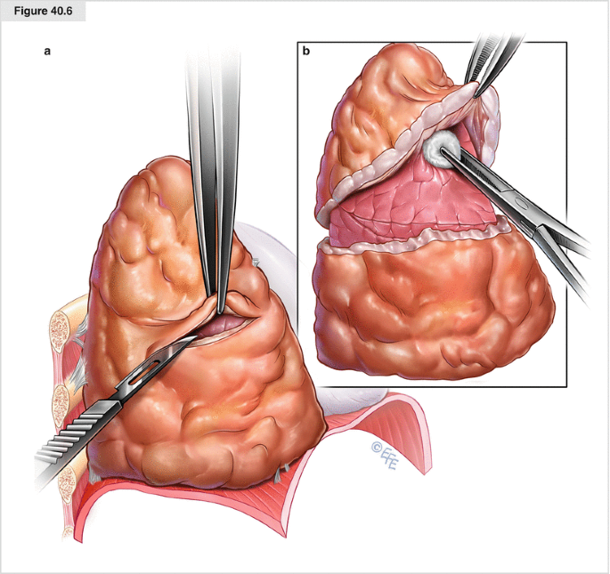
Decortication. Once the parietal pleura has been freed, attention is taken toward separating the visceral pleura from the underlying lung parenchyma. With a scalpel, an incision is made in the tumor lying over the area of the sixth rib (a). The incision is brought down to healthy lung tissue while avoiding injury to the parenchyma. A plane is created between the visceral pleura (adherent to the tumor) and the lung parenchyma with gentle blunt dissection with a peanut sponge (b). The plane of dissection is brought superiorly over the apex and then inferiorly
Repair of the diaphragm. After the diaphragm is dissected, care must be taken to repair it by attaching the diaphragmatic patch to the pericardium. In addition, the patch must be pulled tight to protect the residual lung from loss of domain and atelectasis, which is caused by upward motion of the abdominal organs. On the right, a double-layer Dexon mesh (Covidien, Norwalk, CT) is adequate. On the left, 2-mm thick Gore-Tex is used (thicker nonabsorbable material is needed to prevent herniation of the abdominal contents). The prosthesis is secured laterally by sutures placed around the ribs through the soft tissue of the chest wall
Obtaining hemostasis. An argon beam coagulator is helpful in controlling diffuse chest wall bleeding. Another, often more effective, device is the Aquamantys System bipolar coagulator (Salient Surgical Technologies, Portsmouth, NH). Three 28 F chest tubes are placed: one anteriorly, one posteriorly, and one right-angled. The right-angled tube is placed from the most anterior incision so it rests along the diaphragm toward the most dependent area of the chest. Evacuation of blood and control of air leaks can be accomplished, and full expansion of the lung is expected. Air leaks usually seal within 72 h if the lung is fully expanded
Conclusion
Most studies have shown that the results of surgery alone are poor, and surgery combined with some form of adjuvant therapy, or with a combination of adjuvant therapies, is preferred. These therapies have included external radiation, brachytherapy, systemic chemotherapy, intrapleural chemotherapy, and photodynamic therapy. The question as to whether to treat patients who are diagnosed with mesothelioma with surgical intervention outside of palliative care remains controversial. Until a conclusion is reached with a randomized controlled trial, the decision to perform EPP or P/D is still be based on a combination of patient and disease characteristics as well as on the surgeon’s discretion. In our opinion, future trials should focus on novel agents combined with P/D to improve the treatment of mesothelioma.
References
Butchart EG, Ashcroft T, Barnsley WC, Holden MP (1976) Pleuropneumonectomy in the management of diffuse malignant mesothelioma of the pleura. Thorax 31:15–24
Flores RM, Akhurst T, Gonen M et al (2003a) PDG-PET predicts survival in patients with malignant pleural mesothelioma. Proc Am Soc Clin Oncol 22:620
Flores RM, Akhurst T, Gonen M et al (2003b) Positron emission tomography defines metastatic disease but not locoregional disease in patients with malignant pleural mesothelioma. J Thorac Cardiovasc Surg 126:11–16
Flores RM, Akhurst T, Gonen M, Zakowski M, Dycoco J, Larson SM, Rusch VW (2006) Positron emission tomography predicts survival in malignant pleural mesothelioma. J Thorac Cardiovasc Surg 132:763–768
Flores RM, Pass HI, Seshan VE et al (2008) Extrapleural pneumonectomy versus pleurectomy/decortication in the surgical management of malignant pleural mesothelioma: results in 663 patients. J Thorac Cardiovasc Surg 135:620–626
Ismail-Khan R, Robinson LA, Williams CC et al (2006) Malignant pleural mesothelioma: a comprehensive review. Cancer Control 13:255–263
Wagner JC, Sleggs CA, Marchand P (1960) Diffuse pleural mesothelioma and asbestos exposure in the North Western Cape Province. Br J Ind Med 17:260–271
Author information
Authors and Affiliations
Corresponding author
Editor information
Editors and Affiliations
Rights and permissions
Copyright information
© 2015 Springer-Verlag Berlin Heidelberg
About this chapter
Cite this chapter
Flores, R.M. (2015). Pleurectomy and Decortication for Mesothelioma. In: Dienemann, H., Hoffmann, H., Detterbeck, F. (eds) Chest Surgery. Springer Surgery Atlas Series. Springer, Berlin, Heidelberg. https://doi.org/10.1007/978-3-642-12044-2_40
Download citation
DOI: https://doi.org/10.1007/978-3-642-12044-2_40
Published:
Publisher Name: Springer, Berlin, Heidelberg
Print ISBN: 978-3-642-12043-5
Online ISBN: 978-3-642-12044-2
eBook Packages: MedicineMedicine (R0)


