
Abstract
Tissue engineering and regenerative medicine (TERM) is a biomedical engineering discipline that uses a combination of cells, engineering, materials, methods, and suitable biochemical and physicochemical factors to restore, maintain, improve, or replace different types of biological tissues. TERM often involves the use of cells placed on tissue scaffolds in the formation of new viable tissue for a medical purpose but is not limited to applications involving cells and tissue scaffolds. This chapter covers the rationale of tissue engineering; the latest innovations; applications of nanoparticles; use of nanomaterials; delivery of bioactive agents/molecules (growth factors, chemokines, inhibitors, cytokines, genes, etc.); use of contrast agents in a controlled manner; their biocompatibility; toxicity; and safety aspects that are important implements to exert control over and monitor during TERM.
Access provided by Autonomous University of Puebla. Download chapter PDF
Keywords
- Hydrogel
- Nanocomposite
- Tissue engineering
- Regenerative medicine
- Bioactive agents
- Delivery systems
- Nanoparticles
- Scaffolds
- Growth factors
- Nanomaterials
- Biocompatibility
- Nanotoxicity
- Safety of nanoparticles
- Toxicity
- Applications
8.1 Introduction
There is an increasing demand for tissue engineering and regenerative medicine (TERM) solutions, which is a rapidly growing multidisciplinary field. The impact of nanotechnology has altered traditional and simple approaches in TERM toward more complex and efficient systems. TERM is an interdisciplinary field dedicated to the regeneration of functional human tissues. Despite the body having intrinsic self-healing properties, the extent of repair varies among different tissues, and may also be undermined by the severity of injury or disease. Therefore, the management of the health of tissues is a prime requirement in TERM. Along with nanoparticles (NPs), other products of nanoscale technology such as nanofibers and nano-patterned surfaces have been used for directing cell behavior in the TERM field. This chapter covers the rationale of tissue engineering, the latest innovations, and the toxic potential of nanomaterials (NMs) used in the discipline of TERM.
8.2 Nanomaterials Used in TERM
The developing field of tissue engineering (TE) aims to regenerate damaged tissues by combining cells from the body with highly porous scaffold biomaterials, which act as templates for tissue regeneration, to guide the growth of new tissue. The aim of regenerative medicine (RM) is to replace degenerated or damaged tissues by combining stem cells, biomaterials, and physiochemical factors, such as growth factors. Applications and use of nanoscale technology such as nanofibers and nano-patterned surfaces have been used for directing cell behavior in the TERM field. Examples of different types of NPs which can be utilized for various applications in TERM include tissue targeting and imaging, bioactive agent delivery, modulating mechanical properties of scaffolds, and providing antimicrobial and antitumor properties.
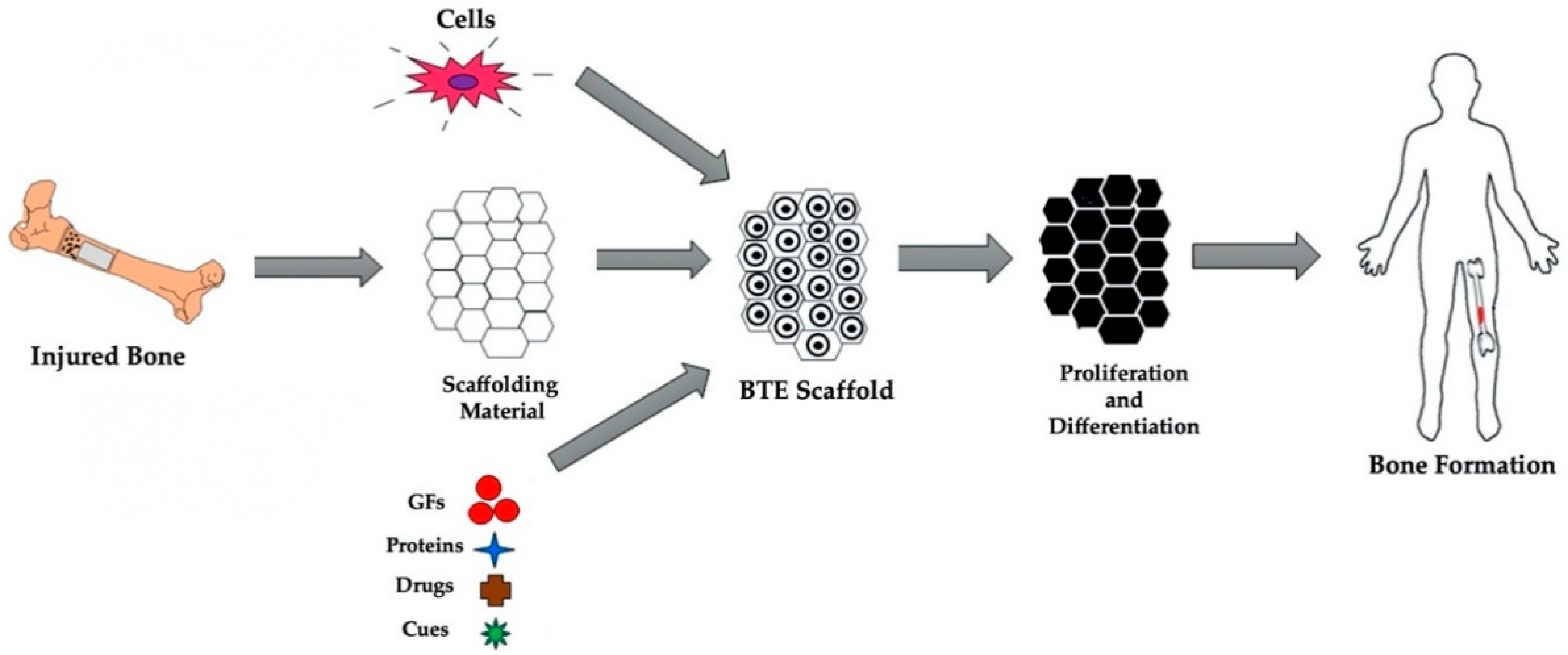
Bone defects are commonly healed through the use of scaffolds which are comprised of osteoprogenitor cells, relevant biomaterials, and biochemical cues, such as growth factors. In bone tissue engineering (BTE), bone tissue scaffolds a 3D porous matrix consisting of cells, biocompatible materials, and essential biological and biochemical cues. The typical process for scaffold-based approaches for BTE can be seen in Fig. 8.1. The scaffold biomaterials have characteristic advantages like high penetration ability and high surface area with tunable surface properties, making them one of the widely preferred candidates in different fields of TERM. Classification and synthesis based on different criteria such as composition, structure, and manufacturing process have already been dealt with in Chap. 2.

Process for scaffold-based approaches for bone tissue engineering
https://www.mdpi.com/jfb/jfb-13-00001/article_deploy/html/images/jfb-13-00001-g001.png
A few commonly used NPs/NMs for TERM are summarized as under:
8.2.1 Metallic NPs
The unique physiochemical properties of metal NPs, such as antibacterial effects, shape memory phenomenon, low cytotoxicity, stimulation of the proliferation process, good mechanical and tensile strength, acceptable biocompatibility, significant osteogenic potential, and ability to regulate cell growth pathways, suggest that they can perform as novel types of scaffolds for BTE. Metallic NPs such as gold (Au) and Ag(Ag) NPs can be manufactured and modified by utilizing different functional groups that provide conjugation of antibodies, ligands, and drugs as delivery systems.
Ag NPs
Ag NPs can also be described as a colloid of nanometer-sized particles of Ag and are one the most widely used metallic NPs in the biomedical field, mainly for their antimicrobial properties. Agions have been used for a long time for their antimicrobial properties toward a wide range of microorganisms. It has been shown that Agions are able to block the microbial respiratory chain system and precipitate bacterial cellular protein. NPs in the range of 1–10 nm can act differently against Gram- bacteria by (i) attaching to cell membrane affecting permeability and respiration, (ii) penetrating inside bacteria and damaging them, or (iii) releasing Agions (Chap. 3). Different scaffolds of Ag NPs can be produced in different formats including bulk materials, electrospun fibers, fibers mats, and nanofibrous or porous scaffolds. Delivery of Ag NPs not only has an antimicrobial effect but it also accelerates the rate of healing, and has the ability to regulate the cytokines associated with burn wound healing. Ag NPs also can show cytotoxic effects on cancer cells. A porous chitosan-alginate with biosynthesized Ag NPs also have cytotoxic effects against breast cancer cells. Therefore, it is very likely that these NPs have a potential for TERM.
Au NPs
Colloidal Au NPs solutions present different properties compared to the bulk gold because of their optical property due to their unique interaction with light. Due to their strong affinity to gold, it is possible to conjugate various ligands including polypeptide sequences, antibodies, and proteins with various moieties such as phosphines, amines, and thiols including nuclear targeting of cancer cells. Therefore, Au NPs play a crucial role in the success of cancer treatment. Therefore, applying Au NPs prior to implantation can provide a safety measurement toolbox to minimize the recurrence of tumor through targeted delivery to cancer cells, and consequently increase the chance of effective implantation for various TERM applications.
8.2.2 Magnetic NPs
Magnetic nanoparticles (MNPs) are iron oxide (Fe3O4) NPs (or Fe2O3), which are widely studied in the biomedical field because of their low toxicity. Recently, superparamagnetic Fe3O4(SPIO)-Au core-shell NPs decorated with nerve growth factor (NGF) with low toxicity have been developed for neuron growth and differentiation. NGF functionalized NPs have provided higher neuronal growth, and orientation on PC-12 cells under dynamic magnetic fields utilizing rotation has been obtained compared to static magnetic fields. Magnetic NPs also have been used for controlling collagen fiber orientation dynamically and remotely in situ during the gelation period through an applied external magnetic field. Iron oxides also have the ability to pass the blood–brain barrier, which could be used for the conjugation of various peptides and growth factors to cure and regenerate brain tissue.
8.2.3 Ceramic NPs
Ceramic nanoparticles (CNPs) are primarily made up of oxides, carbides, phosphates, and carbonates of metals and metalloids such as calcium, titanium, silicon, etc. They have a wide range of applications due to a number of favorable properties, such as high heat resistance and chemical inertness. CNPs can be classified according to their tissue response as being (a) bioinert, (b) bioactive, or (c) resorbable ceramics and (d) magnetic NPs. In general, CNPs can be used in the production of nanoscale materials of various shape, size, and porosity.
Bioinert Nanoceramics
Bioinert nanoceramics including TiO2, ZnO are utilized for different medical applications as they show positive interactions with body tissues. TiO2 NPs can be synthesized with different manufacturing processes including hydrothermal, solvothermal, sol-gel process, and emulsion precipitation methods. It is possible to manufacture uniformly distributed (in size) bioceramics in targeted size range. With the advancement of nanotechnology, TiO2 NPs, nanotubes, or nanoprobes labeled with fluorescent dye or magnetic resonance contrast agents have been successfully prepared for cell imaging through fluorescent analysis or magnetic resonance imaging (MRI). Similarly, Au NPs and Ag NPs, metal oxide NPs, and nanocomposite of chitosan/hydroxyapatite-zinc oxide (CTS/HAp-ZnO) supporting organically modified montmorillonite clay (OMMT) can be prepared for bone TE applications. Nanocomposite has shown strong antibacterial activities for both Gram+ and Gram- bacteria.
Bioactive Glass Ceramic NPs (n-BGC)
n-BGC with SiO2-CaO–P2O5-Na2O core structures can be formed from various elements such as silicone, sodium, potassium, magnesium, phosphorous, oxygen, and calcium which can be absorbed by the cells. Antibacterial and angiogenic properties and excellent bioactivity of nBGC have made them a suitable candidate for dentin regeneration applications. The incorporation of boron-modified nBGC in the cellulose acetate/oxidized pullulan/gelatin-based constructs has shown promising results for dentin regeneration through an increase in cellular viability.
Bioresorbable Nanoceramics
Bioresorbable nanoceramics have a calcium phosphate (CaP)–based composition which includes a variety of materials such as hydroxyapatite (HAp), calcium aluminate, tricalcium phosphate, calcium phosphate dicalcium phosphate dehydrate, calcium carbonate (CaCO3), calcium sulfate hemihydrate, octacalcium phosphate, and biphasic calcium phosphate. These materials have been applied in orthopedics, such as bone substitutes. HAp is commonly used in TERM applications. The combination of HAp with various forms of carriers such as electrospun fibers, porous scaffolds, and hydrogels can be used for the preparation of nanocomposite materials to modulate the desired cellular activities. Similarly, CaPs can be prepared with different types of polymers to produce nanocomposite materials. The advantages of these nanocomposites such as excellent mechanical characteristics could be utilized for bone tissue regeneration through enhancing scaffolds’ performance.
8.2.4 Polymeric Nanoparticles
Low cytotoxicity of polymeric nanoparticles (PNPs), good biocompatibility, higher permeation and retention (EPR) effect, ability to deliver poorly soluble drugs and sustained release of them, and retaining bioactivity of bioactive agents from enzymatic degradation for TE applications make PNPs one of the fastest growing platforms to overcome obstacles in TERM. Most of the new PNP systems are designed to be sensitive to different physicochemical stimuli such as magnetic field, temperature, enzymes, pH, light, and reducing/oxidizing agents, which helps the delivery or targeting systems with high specificity and efficiency for TERM applications.
8.3 Scaffolds for TERM
Scaffolds are materials that have been engineered to cause desirable cellular interactions to contribute to the formation of new functional tissues for medical purposes. Cells are often “seeded” into these structures capable of supporting three-dimensional tissue formation. As such scaffolds represent important components for TE. However, researchers often encounter an enormous variety of choices when selecting scaffolds for TE. Apart from blood cells, most, if not all other, normal cells in human tissues are anchorage-dependent residing in a solid matrix called extracellular matrix (ECM). There are numerous types of ECM in human tissues, which usually have multiple components and tissue-specific composition. As for the functions of ECM in tissues, they can be generally classified into five categories. These supports include: structural support, mechanical support, providing bioactive cues for cells, acting as the reservoir of growth factors, and finally providing a flexible physical environment (Table 8.1).
8.4 Scaffolding Approaches
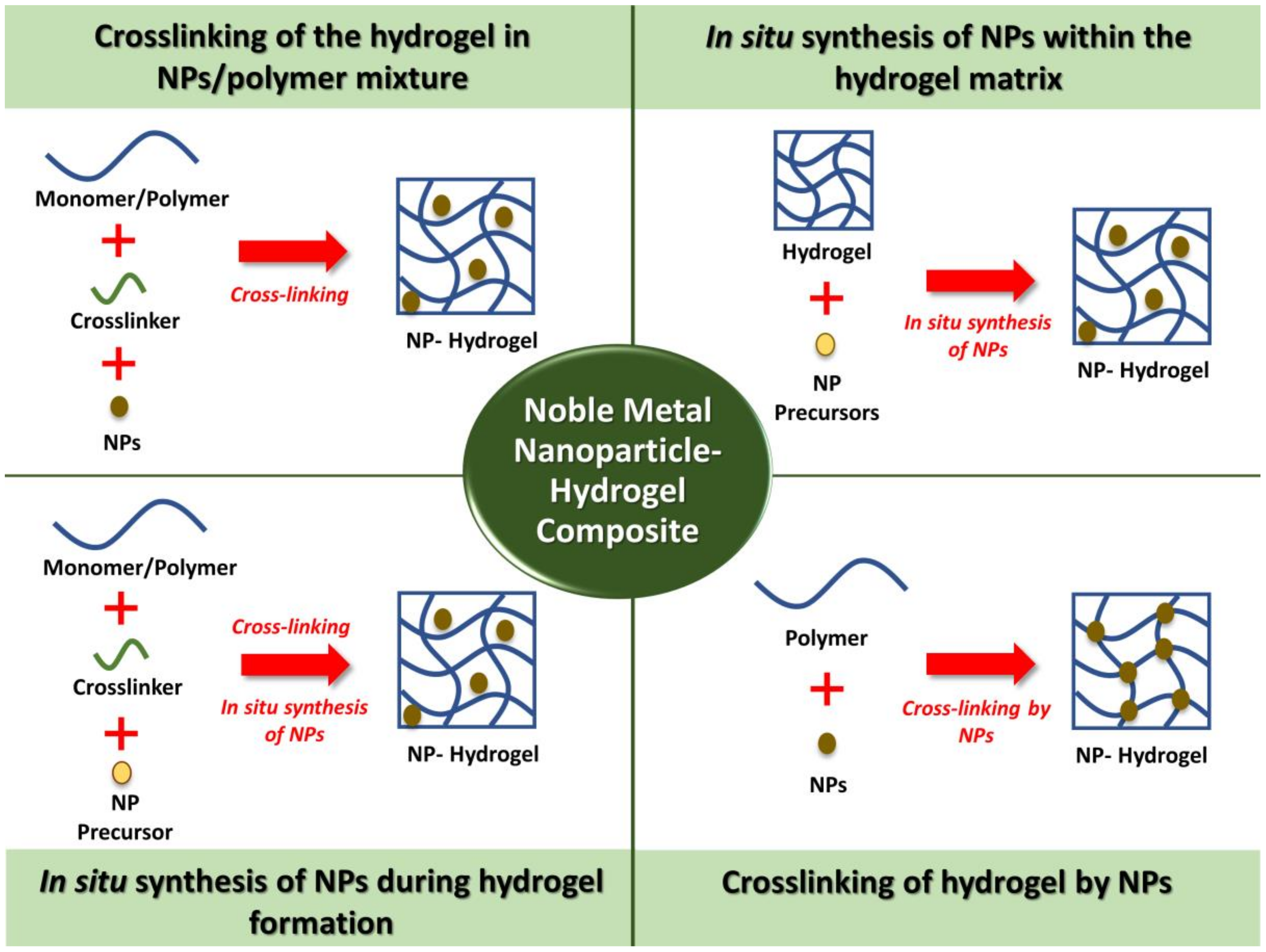
The application of hydrogel incorporated with metal NPs has become a new emerging research area in TERM. Disease, injury, and trauma often results in tissue damage and degeneration. By combining the hydrogel and NPs, the property enhancement of the materials can be achieved. This noble metal NPs have shown to have potential in TE applications. There are a few main approaches that have been adopted for the preparation of NP–hydrogel composites. Common examples of the preparation methods for biomedical applications (Fig. 8.2) include: (a) crosslinking of the hydrogel in NPs/polymer mixture, (b) in situ synthesis of NPs within the hydrogel matrix, (c) in situ synthesis of NPs during hydrogel formation, and (d) application of noble metal NP–hydrogel composites in TE. The potential of noble metal NPs and hydrogel in tissue regeneration have stimulated the high interest of researchers to further characterize the property of the hybrid of these materials. To address this, the studies on NP–hydrogel composites for the regeneration of tissues such as soft tissues, bone tissues, and cardiac tissues are being explored. The details of the synthesis of various scaffolds are out of the scope of this chapter.

Schematic diagram showing noble NP-hydrogel composite scaffolding approaches
Bioengineering, https://www.mdpi.com/bioengineering/bioengineering-06-00017/article_deploy/html/images/bioengineering-06-00017-g001.png
8.5 Properties of Scaffolds/Matrices for TERM
As indicated previously, there are several scaffolding approaches in TERM. Each approach has its own pros and cons and preferred TE applications. In planning for TE for a complex tissue such as an intervertebral disc (IVD), these scaffolding approaches serve as important guidelines and can be used in combinations. Moreover, tissue-specific considerations in relation to the extent of injury, the unique structural functional relationship, multiple tissue composition, and interfaces in IVD deserve special attention. Scaffolds are three-dimensional (3D) porous solid biomaterials designed having the following properties:
-
(a)
Provide a spatially correct position of cell location.
-
(b)
Promote cell-biomaterial interactions, cell adhesion, and ECM deposition.
-
(c)
Permit sufficient transport of gases, nutrients, and regulatory factors to allow cell survival, proliferation, and differentiation.
-
(d)
Biodegrade at a controllable rate that approximates the rate of tissue regeneration.
-
(e)
Provoke a minimal degree of inflammation or toxicity in vivo.
Apart from blood cells, most of the normal cells in human tissues are anchorage-dependent residing in a solid matrix (ECM). The best scaffold for an engineered tissue should be the ECM of the target tissue in its native state.
Generally, a scaffold, to serve as a suitable matrix to the reconstruction of tissue, should exhibit some of the following important features:
-
(a)
A high porosity and an adequate pore size are necessary to facilitate cell seeding and diffusion throughout the whole structure of both cells and nutrients.
-
(b)
Should allow effective transport of nutrients, oxygen, and waste.
-
(c)
Biodegradability is essential, since scaffolds need to be absorbed by the surrounding tissues without the necessity of surgical removal.
-
(d)
The rate at which degradation occurs has to coincide with the rate of tissue formation.
-
(e)
Should be biocompatible.
-
(f)
Should have adequate physical and mechanical strength. For example, the scaffolds in dentistry have an important distinction. In the bone, TERM requires a rigid scaffold that reproduces the size and architecture of the tissue to be rebuilt. In the pulpodentinal complex and in the periodontal apparatus of the TERM, due to the small size and difficulty to reach, the receiving site requires soft and injectable scaffolds. For this reason, the biomaterials used in scaffold formation can be classified according to the natural and synthetic sources or depending on the physical consistency, either rigid or soft.
8.6 Applications and Use in TERM
Novel cell sources, engineering materials, and tissue architecture techniques have helped to restore, maintain, improve, or replace biological tissues (Fig. 8.3). A number of therapies utilize NPs for the treatment of cancer, diabetes, allergy, infection, and inflammation. The surface conjugation and conducting properties of Au NPs, the antimicrobial properties of Ag and other metallic NPs and metal oxides, the fluorescence properties of quantum dots, and the unique electromechanical properties of carbon nanotubes (CNTs) have made them very useful in numerous TERM applications. In addition, magnetic NPs have been applied in the study of cell mechano-transduction, gene delivery, controlling cell patterning, and construction of complex 3D tissues. The use of the right type of NPs can significantly enhance the biological, mechanical and electrical properties of scaffolds as well as can serve various functions in TERM.

Novel cell sources, engineering materials, and tissue architecture techniques have provided engineering tissues to restore, maintain, improve, or replace biological tissues
8.6.1 Biological Properties
Au NPs and titanium dioxide (TiO2) NPs have been used to enhance cell proliferation rates for bone and cardiac tissue regeneration, respectively. Au NPs have shown superior biocompatibility and the ability for surface modification, which has resulted in interesting biomedical applications. These NPs can be described as a colloid of nanometer-sized particles of Au. Colloidal Au solutions present different properties compared to bulk Au because of their optical property that provides unique interaction with light. On Au surface it is possible to conjugate various ligands including polypeptide sequences, antibodies, and proteins with various moieties such as phosphines, amines, and thiols because of their strong affinity to Au. In bone, Au NPs promote osteogenic differentiation of an osteoblast precursor cell line, MC3T3-E1. In addition, these NPs also influence osteoclast (or bone resorbing cell) formation from hematopoietic cells while providing protective effects on mitochondrial dysfunction in osteoblastic cells. Therefore, Au NPs present themselves as excellent candidates for TERM. They seem to be perfect candidates to replace bone morphogenetic proteins (BMPs). Although BMPs have beneficial effects on bone regeneration and repair, they also have many disadvantages such as high cost and susceptibility to result in unwanted bone formation and local inflammatory reactions.
8.6.2 Mechanical Properties
Change in the mechanical properties of some NPs leads to superior mechanical properties for TERM applications compared to scaffolds without NP reinforcements. For example, NP-embedded nanocomposite polymers both in the form of hydrogels and electrospun fibers exhibited an enhanced response than simple NPs. Likewise, a TiO2-embedded biodegradable patch showed a higher tensile strength in reinforcing the scar after myocardial infarction. Hydrogel microfibers with poly(N-isopropylacrylamide) (PNIPAm) and MNPs increased their mechanical strength. CNTs have also been used to enhance the mechanical properties of polymers for TERM applications. CNTs reinforce the polymers especially due to their remarkable mechanical properties, tensile strength, and fiber-like structure.
8.6.3 Electrical Properties
Nanoparticles have also been used to enhance the electrical properties of scaffolds, which can be highly beneficial in cardiac TERM. Au NP-based electrospun fibrous scaffolds are excellent for cardiac tissue regeneration. Au NPs are deposited on the surface of the gelatin and PCL–gelatin fibers, creating nanocomposites with a nominal Au thickness. Cardiac tissues engineered within these Au NP scaffolds can be used to improve the function of the infarcted heart. Likewise, Au nanowires have also been used as conductive materials alongside scaffolds to enhance the electrical coupling between the cells. With time, cardiac muscle cells start growing within the 3D porous scaffolds and result in synapse formation. The use of CNTs in polymer composites can significantly improve conductivity to promote cardiomyocyte functions, therefore conductive nanomaterials have a promising future in cardiac applications.
8.6.4 Antibacterial Properties
Some metal oxides, particularly Ag NPs, have shown great antimicrobial effects, as well as wound-healing capabilities. Likewise, Ag-containing poly(3-hydroxybutyrate-co-3-hydroxyvalerate) (PHBV) nanofibrous scaffolds had high antibacterial properties, and they exhibit excellent in vitro cell compatibility. This shows that PHBV nanofibrous scaffolds containing Ag NPs have prospects to be used in joint arthroplasty. Biocomposite scaffolds containing nano Ag could regulate bacterial infection during reconstructive bone surgery. Thus, the presence of Ag NPs in the scaffolds acts as an affixed coating for protection against infection, sepsis, and malfunctioning of implants. Specifically, Fe3O4 NPs also have much promise in killing post-biofilm formation in bacteria (especially when functionalized with sugars such as fructose and sucrose) and can penetrate biofilms, whereas antibiotics cannot (magnetic field disrupts and kills bacteria). This has enormous consequences in TERM since currently if a biomaterial becomes infected, it needs to be removed and adjacent tissue cleaned. Strategies that do not rely on implant removal and cleaning can have a bright future in TERM.
8.6.5 Gene Delivery
Gene delivery has been used for regenerative medicine applications to create or restore normal function at the cell and tissue levels. For effective gene therapy applications, it is vital to build up a suitable vector system with a high gene transfection efficiency, low cytotoxicity, and high specificity to unhealthy cells. Gene delivery is classified into two categories: nonviral and viral.
The advantages of nonviral methods are their simplicity and the absence of an immune response, while the disadvantage is low efficiency due to low transfection rate. The promise for gene delivery has been seen through the use of NPs and self-assembled NMs. Magnetofection is a new method for gene delivery in which gene transfection is accomplished using magnetic NPs. To achieve magnetofection using plasmid DNA, cationic lipids or polymers with complexes of DNA interact with magnetic beads and then through a magnetic force are attracted onto target cells so that they can accumulate on the surface. For example, Fe3O4 magnetic particles can attach to the gene and improve transfection efficiency. These particles are distributed within a polymer matrix or internalized in a polymer or metallic case, which binds DNA through charge contact. CNTs, which have shown numerous applications and can be synthesized through several different approaches, have also shown remarkable potential as nonviral gene delivery agents. A technique called nanotube spearing can be used to prepare nickel-embedded, magnetic nanotubes where DNA is attached.
Viral transduction methods have also been used in TERM applications; however, one problem associated with this method is the difficulty in preparing viral vectors with a high titer.
8.6.6 Mechano-Transduction
In addition to various bioactive molecules and growth factors that regulate cell functions in the human body, mechanical forces play a major role in determining cell functions by affecting mechano-transduction pathways. Numerous approaches such as the introduction of shear stress by bioreactors and stiffness of patterned substrates to mechanically control cell functions have been used. However, magnetic NPs have proven to be superior to all these methods since they can be controlled remotely, spatially, and temporally through a magnetic field. In this process, first, the MNPs are coated with a certain targeting antibody. Once the magnetic field is applied, the cells are clustered in the direction of the magnetic field. Based on the antibody used, receptor-mediated cell function is affected.
8.6.7 Magnetic Cell Patterning
TERM requires the fabrication of tissue architecture similar to in vivo conditions. To get cell adhesion within a specified design pattern magnetic cell-patterning technique is used. A cell-patterning technique has been developed using magnetite cationic liposome (MCL), where a magnet with a magnetic field concentrator is laid under a cell culture surface. Various cell patterns can be successfully fabricated using this technique by manipulating the line patterns of the magnetic field concentrators. When human umbilical vein endothelial cells are used, the cells connect and form capillary-like structures with patterned in a line.
8.7 Constructing 3D Tissues
Three-dimensional bioprinting is a rapidly growing technology that has been widely used in TE, disease studies, and drug screening. It provides the unprecedented capacity of depositing various types of biomaterials, cells, and biomolecules in a layer-by-layer fashion, with a precisely controlled spatial distribution. There are three categories of 3D bioprinting strategies: inkjet-based printing (IBP), extrusion-based printing (EBP), and light-based printing (LBP).
Bioinks are formed by combining cells and various biocompatible materials, which are subsequently printed in specific shapes to generate tissue-like, 3D structures. These bioinks mimic the ECM environment, support cell adhesion, proliferation, and differentiation after printing. In contrast to traditional 3D printing materials, bioinks must have:
-
Print temperatures that do not exceed physiological temperatures.
-
Mild cross-linking or gelation conditions.
-
Bioactive components that are non-toxic and can be modified by the cells after printing.
Three-dimensional bioprinting allows for the spatially controlled placement of cells in a defined 3D microenvironment. Due to the high degree of control on structure and composition, 3D bioprinting has the potential to solve many critical unmet needs in medical research, including applications in cosmetics testing, drug discovery, regenerative medicine, and functional organ replacement. Personalized models of disease can be created using patient-derived stem cells, such as induced pluripotent stem cells (iPS cells) or mesenchymal stem cells. Depending on the application, a range of materials, methods, and cells can be used to yield the desired tissue construct. 3D bioprinting allows for the spatially controlled placement of cells in a defined 3D microenvironment. This technology is expected to address the organ-shortage issue in the future. The combination of stem cell technology and 3D bioprinting is expected to allow the construction of better-functioning tissues/organs and organs-on-chips. For the longevity and functionality of printed architectures, vascularization and innervation need to be further investigated.
8.8 Bioactive Agents/Molecules
Bioactive agents act as a scaffolding frame to deliver cells to the appropriate site, define a space for tissue development, and direct the shape and size of the engineered tissue. Agents including proteins and small molecules involved in TERM play an important role in controlling the microenvironment in vivo. Chemotactic signals from bioactive molecules are responsible to regulate host cell migration, proliferation, and differentiation and allow cells to interact via specific receptors for chemical recognition with their surrounding microenvironment. Moreover, an anatomic destination is identified according to certain concentration gradients of chemicals produced at injured sites within the microenvironment. Thus incorporation of a suitable bioactive molecule through the design of a tissue-engineered scaffold can promote tissue regeneration by stimulating the transplanted cells or adjacent host cells. The mode of release is especially relevant when the bioactive agent is a growth factor (GF) because the dose and the spatiotemporal release of such agents at the site of injury are crucial to achieve a successful outcome. Strategies that combine scaffolds and drug delivery systems have the potential to provide more effective tissue regeneration relative to current therapies. NPs can protect the bioactive agent, control its profile, decrease the occurrence and severity of side effects, and deliver the bioactive agent to the target cells maximizing its effect. Scaffolds containing NPs loaded with bioactive agents can be used for their local delivery, enabling site-specific pharmacological effects such as the induction of cell proliferation and differentiation, and, consequently, neo-tissue formation. Recently, plasma protein–based NPs have gained attention due to their high bioavailability, non-toxicity, biodegradability, ease of manipulation, long in vivo half-lives, and long shelf lives. There are more than 100,000 proteins in human plasma, but just a couple of these proteins have been used in TERM as a nanocarrier platform for imaging, drug delivery, and tissue regeneration. High density lipoproteins (HDL) NPs are among candidates for enhancing photodynamic therapy applications through presenting excellent tumor targeting and internalization capacity. Fibrin is another plasma protein that has been used for encapsulation of vascular endothelial growth factor (VEGF) for promoting angiogenesis for wound-healing applications. NPs from albumin, as the most abundant plasma protein, are being used for bone regeneration through a sustained release of bone morphogenetic protein-2 (BMP-2). Bone tissue–related diseases such as a tumor or trauma generally are treated with bone grafts and substitutes. Nowadays TERM has provided an alternative approach for bone tissue regeneration by offering a variety in forms of 3D scaffolds. Bone scaffolds containing stem cells have the advantage of controlling the cellular activity such as differentiation, if appropriate bioactive agents such as drugs (e.g., dexamethasone) or growth factors [e.g., bone morphogenetic proteins (BMPs)] are incorporated in them.
Ascorbic acid is an important water-soluble bioactive molecule in the bone formation process. It is also called vitamin C, acts as a cofactor for the key enzymes involved in collagen biosynthesis, and demonstrates a major function in stabilizing the helical structure of collagen. Bone tissue–related diseases such as a tumor or trauma generally are treated with bone grafts and substitutes. Nowadays TERM has provided an alternative approach for bone tissue regeneration by offering a variety in forms of 3D scaffolds. Natural biodegradable multi-channeled scaffolds composed of ordered electrospun nanofibers with neurotrophic gradient have been designed to control axon outgrowth. Likewise, various NP-based dressings have been developed for delivering bioactive agents with spatiotemporal control for enhancing the wound-healing process.
8.9 Imaging and Contrast Agents
Imaging strategies, in conjunction with exogenous contrast agents, can help in assessing in vivo therapeutic progress of TE. Proper use of these monitoring/imaging and regenerative agents (MIRAs) can help increase TERM therapy successes and allow for clinical translation. MIRA research is still in its beginning stages with much of the current research being focused on imaging or therapeutic applications, separately. Advancing MIRA research will have numerous impacts on achieving clinical translations of TERM therapies.
8.10 Biocompatibility
Materials obtained using nanotechnology and currently used in TERM include a wide range of products. In order to overcome the challenges of high organ demand and biocompatibility issues, scientists in the field of TREM are working on the use of scaffolds as an alternative to transplantation. For complex scaffolds of different compositions and structures, the scaffolds are being developed to mimic the ECM, act as structural support, and define the potential space for new tissue development as well as enhance cell attachment, proliferation, and differentiation. Tissue-engineered products (TEPs) containing either cells or growth factors or both cells and growth factors may be used as an alternative to the autografts taken directly from the bone of the patients. Nevertheless, the use of TEPs needs much more understanding of biointeractions between biomaterials and eukaryotic cells. Despite the possibility of the use of in vitro cellular models for the initial evaluation of the host response to the implanted biomaterial, it is observed that most researchers use cell cultures only for the evaluation of cytotoxicity and cell proliferation on the biomaterial surface, and then they proceed to animal models and in vivo testing of bone implants without fully utilizing the scientific potential of in vitro models. However, nanostructured hydroxyapatite (nano-HA) and nano CaP have received considerable attention. The nano-HA has shown excellent biological performances compared to conventional HA because it has better biocompatibility and bioactivity in respect of bone components (probably as a result of its similarity with the chemical component and mineral structure of bone tissue). Moreover, due to their small size and large specific surface, nano-HAs may not only promote ion exchange within a physiological environment but also increase protein absorption and cellular response, especially if stressed by physical means.
8.11 Nanotoxicity of NMs
The main risks in TE are tumorigenity, graft rejection, immunogenity, and cell migration. Secondly, the toxicity of NPs is highly dose- and exposure-dependent. In many applications, the NPs are used below their threshold concentrations at which they are considered not harmful. However, bioaccumulation of NPs inside the body over a large period of time is well known. Thus, any NP used in the human body has the potential to accumulate over a long period of time to reach a concentration that can cause toxicity to cells, cancers, or harmful effects on reproductive systems and fetuses before their birth. In addition, even though numerous products containing NPs/NMs are already on the market, there are still some scientific and methodological gaps in the knowledge of specific hazards of NMs. As has been indicated previously (Chap. 5), currently there are no international standards yet for nano-specific risk assessments, including specific data requirements and testing strategies. The risk assessments of NMs are laborious and costly. Currently, manufacturers are committed to assess the safety of their NP-based products and to implement the necessary safety measures (self-supervision). To date, the regulatory tools are not nano-specific; for example, the data requirements for notification of chemicals, criteria for classification, and labeling requirements for safety data sheets are still not widely available. Thus, there is a need for precautionary measures for the applications of NPs wherever there is a possibility of chronic bioaccumulation.
8.12 Conclusion
Nanoparticles exhibit superior biocompatibility and well-established strategies for surface modification, which have made them highly effective in numerous biomedical applications. The inability to deliver bioactive agents locally in a transient but sustained manner is one of the challenges in the development of bio-functionalized scaffolds for TERM. In vivo maturation of engineered tissues requires a well-synchronized series of events involving the host immune system, circulatory system, and cellular components of the implanted material. The physicochemical properties of the scaffolds and the presence/immobilization of bioactive agents within the scaffolds can help in achieving precise control over maturation. NPs have been shown to develop optical, electrical, and electrochemical biosensors for molecules, proteins, and DNA detection with highly accurate results. By optimizing these functions, these sensors could have a great influence on medical use. Although NPs show a promising future in TERM applications, there is still a lack of in vivo experimentation that needs to be done in order to verify the wide variety of successful results from in vitro studies. However, the numerous existing applications in the literature reiterate the great potential NPs can have on TERM through bioinks, imaging, and contrast biosensors. At the same time, it is important to look into the risks associated with TERM which are tumorigenity, graft rejection, immunogenity, and cell migration. Therefore, our aim is to understand the risks, how to minimize them and, especially, how to predict and prevent them.
Further Reading
Fang YL, Chen XG, Godbey WT. Gene delivery in tissue engineering and regenerative medicine – a review. J Biomed Mater Res B Appl Biomater. 2015 Nov;103(8):1679–99. https://doi.org/10.1002/jbm.b.33354.
Fathi-Achachelouei M, Knopf-Marques H, Ribeiro da Silva CE, Barthès J, Bat E, Tezcaner A, Vrana NE. Use of nanoparticles in tissue engineering and regenerative medicine. Front Bioeng Biotechnol. 2019;7:113. https://doi.org/10.3389/fbioe.2019.00113.
Gupta PK. Chapter 15: Toxic effects of nanoparticles. In: Toxicology: resource for self study questions. 2nd ed. Seattle: Kinder Direct Publications; 2020a.
Gupta PK. Chapter 14: Toxicology of nanoparticles. In: Problem solving questions in toxicology – a study guide for the board and other examinations. 1st ed. Cham: Springer; 2020b.
Gupta PK. Toxic effects of nanoparticles. In: Brain storming questions in toxicology. 1st ed. Boca Raton: Taylor & Francis Group, LLC, CRC Press; 2020c. p. 297–300.
Gupta PK. Fundamentals of Nanotoxicology. 1st ed. New York: Elsevier Inc.; 2022.
Hasan A, Morshed M, Memic A, Hassan S, Webster TJ, Marei HE. Nanoparticles in tissue engineering: applications, challenges and prospects. Int J Nanomed. 2018;13:5637–55. Published 2018 Sept 24. https://doi.org/10.2147/IJN.S153758.
Khanna P, Ong C, Bay BH, Baeg GH. Nanotoxicity: an interplay of oxidative stress, inflammation and cell death. Nanomaterials. 2015;5:1163–80. https://doi.org/10.3390/nano5031163.
Rychter M, Baranowska-Korczyc A, Luleka J. Progress and perspectives in bioactive agent delivery via electrospun vascular grafts. RSC Adv. 2017;7:32164.
Tan H-L, Teow S-Y, Pushpamalar J. Application of metal nanoparticle–hydrogel composites in tissue regeneration. Bioengineering. 2019;6(1):17. https://doi.org/10.3390/bioengineering6010017.
Walmsley GG, McArdle A, Tevlin R, Momeni A, Atashroo D, Hu MS, Feroze AH, Wong VW, Lorenz PH, Longaker MT, Wan DC. Nanotechnology in bone tissue engineering. Nanomedicine. 2015;11(5):1253–63. https://doi.org/10.1016/j.nano.2015.02.013.
Zhang B, Gao L, Ma L, Luo Y, Yang H, Cui Z. 3D bioprinting: a novel avenue for manufacturing tissues and organs—review. Res 3D Bioprint. 2019;5(4):777–94. https://doi.org/10.1016/j.eng.2019.03.009.
Author information
Authors and Affiliations
Rights and permissions
Copyright information
© 2023 The Author(s), under exclusive license to Springer Nature Switzerland AG
About this chapter

Cite this chapter
Gupta, P.K. (2023). Tissue Engineering and Regenerative Medicine. In: Nanotoxicology in Nanobiomedicine. Springer, Cham. https://doi.org/10.1007/978-3-031-24287-8_8
Download citation
DOI: https://doi.org/10.1007/978-3-031-24287-8_8
Published:
Publisher Name: Springer, Cham
Print ISBN: 978-3-031-24286-1
Online ISBN: 978-3-031-24287-8
eBook Packages: Biomedical and Life SciencesBiomedical and Life Sciences (R0)