Abstract
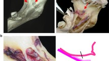
Currently panoramic radiography is used for diagnosis of dental and bone lesions, but anatomical structures also can be seen and may be useful in dental managements. This chapter study aimed to investigate the visibility of some important mandibular features relating to neurovascular structures and also to indicate common anatomical variations and pathologies.
Access provided by Autonomous University of Puebla. Download chapter PDF
Keywords
5.1 Anatomical Landmarks
A panoramic radiography taken from the patient should be evaluated by the physician as described below;
When evaluating the radiography, first the right-left half segment of the maxilla, midline, nasal cavity, and sinuses are evaluated, then the shadows of the tongue and soft palate are evaluated at this stage. Following this, cervical vertebra and related structures are evaluated. Then start from the midline of the mandible and continue towards the back. The last teeth should be examined. First, it starts from the right or left half of the maxilla or mandible, then passes to the other half, and then the other jaw teeth are examined ([1], p. 168).
5.1.1 Normal Anatomy
Normal anatomical shadows may vary from one radiograph taken with a panoramic device to another. However, it is generally divided into subgroups:
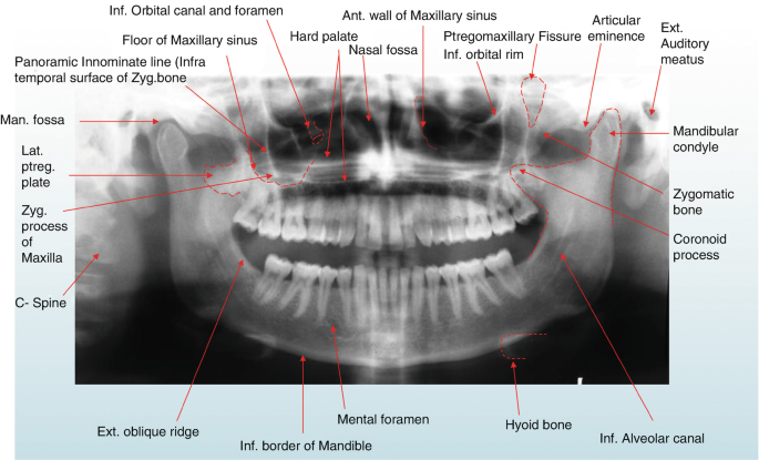
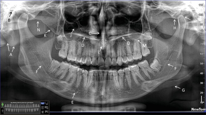
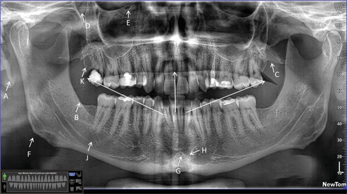
5.1.1.1 Hard Tissue (Figs. 5.1, 5.2, 5.3, 5.4, 5.5, 5.6, 5.7, 5.8, 5.9, 5.10, 5.11, 5.12, 5.13, 5.14 and 5.15)
-
Teeth.
-
Mandible.
-
Base of maxilla and antrum, medial and posterior wall (A).
-
Hard palate (HP).
-
Zygomatic arch (Z).
-
Styloid process (SP).
-
Hyoid bone (H).
-
Nasal septum (NS).
-
Orbita (O).
-
Skull base.
-
Meatus acusticus externus (EAM).
Representative panoramic Images showing anatomical landmarks. A—Ear Lobe, B—External Auditory Meatus, C—Nasal Septum, D—Palate, E—Mental Foramen, F—Mandibular Canal, G—Hyoid Bone, H—Articular Eminence, I—Mandibular Condyle, J—Maxillary Sinus, K—Coronoid Process, L—Spina Nasalis Anterior, M—Inferior Border of Maxillary Sinus, N—Zygomatıc Arch
Representative panoramic Images showing anatomical landmarks. A—Styloid Process, B—External Oblique Ridge, C—Pterygoid Plates, D—Pterygomaxillary Fissure, E—Infraorbital Foramen, F—Angulus Mandibula, G—Lingaul Foramen, H—Genial Tubercle, I—Palatoglossal Air Space, J—Submandibular Gland Fossa, K—Tuber Maxilla
5.1.1.2 Airway Shadows
-
Nasal fossa.
-
Oral cavity.
-
Oropharynx.
-
Nasopharynx.
5.1.1.3 Soft Tissue Shades
5.2 Panoramic Radiography: Diagnosing Dentition Anomalies
Dental anomalies are generally anomalies that affect the shape, structure, number, and size of teeth during tooth development. These dental anomalies that occur during tooth development can affect the organization of the dental arches and cause malocclusion.
Accordingly, it affects surgical treatment, orthodontic treatment, endodontic treatment, and restorative dental treatment procedures. Dental anomalies affect both primary dentition and permanent dentition [2].
Panoramic radiography can be used to diagnose these dentition anomalies. In cases where there is hypodontia, hyperdontia, gemination or fusion in primary dentition, anomaly development in permanent dentition is under the high risk group. Due to this close relationship between primary dentition and permanent dentition, early diagnosis of anomalies helps the physician to examine for the future and determine the best treatment plan at the earliest stage.
Panoramic radiography has a very important place in the identification, localization, and surgical removal of supernumerary teeth. In addition, anomalies in the permanent dentition, supernumerary teeth and morphological anomalies of maxilla and mandible seen in cleiocranial dysplasia can diagnosed by panoramic radiography.
Gardner syndrome, which is seen with multiple embedded supernumerary teeth, osteomas in the skull and jaws in long bones, multiple polyps in the intestines, and epidermoid or dermoid cysts, can be diagnosed by panoramic radiography. Fatal malignancy can be prevented by early diagnosis of Gardner syndrome when osteomas and multiple supernumerary teeth are seen in the jaws in panoramic radiography ([3], p. 48).
Early diagnosis of macrodontia is an important place in orthodontic treatment planning. If there is macrodontia and tooth development is not possible due to this reason, if it is impacted or maloccluded, panoramic radiography helps early diagnosis ([3], p. 41). Taurodontism is hereditary morphology anomalies seen in multi-root teeth due to invagination error of the Hertwig epithelial sheath. Taurodontism is the enlargement of the pulp chamber as a result of the displacement of the pulp base and bifurcation towards the apical [4]. Taurodontism is usually seen symmetrically and bilaterally. It is rarely seen when a single tooth is affected. No anomaly is encountered in the relevant teeth during clinical examination. Taurodontism is diagnosed by radiography. Panoramic radiography has been found to be reliable for the diagnosis of taurodontism ([3], p. 53).
Whittington and Durward examined the anomalies of primary dentition and permanent dentition using panoramic radiography in 1680 people aged 5 years. They found hypnodontics in 6 children, supernumerary teeth in 3 children, and fusion and gemination in 14 children. In 14 of these children with primary dentition anomalies, anomalies were also found in permanent dentition. Due to this close relationship between primary dentition and permanent dentition, early diagnosis of anomalies helps the physician to examine for the future and determine the best treatment plan at the earliest stage ([3], p. 41).
Patil et al. conducted a research to determine the diagnosis and prevalence of dental anomalies on panoramic films taken from 4133 patients. As a result of the research, 36.7% of them have dental anomalies. Of these, 16.3% are congenital tooth deficiency, 15.5% are impacted teeth, 1.2% are supernumerary teeth, and 1% are microdontia [5].
Panoramic radiography is very effective and useful in diagnosing dental anomalies in addition to clinical examinations. Since dental development anomalies can affect patients’ esthetics and self-worth perception, early diagnosis of anomalies enables us to make the necessary treatment planning. Although the treatment of anomalies that are not diagnosed early is more difficult, the prognosis is not good ([3], p. 66) (Fig. 5.18).
5.3 Panoramic Radiography: Evaluation of Tooth Eruption and Impaction
Radiography is of great importance in evaluating whether the tooth eruption process is normal in terms of both position and timing. This is especially important in the evaluation of clinically undetectable teeth with eruption delay or impacted teeth. The impacting of a tooth may develop due to either the surrounding tissues or a pathology.
Eruption delays, affecting both primary dentition and permanent dentition, may be associated with a systemic disease such as rickets, cretinism or cleidocranial dysplasia ([3], p. 73).
Neonatal diseases or postnatal nutrition are closely related to the eruption timing of primary teeth. Delays in tooth eruption are observed in fibromatosis gingiva due to enlarged gingiva. If eruption delays are due to a local cause (fibromatous gingiva, supernumerary teeth, odontome, etc.), eruption can be controlled with early treatment (Fig. 5.19).
In cases where eruption delays are prolonged, they should be examined with panoramic radiography or intraoral radiography. Since panoramic radiology can cooperate during patient exposure, it provides the most appropriate view. Periodic panoramic radiology is useful in the diagnosis of conditions such as tooth eruption anomalies caused by dentigerous cysts around 20-years-old teeth.
The impaction of the third molar teeth is classified according to the Winter Method, depending on the position. Panoramic X-rays show the mesiodistal and vertical position of the impacted tooth, but do not provide information about the buccolingual position or buccolingual angulation. Therefore, when deciding on the treatment plan, we may need to support panoramic radiography with occlusal films or three-dimensional images created using an intraoral detector ([3], p. 74). As a result, although there are some discussions about the monitoring or removal of the third molar tooth with panoramic radiography, panoramic radiography is an important pre-diagnosis device used in the evaluation of these teeth. In order to evaluate the relationship of the mandibular posterior tooth roots with the mandibular canal, it should be supported by other additional radiographs that provide information bucco-lingually [6] (Fig. 5.20).
5.4 Panoramic Radiography: Evaluations in Orthodontics
Today, panoramic radiography has become an indispensable diagnostic method in evaluating the success and failure of orthodontic treatment. Panoramic radiography provides information about the presence and absence of the tooth, its morphological and structural variations, and the eruption pattern. In addition, panoramic radiography has become the standard in the evaluation of tooth parallels, especially for orthodontics.
Panoramic radiography is one of the first preferred 2-dimensional imaging methods for patients who will undergo orthodontic treatment. The lack of teeth or the presence of supernumerary teeth in some patients can be understood as a result of clinical examination. However, panoramic radiography provides a wide examination opportunity including all maxillary and mandibular arches of the patient together with the temporomandibular joint. Panoramic radiographs are useful in showing root morphology deviations, eruption times or changes in development, impactedness, loss or supernumerary teeth, as well as any pathological lesions or mandibular asymmetry, while they also provide limited information about sinuses, root parallelism, and periodontal health ([7], p. 35; [8]). It also helps to assess the quality and quantity of alveolar bone for the placement of temporary anchoring devices or implants and to determine their distance to vital structures ([7], p. 35).
5.5 Panoramic Radiography: Mandibular Canal Variations
The mandibular canal, which neurovascular structures pass through, feeding the lower teeth and adjacent anatomical structures, is a bone-surrounded space that starts from the foramen mandible at the back and extends to the foramen mentale in front (Fig. 5.21). It is important to determine the position and course of the canal correctly in terms of evaluating whether there is an anatomical variation and whether there is a bifid mandibular canal. In addition, it provides protection against complications that may occur during surgical operations such as tooth extraction or implant placement [9]. Bifid mandibular canal, rather than being a projection artifact, are real variations affecting approximately 0.5–1% of the population (Fig. 5.22). Although they are usually asymptomatic, they sometimes cause pain and TMJ dysfunction [10].
As a result of the study performed on 75 CBCTs from data obtained from private clinics near São Paulo, bifid mandibular canals were classified into four different groups.
-
1.
Type 1; It is divided into two subgroups.
-
(a)
Unilateral bifurcation extends to the third molar and its surrounding tissues,
-
(b)
The double-sided bifurcation extends to the third molar and its surrounding tissues.
-
(a)
-
2.
Type 2; It is divided into four subgroups.
-
(a)
Unilateral bifurcation extends to the main canal and converges in the ramus of the mandible,
-
(b)
Unilateral bifurcation extends to the main canal and converges at the mandible corpus,
-
(c)
The bilateral bifurcation extends to the main canal and converges in the ramus of the mandible,
-
(d)
Bilateral bifurcation extends to the main canal and converges at the mandible corpus.
-
(a)
-
3.
Type 3; It is seen as a combination of type 1 and type 2 bifurcation types.
-
4.
Type 4; The two channels start from two completely different foramen mandible and then merge to form a single and wide mandibular canal [11].
The presence of a bifid mandibular canal is large enough to be diagnosed using panoramic radiography. However, since the observation that can be made in panoramic radiography is limited, there is a doubt about the actual existence of the bifid mandibular canal [9].
Orhan et al. examined a total of 484 mandibular canals (right and left) on CBCT scans taken from 242 patients for various reasons and 225 bifid mandibular canals were found. In the studies performed on conventional panoramic radiographs, they stated that, since the bifid mandibular canal can be seen much less, it is necessary to examine with 3D imaging methods before implant placement, 20-year-old tooth extraction and similar surgical interventions [12].
A study was conducted at Chonnam National University on 1000 panoramic radiography and on 40 dry mandibles whether there was a bifid mandible canal. Bifid mandibular canal was found in 1 of them on panoramics taken from 40 dry mandible. According to the results of this study, it was argued that the bifid mandibular canal diagnosed in panoramic radiography should be supported with CBCT before a surgical treatment and should be evaluated in terms of whether it contains neurovascular tissue in both channels [9].
Orhan et al. stated that the course and localization of the incisive canal should be evaluated with 3D imaging methods before surgical interventions such as implant placement in the anterior region in order to prevent complications that may develop due to the incisive canal variations they detected as a result of their study on CBCT images of 356 patients [13].
5.6 Panoramic Radiography: Common Pathological Conditions
It has been observed that the disease process can affect the panoramic radiographic view of the mandibular canal in various ways. While localized losses of the mandibular canal cortex may be associated with chronic apical periodontitis, chronic periodontitis and, rarely, large Stafne bone cavity, generalized losses are usually associated with severe infections and aggressive neoplasms, osteomyelitis, invasive squamous cell carcinoma, multiple myeloma, osteogenic sarcoma, and rarely it may be related with ameloblastoma.
In cases of large radicular cysts, residual cysts, dentigerous cysts, Cemento-ossifying fibroma, benign cystic or neoplastic cases, the displacement of the mandibular canal may occur.
While benign lesions such as neurilemmomas that develop inside the mandibular canal cause the expansion of the mandibular canal, although they are generally of neurovascular origin, they cause the canal to be displaced towards the top or the bottom, while malignant lesions such as leiomyosarcoma cause destruction in the canal cortex ([3], p. 110).
As a result, it is very important for the dentist to be aware of normal anatomy and the changes that various diseases will make to normal anatomy. While some diseases cause the formation of permanent and distinctive radiological changes, others cause variable nonspecific formations. Considering the distinct panoramic radiological features of the lesions, the changes that may occur in the channel are accurately predicted and a diagnosis list can be created based on this information ([3], p. 115).
5.7 Panoramic Radiography: Maxillary Sinus
Panoramic radiography and waters radiography show the upper, lower, lateral walls, soft tissue, and fluid contents, but not the anterior and posterior wall of the maxillary sinus. Therefore, magnetic resonance imaging or computerized tomography is used as an alternative in the examination of the maxillary sinus. Although computerized tomography and magnetic resonance imaging are considered appropriate for examining the maxillary sinuses, these options should be preferred only in cases where there are signs and symptoms. Since most maxillary sinus lesions are asymptomatic, panoramic radiographs taken for dental observations are usually the first cases where maxillary sinus lesions are diagnosed. Although panoramic radiography helps to detect maxillary sinus lesions, it should never be used alone to exclude sinus pathology, because the lesion will only be seen when it remains within the image layer ([3], p. 111) (Fig. 5.23).
5.7.1 Inflammations Involving the Maxillary Sinus
The most common inflammatory condition involving the maxillary sinus is mucus retention cyst. Mucus retention cyst usually affects 5.8% of the population and is usually two times more common in men. Mucus retention cyst is seen on panoramic radiography as a smooth dome-shaped swelling with homogeneous radiodensity. Sinus polyps can only be seen on panoramic radiography if they are located in the image layer. The mucus retention cyst is usually located at the sinus floor and does not cause the sinus floor to displace. It is rarely symptomatic. Usually does not require treatment [14] (Fig. 5.24).
Aspergilloma, which was formed by the aspergillus organism, which was first described at the end of the eleventh century, are the most opportunistic fungal infections in the body after candida infections. The most frequently affected area in the head and neck region is the paranasal sinuses. Although aspergilloma is rarely seen in healthy individuals, it can be seen more in immunosuppressive cancer patients. These patients usually present to us with moderate pain in the area where the lesion is localized and subfertile fever. Since aspergilloma may be invasive, CBCT should be preferred over conventional panoramic radiographs to evaluate lesion margins [15].
Chronic dental abscesses involving the tooth roots in the maxillary posterior region cause loss of continuity of the lower wall of the maxillary sinus and sometimes thickening of the mucosa ([3], p. 120). Benign cysts and neoplasms involving the maxillary sinus cause displacement of the sinus floor and cause maxillary expansion. Apical cysts and residual cysts may cause an upward displacement of the maxillary sinus floor, but do not disrupt the continuity of the maxillary sinus wall. Benign neoplasms such as keratocystic odontogenic tumors are in the form of uniocular or multiloocular radiolucency, causing displacement of the sinus floor and jaw expansion. Primary malignancies involving the maxillary sinus are squamous cell carcinoma, adenoid cystic carcinoma, and adenocarcinoma. Early diagnosis of invasion to the maxillary sinus in malignant neoplasms is very important for the prognosis of the patient.
In conclusion, panoramic radiography alone is insufficient in the diagnosis of maxillary sinus lesions, but it is very important for the initial diagnosis. Experimental studies have shown that computerized tomography is more successful in diagnosing osteolytic lesions involving the posterior wall of the maxillary sinus. However, panoramic radiography is more successful than waters radiography in the diagnosis of lesions affecting the floor of the maxillary sinus and determining their localization [14] (Fig. 5.25).
5.8 Panoramic Radiology: Endodontics
While panoramic imaging can serve as an important endodontic diagnostic tool, it is also used as an important diagnostic tool in evaluating the success of endodontic treatment.
Radiology is particularly important in detecting periapical lesions and evaluating treatment success, including recovery after endodontic therapy. There are no radiological signs in irreversible pulpitis in the early stages. With the spreading of the pulpal inflammation to the periapical tissues, the pain of the patient causes localized and sensitivity in percussion. At this stage, the periodontal interval is seen on the radiograph as enlarged.
In the acute phase of periapical periodontitis, cell activation that will cause bone destruction has not started yet. If the irritant is strong enough to cause tissue detraction, pus formation and after that, abscess formation will occur. After that, abscess formation will occur. If the lesion remains long enough to form chronic inflammation cells, granulation tissue forms and results in periapical radiolucency. In cases with periapical radiolucencies larger than 1 cm in radiography, the lesion probably contains a cystic component, while smaller lesions may be cystic. Panoramic radiology is more advantageous when the margins of the lesion are too large to be seen on periapical radiography. Therefore, panoramic radiography is more useful when it is necessary to examine the lesion borders. Panoramic radiography is an excellent diagnostic tool that provides information to clinicians about the general appearance of dentoalveolar structures [16].
Ahlqwist et al. conducted an epidemiological study on women to evaluate oral health and compare panoramic and intraoral radiographs. In the study conducted on 75 women, the distribution of teeth, missing teeth, restorations, endodontic treatments with osteolytic lesions in the periapical area, and caries were evaluated. In the comparison between panoramic radiography and intraoral radiographs, 100% correlation was detected for all units, and it was seen that intraoral radiography was more suitable only for determining the caries depth ([3], p. 136). As a result, although periapical radiographs are necessary for the evaluation of root canal morphology, panoramic radiography is useful for the first diagnostic film in the comprehensive evaluation of large periapical lesions, but it is not suitable for the diagnosis of endodontic pathologies ([3], p. 141).
5.9 Panoramic Radiography: Diagnosis of Pericoronal Pathologies
Panoramic radiography has a great role in the diagnosis and evaluation of the pathological conditions present in the jaws. Crowns of unerupted teeth are normally found surrounded by a dental follicle. This dental follicle surrounds the homogeneous radiolucent dental crown in the form of a halo, starting from the enamel-cementum junction. This follicle has a thin radiopaque wall and continues with the lamina dura surrounding the periodontal ligament space. In order to distinguish the normal and abnormal dental follicle area:
Except for the maxillary canine teeth, all teeth have a periodontal space of 2.5 mm in the periapical film and 3 mm in the panoramic. In the absence of clinical symptoms, in cases of large or growing follicles and growth delays, it should be followed up every 6 months. This can be accomplished with the use of panoramic radiography. If the dental follicle undergoes cyst formation, it can turn into a “follicular cyst” If the follicular cyst covers the tooth crown completely and originates from the bone, it is called “dentigerous cyst,” and in cases of soft tissue origin, it is called “eruption cyst” [17]. Although dentigerous cyst is the most common development among developmental odontogenic cysts, it is frequently seen in the mandibular third molar teeth, maxillary canine teeth, mandibular premolars followed by the maxillary third molar region in regions where there is a high probability of impacted teeth. While the dentigerous cyst is asymptomatic except for the impacted tooth, it sometimes causes jaw expansion in the locally affected area. While dentigerous cyst usually causes the displacement of the affected tooth, it also causes resorption in the roots of neighboring teeth [18]. Dentigerus cysts are seen on panoramic radiography as uniocular, in different sizes, with well sclerotic margins. While panoramic radiography is sufficient for the diagnosis of dentiginous cysts, more advanced imaging methods should be preferred for treatment planning [19] (Fig. 5.26).
Keratocystic odontogenic tumor is an odontogenic cyst originating from the dental lamina that usually occurs in the second and third decade of life. While it is generally seen as uniocular, well-defined radiolucency on panoramic radiography, it has a thin radiopaque smooth border. Multilocular lesions are seen less frequently and have higher recurrence [20].
Ameloblastomas are benign tumors originating from Malassez epithelial residues that have significant growth potential in bone and cause bone deformity. Ameloblastomas are usually asymptomatic and can sometimes cause jaw expansion, tooth displacement, and root resorption of the affected teeth as they are diagnosed during routine dental radiographs [21]. Ameloblastomas are seen on panoramic radiography as uniocular or multiocular, well-circumscribed. The boundaries of the lesions seen in the mandible are generally well-circumscribed, often cortical and sometimes scalloped. On the contrary, lesions in the maxilla are seen with irregular borders. The lesion may give a mixed image radiographically either completely radiolucent or depending on the presence of bone septa.
Calcified epithelial odontogenic tumor (Pindborg tumor) accounts for approximately 1% of odontogenic tumors. Although calcified epithelial odontogenic tumor is less aggressive than ameloblastoma, its only symptom is expansion in the jaws. Similar to ameloblastoma, it is mostly seen in the molar and premolar region in the mandible and is associated with 52% of the impacted teeth. These lesions are seen in uniocular or multiolcular radiolucency with different numbers of radiopaque formations in various shapes and densities. The most characteristic finding is that radiopacities are seen close to the tooth crown. Radiographically, the lesion is called “driven snow” ([22], p. 377).
Adenomatoid odontogenic tumor (AOT) is an epithelial origin, a rare, benign, painless, non-invasive, slow-growing odontogenic tumor. AOT often affects maxillary canine teeth. AOT is a pericoronal well-circumscribed, uniocular, radiopaque-radiolucent, mixed lesion that usually affects an impacted tooth, sometimes involving a few teeth. There are radiopaque foci in 78% of the lesion and they are seen as “conglomerate,” while 22% are present in lesions that do not contain radiopaque foci [23].
Odontoma is the most common odontogenic tumor. It is thought that it may be a hamartamatous lesion due to its slow development, its ability to remain in the same size for a long time and not show recurrence. They do not choose gender and generally take shape during dentition. It is divided into two groups as complex and compound odontoma. Odontomas are well-circumscribed, surrounding radiolucent areas containing radiopaque calcified tissue and irregular mass on panoramic radiography. Often they are with the impacted tooth. Since compound-type odontomas have good morphodifferentiation, radiopaque masses can be observed as tooth-like formations, while complex-type odontomas are observed as an accumulation of calcified masses ([22], p. 378).
Although ameloblastic fibromas are benign, mixed odontogenic tumors, they are generally seen as uniocular or multilocular radiolucencies, which are rounded, surrounded by sclerotic margins and associated with the impacted tooth crown ([1], p. 381). Odontogenic mycosoma is a benign odontogenic tumor affecting the jaws.
Plain radiographs and panoramic radiographs are frequently preferred in the diagnosis of hard tissue pathologies in the dentoalveolar region. It is usually seen in a multilocular, spherical form on the panoramic radiography. Its most prominent radiological feature is that it gives an image in the form of “soap bubbles” or “honeycomb” [24].
Sementoblastoma is a benign neoplasm that is frequently seen in the posterior region of the mandible, related to the tooth root. It is seen radiographically that a speckled radiopaque mass surrounds the resorbed tooth root ([22], p. 387).
Cherubism is an autosomal dominant disorder and is a rare fibro-osseous disorder that causes bilateral swelling of the bones in early childhood. Cherubism is usually in the form of bilateral multilocular radiolucencies and is mostly seen in the mandible. It often starts from the angulus mandible and extends into the ramus and corpus [25].
As a result, panoramic radiographs have an important place in the diagnosis and post-operative follow-up of pericoronal lesions, while more advanced imaging methods are preferred for the precise determination of the localization and contour of the lesion, evaluation of invasion into the soft tissue and treatment planning [26].
5.10 Panoramic Radiology: Maxillofacial Trauma
Panoramic radiography has an important role in the diagnosis of suspected jaw fractures, including teeth, and in the evaluation of corpus mandible and angulus mandible fractures. However, panoramic radiographs should not be considered for the diagnosis of temporomandibular joint and condyle head fractures. Diagnosis of maxillary trauma should be supported by computed tomography in addition to panoramic radiography. Although panoramic radiography is useful in demonstrating changes in teeth and alveolar bone, computed tomography is better in evaluating the maxilla (Fig. 5.27).
Panoramic radiography in maxillofacial surgery is helpful in the presence of the following situations;
-
1.
Examination of impacted teeth,
-
2.
Mandible and tooth fractures,
-
3.
In maxillofacial cysts and tumors,
-
4.
In the evaluation of jaw pathologies ([3], p. 155).
Impacted teeth forming a predisposing factor to mandibular fracture;
Many researchers have confirmed that impacted third molar teeth are a risk factor for angulus mandible fractures. Ma’aita and Alwrikat examined 615 panoramic radiographs with mandibular fractures, whether there was a relationship between mandibular fracture and the presence, absence and degree of impaction of the third molar teeth. Angulus mandible fracture was found in 127 of 426 maxillofacial trauma patients with impacted teeth. As a result of the study, the incidence of mandibular fracture was found to be more common in cases where third molar teeth were present than in cases where it was not [27].
In a retrospective study of Meisami et al. Performed on 413 panoramic films, they encountered mandibular fractures in 214 patients. As a result of the study, it was observed that people with embedded third molar were exposed to mandibular fracture 3 times more and it was more common in men than women [28].
Blaeser et al. examined the injury of the n.alveolaris inferior after the extraction of the third molar tooth in a study on panoramic radiography. In the study, eight patients who faced nerve injury after tooth extraction and 17 control patients without nerve injury were compared.
As a result of the study, it was revealed that n.alveolaris inferior injury was more common after impacted third molar tooth extraction in patients with deviations in the canalis alveolaris inferior on the panoramic film and the roots of the third molar teeth appear dark [29].
Sometimes, even in the presence of minor traumas, mandibular fractures can occur due to pathologies such as cysts and tumors that weaken the mandibular bone. Although panoramic radiography helps us about possible diagnoses of pathology, histopathological examination is required for definitive diagnosis. Another factor that causes mandibular fracture may be foreign bodies such as bullets ([3], p. 164).
5.11 Panoramic Radiography: Diagnosis of Systemic Diseases
Atheroma plaques are plaques formed by fat substances, cholesterol, thrombocytes, cell debris, and calcium in the inner layer of carotid and coronary arteries. Plaques within the carotid artery are clinically asymptomatic and subsequently cause late onset of cerebrovascular and cardiovascular disorders.
Atherosclerotic lesions present in the carotid bifurcation area are seen in the lower corners of the panoramic radiography, near the cervical vertebra and hyoid bone. Atheroma plaques are seen on radiography as radiopaque nodular masses or as double radiopaque lines in the neck region. These calcifications are seen in the lower margin of the third or fourth vertebra, approximately 1.5–2.5 cm inferior-posterior part of the angulus mandible. These lesions affect 3–5% of the general dental population. While calcifications in the carotid artery are seen in the lateral parts of the cervical vertebrae, thyroid gland calcifications and epiglottis are seen superposed on the vertebra in the midline.
Freidlander et al. investigated the relationship between the prevalence of atheroma plaques and sleep apnea patients. Atheroma plaque formation occurs as a result of hypoxia in patients with sleep apnea syndrome (Fig. 5.28).
The central nervous system is stimulated and the sympathetic nervous system is activated due to hypoxia. As a result of hypertension, the vessel wall is fragmented and permeability is impaired. During hypoxia, thrombocytes are activated and progress to the fragmented vessel wall and accordingly, growth factors are activated and cause the growth of the muscles in the vessel wall. Due to hypoxia, low-density lipoproteins move towards the damaged vessel wall as oxidized and are swallowed by macrophages.
Macrophages and smooth muscle cells in the damaged vessel wall subsequently cause atheroma plaque formation, as a result, calcium salts are absorbed by the lesion and observed in panoramic radiography [30].
If the dentist suspects calcifications under the mandibular angle, in the panoramic radiography, they should inform the patient, considering that this may be an atheroma plaque, and consultation with the patient’s doctor should be made due to the risk of heart attack or ischemia [31].
In hyperparathyroidism where parathyroid hormone levels are above normal values, osteoporosis, weakening or disappearance of the lamina wall surrounding the teeth, as well as unilocular or multilocular cystic radiolucencies, which are called brown tumors, are present in panoramic radiography, which are called brown tumors, are present in panoramic radiography [32, 33].
Tuberculosis is a specific infection caused by mycobacterium tuberculosis. It is rare for tuberculosis to affect the oral tissues, but it occurs as a result of direct oral inoculation of the bacteria. In cases where this infection affects the jaws, it is seen that patients have persistent toothache-like pain attacks and swelling in the affected area. When it involves the temporomandibular joint, trismus is seen. However, calcifications are seen in the cervical lymph nodes on panoramic radiography.
It is rare for cancers to metastasize to the jaws, and the most metastasis originates from the lung, prostate, breast, and kidneys. Metastasis affects either the soft tissues in the oral area, the jaw bones, or both. The mandibular molar region is the most common metastatic region.
Oral region cancers cause local pain, swelling, numbness, paresthesia in the jaws and lips, and loss of teeth. The radiographic picture of this condition changes depending on the balance between osteoblastic and osteoclastic activity. While osteoblastic lesions usually occur in metastasis caused by prostate cancer, on the other hand, more osteolytic lesions are seen in kidney, lung, and breast metastases. Lesions in the mandible are generally seen as “moth-eaten.” Resorption is observed in the cortex in areas adjacent to the lesion, such as the mandibular canal, maxillary sinus, and base of the nose [34].
As a result, although panoramic radiography is useful in the diagnosis of systemic diseases, it is of great importance for the dentist to be able to see the changes caused by systemic diseases in panoramic radiography. Early diagnosis of such conditions is of great importance in terms of providing the necessary treatment and increasing the quality of life of the patient.
5.12 Panoramic Radiology and Oncology
Dental oncology is a focus of dentistry dedicated to meet the unique dental and oral health care needs that arise as a result of cancer therapy. It is an area of oral medicine devoted to improving the well-being and quality of life of people battling cancer.
Panoramic radiology plays a role as an important factor in supporting oncological dentistry practice. Panoramic radiography is not only for diagnosing malignancies;
-
1.
Preparing the oral cavity before chemotherapy and radiotherapy to reduce and prevent complications that may occur,
-
2.
Early diagnosis of complications in the maxillofacial region as a result of cancer treatment,
-
3.
It has an important place in the diagnosis of recurrent cancers in the maxillofacial region [35].
Oncological dentistry is a branch of dentistry that deals with the oral and dental health of individuals who have been treated for cancer, especially chemotherapy and radiotherapy.
5.12.1 Oncological Approach
-
Before cancer treatment, to ensure that oral health is ready against potential side effects that may occur due to treatment,
-
Educating sick individuals against complications that may occur in the face of short- and long-term treatment,
-
Providing the necessary oral hygiene training for patients to protect their oral health,
-
It has responsibilities such as keeping the patient under follow-up for a long time due to the possibility of cancer recurrence after treatment ([3], p. 183).
Panoramic radiography is a key diagnostic tool for the oncological dentist (Fig. 5.29). It makes an important contribution to the initial diagnosis of maxillofacial cancers. Panoramic radiography is preferred over intraoral radiographs because the oral cavity of individuals undergoing cancer treatment can be extremely sensitive and painful due to chemotherapy and radiotherapy [36].
References
Whaites E. Essentials of dental radiography and radiology. 3rd ed. China: Elsevier Science; 2002.
Kathariya MD, Nikam AP, Chopra K, Patil NN, Raheja H, Kathariya R. Prevalence of dental anomalies among school going children in India. J Int Oral Health. 2013;5:10–4.
Farman AG. Panoramic radiology. New York: Springer; 2007.
Köşger H, Konarılı M, Ay S. Taurodontizm (vaka raporu). Atatürk Üniv Diş Hek Fak Derg. 2003;13(2):51–5.
Patil S, Doni B, Kaswan S, Rahman F. Prevalence of dental anomalies in Indian population. J Clin Exp Dent. 2013;5(4):e183–6.
Neves FS, Souza TC, Almeida SM, Haiter-Neto F, Freitas DQ, Boscolo FN. Correlation of panoramic radiography and cone beam CT findings in the assessment of the relationship between impacted mandibular third molars and the mandibular canal. Dentomaxillofacial Radiol. 2012;41:553–7.
English JD, Peltomaki T, Pham-Litscel K. Mosby’s orthodontic review. St. Louis, MO: Mosby; 2009.
Quintero JC, Trosien A, Hatcher D, Kapila S. Craniofacial imaging in orthodontics: historical perspective, current status, and future developments. Angle Orthod. 1999;69(6):491–506.
Kim MS, Yoon SJ, Park HW, Kang JH, Yang SY, Moon YH, diğerleri. A false presence of bifid mandibular canals in panoramic radiographs. Dentomaxillofac Radiol. 2011;40:434–8.
Haghnegahdar AA, Bronoosh P, Khojastepour L, Tahmassebi P. Prevalence of bifid mandibular condyle in a selected population in south of Iran. J Dent Shiraz Univ Med Sci. 2014;15(4):156–60.
Correr GM, Iwanko D, Leonardi DP, Ulbrich LM, de Araújo MR, Deliberador TM. Classification of bifid mandibular canals using cone beam computed tomography. Braz Oral Res. 2013;27(6):510–6.
Orhan K, Aksoy S, Bilecenoglu B, Sakul BU, Paksoy CS. Evaluation of bifid mandibular canals with cone-beam computed tomography in a Turkish adult population: a retrospective study. Surg Radiol Anat. 2011;33:501–7.
Orhan K, İçen M, Aksoy S, Ozan O, Berberoğlu A. Cone-beam CT evaluation of morphology, location, and course of mandibular incisive canal: considerations for implant treatment. Oral Radiol. 2014;30(1):64–75. https://doi.org/10.1007/s11282-013-0138-0.
Mathew LA, Sholapurkar AA, Pai KM. Maxillary sinus findings in the elderly: a panoramic radiographic study. J Contemp Dent Pract. 2009;10(6):E041–8.
Orhan K, Kocyigit D, Turkoglu K, Kartal Y, Arslan A. Aspergillosis of maxillary sinus in immunocompromised patient: case report. N Y State Dent J. 2012;78(1):46–9.
Molander B, Ahlqwist M, Grondahl HG, Hollender L. Comparison of panoramic and intraoral radiography for the diagnosis of caries and periapical pathology. Dentomaxillofac Radiol. 1993;22:28–32.
Farah CS, Savage NV. Pericoronal radiolucencies and the significance of early detection. Aust Dent J. 2002;47(3):262–5.
Gonzalez SM, Spalding PM, Payne JB, Giannini PJ. A dentigerous cyst associated with bilaterally impacted mandibular canines in a girl: a case report. J Med Case Rep. 2011;5:230.
Kasat VO, Karjodkar FR, Laddha RS. Dentigerous cyst associated with an ectopic third molar in the maxillary sinus: a case report and review of literature. Contemp Clin Dent. 2012;3(3):373–6.
Srivatsan KS, Kumar V, Mahendra A, Singh P. Bilateral keratocystic odontogenic tumor: A report of two cases. Natl J Maxillofac Surg. 2014;5(1):86–9.
Paikkatt VJ, Sreedharan S, Kannan VP. Unicystic ameloblastoma of the maxilla: a case report. J Indian Soc Pedod Prev Dent. 2007;25(2):106–10.
White SC, Pharoah MJ. Oral radiology: principles and interpretation. St. Louis: Mosby/Elsevier; 2009.
More CB, Das S, Gupta S, Bhavsar K. Mandibular adenomatoid odontogenic tumor: radiographic and pathologic correlation. J Nat Sci Biol Med. 2013;4(2):457–62.
Friedrich RE, Scheuer HA, Fuhrmann A, Zustın J, Assaf AT. Radiographic findings of odontogenic Myxomas on conventional radiographs. Anticancer Res. 2012;32:2173–8.
Wagel J, Łuczak K, Hendrich B, Guziński M, Sąsiadek M. Clinical and radiological features of nonfamilial cherubism: a case report. Pol J Radiol. 2012;77(3):53–7.
More C, Tailor M, Patel HJ, Asrani M, Thakkar K, Adalja V. Radiographic analysis of ameloblastoma: a retrospective study. Indian J Dent. 2012;23:698.
Maaita J, Alwrikat A. Is the mandibular third molar a risk factor for mandibular angle fracture? Oral Surg Oral Med Oral Pathol Oral Radiol Endod. 2000;89:143–6.
Meisami T, Sojat A, Sandor GKB, Lawrence HP, Clokie CML. Impacted third molars and risk of angle fracture. J Oral Maxillofac Surg. 2002;31:140–4.
Blaeser BF, August MA, Donoff RB, Kaban LB, Dodson TB. Panoramic radiographic risk factors for inferior alveolar nerve injury after third molar extraction. J Oral Maxillofac Surg. 2003;61(4):417–21.
Friedlander AH, Altman L. Carotid artery atheromas in postmenopausal women. Their prevalence on panoramic radiographs and their relationship to atherogenic risk factors. J Am Dent Assoc. 2001;132:1130–6.
Barkhuysen R, Berge SJ, van Damme PA. Non ordinary radiopacity on a panoramic radiograph. Ned Tijdschr Tandheelkd. 2006;113:148–9.
Morano S, Cipriani R, Gabriele A, Medici F, Pantellini F. Recurrent Brown tumors as initial manifestation of primary hyperparathyroidism. An unusual presentation. Minerva Med. 2000;91:117–22.
Ganibegovic M. Dental radiographic changes in chronic renal diseases. Med Arh. 2000;54:115–8.
Kumar GS, Manjunatha BS. Metastatic tumors to the jaws and oral cavity. J Oral Maxillofac Pathol. 2013;17(1):71–5.
Huber MA, Terezhalmy GT. The head and neck radiation oncology patient. Quintessence Int. 2003;34(9):693–717.
Marsiglia H, Haie-Meder C, Sasso G, Mamelle G, Gerbaulet A. Brachytherapy for T1-T2 floor-of-the-mouth cancers: the Gustave-Roussy institute experience. Int J Radiat Oncol Biol Phys. 2002;52(5):1257–63.
Author information
Authors and Affiliations
Corresponding author
Editor information
Editors and Affiliations
Rights and permissions
Copyright information
© 2022 The Author(s), under exclusive license to Springer Nature Switzerland AG
About this chapter
Cite this chapter
Orhan, K., Delantoni, A. (2022). Panoramic Radiographic Anatomy. In: Delantoni, A., Orhan, K. (eds) Atlas of Dentomaxillofacial Anatomical Imaging. Springer, Cham. https://doi.org/10.1007/978-3-030-96840-3_5
Download citation
DOI: https://doi.org/10.1007/978-3-030-96840-3_5
Published:
Publisher Name: Springer, Cham
Print ISBN: 978-3-030-96839-7
Online ISBN: 978-3-030-96840-3
eBook Packages: MedicineMedicine (R0)


