
Abstract
The bedside use of ultrasound for both neuromonitoring, and as an aid to diagnosis on the intensive care has not had the same surge in popularity as other modes of ultrasound. Reasons for this include an absence of familiarity, lack of equipment or a perception of difficulty in comparison to more standard measures of intracranial pressure (ICP) and pathology. However, neuro-ultrasound is much less invasive than standard ICP monitors, cost-effective and portable. The assessment of optic nerve sheath diameter with ultrasound compares favourably with other modes of ICP measurement. Ultrasonic assessment of pupillary reflexes provides a valuable alternative to direct clinical examination. Transcranial Doppler has many potential applications on the intensive care unit—as an assessment of intracranial pressure, of vasospasm post subarachnoid haemorrhage or as an ancillary test in diagnosing cerebral circulatory death or brainstem death. Supporting evidence and the basic techniques in practical ultrasound of the optic nerve, pupil and transcranial Doppler are described.
Access provided by Autonomous University of Puebla. Download chapter PDF
Keywords
16.1 Introduction
The bedside use of ultrasound for both neuromonitoring, and as an aid to diagnosis on the intensive care has not had the same surge in popularity as other modes of ultrasound. Reasons for this include an absence of familiarity, lack of equipment or a perception of difficulty in comparison to more standard measures of intracranial pressure (ICP) and pathology. However, neuro-ultrasound is much less invasive than standard ICP monitors, cost-effective and portable.
Supporting evidence and the basic techniques in practical ultrasound of the optic nerve and pupil are described. This chapter should be read in conjunction with Chap. 17 on trans-cranial doppler.
16.2 Ophthalmic Ultrasound
The eye is a particularly rewarding organ to scan—the amount of fluid contained within the globe permits excellent conduction of sound waves and the consequent production of clear images and well-visualised structures.
First performed in 1956 by Fledelius [1], the quality of ophthalmic ultrasound has evolved dramatically in recent decades. B-scan (brightness scans) of the eye produce accurate and fairly detailed images of intraocular pathophysiology.
16.2.1 Anatomy of the Optic Nerve Sheath and Pupillary Aperture
The intraorbital section of the optic nerve extends from the globe, where it inserts medially, to the optic canal located in the lesser wing of the sphenoid bone. It is encased by a meningeal sheath consisting of dura mater, arachnoid mater and pia mater. Cerebrospinal fluid is contained in the trabeculated subarachnoid space and is continuously and slowly filtered. As a result the optic nerve sheath is in direct communication with the intracranial subarachnoid space. It is this relationship that forms the physiological basis for using the optic nerve sheath as a surrogate for intracranial pressure measurement.
After light passes through the transparent cornea in the anterior portion of the eye, a fraction enters the pupillary aperture before entering the crystalline lens. The size of this opening is adjusted by the constriction and dilatation of the iris. The iris is a complicated structure of four layers—a superficial anterior layer, the stroma and sphincter muscle, the anterior epithelium, and a posterior pigmented epithelium which lends colour to the iris. The complexity of this structure and the difference in its density in comparison to surrounding structures means that it is well delineated on ultrasound in most views.
When an ultrasound transducer is placed on the closed eyelid, the globe is seen as a round, dark, fluid filled structure. It is commonly visualized in the antero-posterior plane. The cornea appears as a smoothly arched hyperechoic line (Fig. 16.1).

Ophthalmic ultrasound anatomy on transverse imaging
The anterior chamber—and generally speaking, the lens, if normal—are anechoic. The iris appears bright and echogenic. The choroid and retina may be seen as a thin grey layer at the posterior aspect of the globe. The optic nerve is the ‘black stripe’ running away from the posterior aspect of the globe and optic disc, and should ideally be positioned in the centre of the ultrasound screen. The nerve sheath, as seen on ultrasound examination, has a high reflectivity compared to the homeogenous appearance of the nerve, and should be relatively easy to distinguish.
Assessment of the pupil and pupillary response is made possible by the fact that as well as the antero-posterior planes utilized for visualization of the globe and optic nerve, the eye can also be examined in coronal or near coronal section. This view allows the iris and aperture representing the pupil to be seen clearly and distinctly from the strongly reflective lens, and allows assessment of the consensual pupillary response (Fig. 16.2).

Coronal image of the iris and pupillary aperture before and after consensual response
16.3 Evidence for Ultrasound of the Optic Nerve Sheath
The diameter of the optic nerve sheath (ONS) on ultrasound has been found to be a strong predictor of raised intracranial pressure, with a high sensitivity and specificity in multiple studies and in a systematic review [2]. It also performed well in comparison to computed tomography for both the exclusion and inclusion of raised ICP [3].
Ultrasound measurement of the optic nerve sheath diameter (ONSD) permits repeated noninvasive assessments of intracranial pressure and allows evaluation of the response to treatment. As invasive intracranial pressure monitoring is typically restricted to neurosurgical centres, this mode of investigation is particularly suited to patients suspected of raised intracranial pressure prior to transfer for definitive treatment, as well as patients who continue to be cared for in non-neurosurgical critical care units. In addition to its diagnostic goal, there is some, albeit limited, evidence to suggest that ONSD can also be used for prognostication [4, 5].
The majority of articles on ONSD measurement are in the setting of raised intracranial pressure secondary to traumatic brain injury. However, a few studies have used this measurement to diagnose or assess the severity of other pathologies, including meningitis, stroke, hepatic encephalopathy, epilepsy, and acute mountain sickness [6,7,8,9].
The potential value of this technique is reflected in the significant number of studies performed to date. Unfortunately, most have small patient numbers and hence low power, and could be criticised for potential observer bias. Efforts are ongoing to define the ONSD indicating ‘true’ raised intracranial pressure, the best sonographic approach to visualise the optic nerve sheath, the optimum axis to assess the optic nerve sheath, and the impact of operator experience on measurement variability.
16.3.1 Advantages and Disadvantages of Optic Nerve Ultrasound
Advantages of optic nerve ultrasound include:
-
1.
Reproducibility of measurements
-
2.
Non-invasive nature of the technique
-
3.
Ready availability of equipment
-
4.
Portability of equipment
-
5.
Rapid performance
-
6.
Relatively low costs
-
7.
Avoidance of ionising radiation
-
8.
Avoidance of patient transport for imaging.
Practical disadvantages are few and relate primarily to the need to acquire competence in the scanning technique to optimise accuracy, the potential risk of pressure injury to the globe if technique is poor, and the potential for injury resulting from thermal and non-thermal effects of ultrasound.
The primary clinical disadvantage, given the relative novelty of the technique, lies in the ongoing lack of a uniform cut-off value for the diagnosis of raised intracranial pressure. In the earlier studies conducted on ONSD, a tentative association emerged between an ONSD of 5.0 mm and the presence of raised intracranial pressure. In a study of 59 emergency department patients, [10] Tayal (2007) found an ONSD of 5.0 mm had a 100% sensitivity to detect patients with raised intracranial pressure. Another emergency department study by [11] Qayyum (2013), found a sensitivity and specificity of 100% and 75%, respectively, for a cutoff of 5.0 mm, with positive and negative predictive values of 94.5 and 100%.
However, other studies have found evidence for different optimal cut-offs, using 5.2 [12] or even 5.7 mm [13, 14]. Generally speaking, 5.0 mm is taken to be the cut-off but the higher the figure, the more positive the association.
Of note, ethnic differences may need to be taken into account when measuring ONSD as a surrogate measure of ICP. Asian populations in particular have shown widely varying results—a study in Chinese patients correlated an elevated opening pressure on lumbar puncture with a significantly lower ONSD than in Caucasian populations [15]. In contrast, a confounding article in a similar population showed a median sonographic measurement of 5.1 in a group of 519 healthy Chinese adults [16].
ONSD measurement has not been widely examined in paediatric patients and no strong evidence to diagnose raised intracranial pressure exists. One study, in 64 paediatric patients, reported a very low specificity of ONSD for raised intracranial pressure [17].
16.4 Technique of Optic Nerve Ultrasound
-
Select the high frequency linear array probe on the ultrasound machine as this provides the best compromise between footprint and resolution of superficial structures (Fig. 16.3).
Fig. 16.3 
High frequency linear array probe
-
Apply ultrasound gel liberally to the closed eyelid. If desired, a clear thin dressing (e.g. IV cannula dressing) can be used as a barrier between the closed eyelid and the gel medium although this is not strictly necessary.
-
Resting the probe hand on a bony structure such as the forehead or brow ridge stabilises the image and lowers the risk of inadvertent pressure on the globe.
-
Place the ultrasound probe lightly over the gel in a transverse orientation initially. There should neither be any direct contact of the probe with the eyelid nor pressure exerted on the globe. The probe marker should be orientated laterally (Fig. 16.4a).
Fig. 16.4 
a Transverse imaging. b Parasagittal imaging
-
With small, subtle movements scan from side to side (i.e. temporal to nasal), slowly angling the probe superiorly or inferiorly to bring the optic nerve into view. The nerve will appear as a ‘black stripe’ running posteriorly from the rear of the globe. The goal is to centre this on the monitor. If the lens or iris is not seen in your image, the imaging plane is likely off-axis and may result in an underestimation of ONSD.
-
The globe should also be scanned in the parasagittal plane, with the probe marker superiorly, towards the patient’s forehead (Fig. 16.4b).
-
Both eyes should be scanned, in case of unilateral papilloedema.
-
The time spent in active scanning should be minimised. Once the optimum view has been obtained, store the image either as a frame or a video loop and remove the probe from the eye. Measurements can then be performed without unnecessary exposure of the eye to ultrasound energy.
-
Use the caliper function on the ultrasound to enable precise measurement. First locate a point 3 mm posterior to the optic disk. At this point place the calipers at 90° to the axis of the optic nerve to measure the diameter of optic nerve and optic nerve sheath (Fig. 16.5).
Fig. 16.5 
Caliper positions for ONSD measurement


If the optic nerve sheath is markedly dilated, it may be possible to diagnose this from visual estimation alone. In general, however, the software calipers should be used to ensure accurate measurement and recording. In severely raised intracranial pressure, it may be possible to visualise a ‘crescent sign’ [18], an echolucent circular artefact within the sheath separating the sheath from the nerve due to increased subarachnoid fluid.
16.5 Ultrasonic Examination of the Pupillary Reflex
Assessment of the pupillary light reflex is a common component of clinical examination in the intensive care unit. The iris and its response to light and darkness stimuli offers useful information in a variety of conditions [19].
Designed to control how much light enters through the pupillary aperture, the pathway leading to pupil constriction has an afferent limb within the second cranial nerve, and an efferent limb in the oculomotor nerve (the third cranial nerve). The reflex is consensual—stimuli to one eye results in reaction in both. The pupillary ‘dark response’—dilatation of the pupil when light is removed—involves the relaxation of the iris sphincter and contraction of the iris dilator, the latter is controlled by the sympathetic nervous system.
The common method of examination of the pupillary light reflex is the shining of a focused light into one eye and observing the response in both pupils. In some patient groups—for example, those with substantial orbital trauma and associated oedema, or hyphaema, this can be a challenging examination. However it has prognostic significance and is important to perform—absence of the pupillary light reflex (PLR) is directly associated with increased mortality in head trauma [20]. It is useful to have an alternative to direct clinical examination, and ophthalmic ultrasound readily fills this niche. In patients with severe head injuries, serial ultrasound examinations could be used to monitor for non-reactive pupils [21]. The technique described below is assessment of the consensual PLR which is similar to direct PLR in terms of the degree of constriction of the pupil [22].
Antero-posterior planes of the eye, coronal or near coronal sections of the eye are obtainable with ultrasound. It is a relatively simple procedure—in one study, imaging of the iris was achieved in all subjects in less than 2 min with an average time of 1 min 10 s [23].
16.5.1 Technique of Ultrasonic Examination of Consensual Pupillary Light Reflex
-
The patient should be supine. Similar to optic nerve sheath ultrasound, a high resolution linear array ultrasound transducer should be employed. The iris is ideal to scan as it has a complex structure, irregular surface due to its multiple layers, and returns many echoes back to probe allowing visualisation.
-
Place a large amount of ultrasound gel over the closed eyelid. As with the optic nerve sheath diameter ultrasound, a clear plastic occlusive dressing may be employed to protect the eye from inadvertent intrusion of the gel.
-
The transducer is placed transversely across and below the orbit. The globe is visualised and identification of the anterior chamber is performed as in the first example.
-
The transducer is then redirected caudally. The probe should be resting on the zygomatic bone. The probe is tilted 45°.
-
The probe is manipulated until the iris and pupil are seen in cross section as in Fig. 16.6. It is important that a coronal or near coronal section of the eye is attained so that the pupil may be visualised separately from the strongly reflective lens.
Fig. 16.6 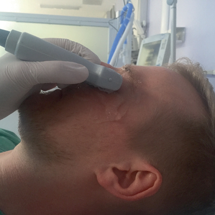
Transverse imaging of the iris and pupillary aperture
-
A light is shone in the other eye in order to perform a sonographic consensual pupillary reflex. The globe axis may change with the patient looking to right or left.
-
The diameter of the pupil can be measured with calipers before and after light stimulation in the contralateral eye. This is best performed in a darkened area.
-
M-mode may be utilized to better visualize the constriction and dilatation of the pupil over time.

Constriction of the pupil on the screen confirms function of the second and third cranial nerves. It also assesses the integrity of the retina, optic nerve and a portion of the midbrain.
16.6 Conclusion
There is scope to advance our practice with regard to neuro-ultrasound—the diagnosis and monitoring of response to treatment in raised intracranial pressure, for example. ONSD ultrasound is promising in this regard, and transcranial Doppler which is non-invasive, does not involve allergenic contrast mediums or exposure to ionizing radiation, or transport outwith the intensive care unit, could also add some valuable data to our day to day assessment of selected patient groups. The assessment of the iris and pupil, as well as the direct and indirect pupillary responses to light, is a useful adjunct to clinical examination in cases of ocular trauma.
With all ultrasound modalities, the pitfall lies principally in the operator’s potential lack of familiarity with the technique and the inability to interpret findings in light of existing comorbidities and factors affecting scanning conditions.
See also:
Chapter 17 Transcranial Doppler.
References
Fledelius HC. Ultrasound in ophthalmology. Ultrasound Med Biol. 1997;23(3):365–75.
Dubourg J, Javouhey E, Geeraerts T, Messerer M, Kassai B. Ultrasonography of optic nerve sheath diameter for detection of raised intracranial pressure: a systematic review and meta-analysis. Intensive Care Med. 2011;37(7):1059–68. http://dx.doi.org/10.1007/s00134-011-2224-2.
Ohle R, McIsaac SM, Woo MY, Perry JJ. Sonography of the Optic nerve sheath diameter for detection of raised intracranial pressure compared to computed tomography: a systematic review and meta-analysis. J Ultrasound Med. 2015;34(7):1285–94.
Hwan Kim Y, Ho Lee J, Kun Hong C, Won Cho K, Hoon Yeo J, Ju Kang M, et al. Feasibility of optic nerve sheath diameter measured on initial brain computed tomography as an early neurologic outcome predictor after cardiac arrest. Acad Emerg Med. 2014;21(10):1121–8. http://dx.doi.org/10.1111/acem.12477.
Legrand A, Jeanjean P, Delanghe F, Peltier J, Lecat B, Dupont H. Estimation of optic nerve sheath diameter on an initial brain computed tomography scan can contribute prognostic information in traumatic brain injury patients. Crit Care. 2013;17(2):R61. https://doi.org/10.1186/cc12589.
Nabeta HW, Bahr NC, Rhein J, Fossland N, Kiragga AN, Meya DB, Dunlop SJ, Boulware DR. Accuracy of noninvasive intraocular pressure or optic nerve sheath diameter measurements for predicting elevated intracranial pressure in cryptococcal meningitis. Open Forum Infect Dis. 2014 Oct 11;1(3):ofu093
Shirodkar CG, Rao SM, Mutkule DP, Harde YR, Venkategowda PM, Mahesh MU. Optic nerve sheath diameter as a marker for evaluation and prognostication of intracranial pressure in Indian patients: An observational study. Indian J Crit Care Med. 2014 Nov;18(11):728–34
Fonseca P, Manno RL, Miller NR. Bilateral sequential trochleitis as the presenting feature of systemic lupus erythematosus. J Neuroophthalmol. 2013 Mar;33(1):74–6
Fagenholz PJ, Gutman JA, Murray AF, Noble VE, Camargo CA Jr, Harris NS. Optic nerve sheath diameter correlates with the presence and severity of acute mountain sickness: evidence for increased intracranial pressure. J Appl Physiol (1985). 2009 Apr;106(4):1207–11
Tayal VS, Neulander M, Norton HJ, Foster T, Saunders T, Blaivas M. Emergency department sonographic measurement of optic nerve sheath diameter to detect findings of increased intracranial pressure in adult head injury patients. Ann Emerg Med. 2007;49(4):508–14. http://dx.doi.org/10.1016/j.annemergmed.2006.06.040.
Qayyum H, Ramlakhan S. Can ocular ultrasound predict intracranial hypertension? A pilot diagnostic accuracy evaluation in a UK emergency department. Eur J Emerg Med. 2013;20(2):91–7. http://dx.doi.org/10.1097/MEJ.0b013e32835105c8.
Rajajee V, Vanaman M, Fletcher JJ, Jacobs TL. Optic nerve ultrasound for the detection of raised intracranial pressure. Neurocrit Care. 2011;15(3):506–15. http://dx.doi.org/10.1007/s12028-011-9606-8.
Soldatos T, Karakitsos D, Chatzimichail K, Papathanasiou M, Gouliamos A, Karabinis A. Optic nerve sonography in the diagnostic evaluation of adult brain injury. Crit Care. 2008;12(3):R67. http://dx.doi.org/10.1186/cc6897.
Bäuerle J, Nedelmann M. Sonographic assessment of the optic nerve sheath in idiopathic intracranial hypertension. J Neurol. 2011;258(11):2014–9. https://doi.org/10.1007/s00415-011-6059-0.
Wang L, Feng L, Yao Y, Wang Y, Chen Y, Feng J, et al. Optimal optic nerve sheath diameter threshold for the identification of elevated opening pressure on lumbar puncture in a Chinese population. PLoS One. 2015;10(2):e0117939. http://dx.doi.org/10.1371/journal.pone.0117939.
Chen H, Ding G, Zhao Y, Rong-Guo Y, Zhou J. Ultrasound measurement of optic nerve diameter and optic nerve sheath diameter in healthy Chinese adults. BMC Neurology 2015;15(106).
Le A, Hoehn ME, Smith ME, Spentzas T, Schlappy D, Pershad J. Bedside sonographic measurement of optic nerve sheath diameter as a predictor of increased intracranial pressure in children. Ann Emerg Med. 2009;53(6):785–91. http://dx.doi.org/10.1016/j.annemergmed.2008.11.025.
Marchese RF, Mistry RD, Scarfone RJ, Chen AE. Identification of optic disc elevation and the crescent sign using point-of-care ocular ultrasound in children. Pediatr Emerg Care. 2015;31(4):304–7. http://dx.doi.org/10.1097/PEC.0000000000000408.
Kardon R. Pupillary light reflex. Curr Opin Ophthalmol. 1995;6:20–6.
Schreiber MA, Aoki N, Scott BG, Beck JR. Determinants of mortality in patients with severe blunt head injury. Arch Surg. 2002;137(3):285–90.
Harries A, Shah S, Teismann N, Price D, Nagdev A. Ultrasound assessment of extraocular movements and pupillary light reflex in ocular trauma. Am J Emerg Med. 2010;28(8):966–9.
Smith SA, Ellis CJ, Smith SE. Inequality of the direct and consensual light reflexes in normal subjects. Br J Ophthalmol. 1979;63:523–7.
Sargsyan AE, Hamilton DR, Melton SL, Amponsah D, et al. Ultrasonic evaluation of pupillary light reflex. Crit Ultrasound J. 2009;1(2):53–7.
Author information
Authors and Affiliations
Corresponding author
Editor information
Editors and Affiliations
Rights and permissions
Copyright information
© 2022 Springer Nature Switzerland AG
About this chapter

Cite this chapter
Shevlin, C. (2022). Neuro-ophthalmic Ultrasound. In: Walden, A., Campbell, A., Miller, A., Wise, M. (eds) Ultrasound in the Critically Ill. Springer, Cham. https://doi.org/10.1007/978-3-030-71742-1_16
Download citation
DOI: https://doi.org/10.1007/978-3-030-71742-1_16
Published:
Publisher Name: Springer, Cham
Print ISBN: 978-3-030-71740-7
Online ISBN: 978-3-030-71742-1
eBook Packages: MedicineMedicine (R0)