
Abstract
Percutaneous femoral catheterization is complicated by intimal dissection with or without associated arterial thrombosis in 0.5 % of the cases. Fortunately, in most cases, subintimal passage of the guidewire or catheter is immediately recognized, and another arterial access is selected, or the procedure is postponed; in such an occasion, usually the vessel patency is not compromised. In a minority of cases, the dissected intima can cause severe stenosis or even acute thrombosis of the arterial lumen.
It is not unusual for arterial dissection or thrombosis to be diagnosed only after completion of the endovascular procedure. A possible mechanism explaining the latter is that the guidewire follows a subintimal route with spontaneous re-entry to the true lumen; this extensive vessel injury, in combination with excessively firm external compression applied after sheath removal, may cause a severe obstruction to flow in the iliofemoral region, resulting in acute thrombosis.
Access provided by Autonomous University of Puebla. Download chapter PDF
Keywords
- Arterial Thrombosis
- Activate Partial Thromboplastin Time
- Stent Deployment
- True Lumen
- Arterial Dissection
These keywords were added by machine and not by the authors. This process is experimental and the keywords may be updated as the learning algorithm improves.
Introduction
Percutaneous femoral catheterization is complicated by intimal dissection with or without associated arterial thrombosis in 0.5 % of the cases. Fortunately, in most cases, subintimal passage of the guidewire or catheter is immediately recognized, and another arterial access is selected, or the procedure is postponed; in such an occasion, usually the vessel patency is not compromised [1]. In a minority of cases, the dissected intima can cause severe stenosis or even acute thrombosis of the arterial lumen [2].
It is not unusual for arterial dissection or thrombosis to be diagnosed only after completion of the endovascular procedure [3]. A possible mechanism explaining the latter is that the guidewire follows a subintimal route with spontaneous re-entry to the true lumen; this extensive vessel injury, in combination with excessively firm external compression applied after sheath removal, may cause a severe obstruction to flow in the iliofemoral region, resulting in acute thrombosis.
Obstructive iatrogenic iliofemoral arterial dissection can cause serious compromise of walking exercise, which is an important component of cardiac rehabilitation in patients with coronary disease, while in some cases the ischemia can even be limb threatening.
Case 1
A 70-year-old lady with a history of minor stroke 1 year ago and coronary artery bypass 5 years ago presented with bilateral critical limb ischemia. Other vascular comorbidities included refractory hypertension, diabetes and chronic obstructive pulmonary disease. Digital subtraction angiography (DSA) was requested in order to accurately depict the status of peripheral arterial tree and renal arteries.
The patient was brought to the interventional suite, and right common femoral access (CFA) was obtained. Immediately following 5-F, 10-cm long sheath insertion into the right CFA, the patient experienced right leg pain, and on clinical examination, the leg was found to be pale, cold and pulseless. A pelvic angiogram through a pigtail catheter at the level bifurcation demonstrated severe stenosis of the right distal external iliac artery tapered down to total occlusion of the ipsilateral CFA (Fig. 93.1).

Pelvic arteriography demonstrates severe stenosis of the right distal EIA tapered down to total occlusion of the ipsilateral CFA
Interventional Approach
-
1.
Maintaining access with a 0.035-in. Teflon J guidewire and under fluoroscopic guidance, the sheath was slowly pulled back at the same time iodinated contrast was continuously injected, so that eventually its tip was placed just at the CFA entrance. This manoeuvre was deemed necessary for the precise depiction of the extension and degree of obstruction caused by the dissection flap, without the obstructive effect of the introducer sheath.
-
2.
Holding steadily the sheath, a smooth manual iodinated contrast injection through the side arm of sheath demonstrated a short (approx. 1 cm long) dissection flap extending from proximal CFA to distal EIA (Fig. 93.2).
Fig. 93.2 
Manual iodinated contrast injection through the side arm of the ipsilateral sheath demonstrates an approx. 1 cm long dissection flap extending from proximal CFA to distal EIA
-
3.
In an attempt to fix the dissection flap, a standard 6 × 40 balloon (Fox Abbott) was inserted through the sheath, positioned at the site of dissection and inflated at 6 atm for 5 min.
-
4.
Following removal of the balloon, a new manual contrast injection through the sheath showed persistence of the dissection flap (Fig. 93.3).
Fig. 93.3 
Following prolonged inflation of a standard balloon at the site of dissection, a new manual contrast injection through the sheath shows persistence of the dissection flap
-
5.
At this point we decided to use a 6 × 40 mm self-expandable nitinol stent (Xpert-Abbott) which was inserted through the right femoral sheath and, under roadmap guidance, was positioned at the site of dissection with an aim to cover its full length (Fig. 93.4). We choose a nitinol stent instead of a balloon expandable or a Wallstent-type self-expandable stent, because nitinol shows high crush resistance to external forces and is ideal for areas of bending such as the groin area. In our experience, nitinol stents, if needed, can safely be extended to the CFA without consequent stent fracture and luminal compromise; however, every effort should be made to avoid stenting of the whole CFA as this prohibits future surgical or percutaneous access to the stented CFA.
Fig. 93.4 
A 6 × 40 mm self-expandable nitinol stent inserted through the right femoral sheath is positioned at the site of dissection
-
6.
The sheath was slightly readjusted in order to confirm that its tip is lying almost at the arterial entry site, so that the proximal part of the stent would not be captured inside the sheath during deployment.
-
7.
The stent was deployed by holding steadily the rod and slowly pulling backwards the covering membrane; at the same time, an assistant was holding firmly the femoral sheath in its marginal position, in order to prevent pulling the sheath completely out from the CFA.
-
8.
In this way the proximal end of the stent was released just above the tip of the femoral sheath and was fully covering the dissection flap at the CFA level.
-
9.
A smooth hand contrast injection through the femoral sheath confirmed successful restoration of lumen patency with some residual stenosis at the site of dissection, which however was not considered haemodynamically significant (Fig. 93.5). This fact together with the anticipated nitinol stent expansion within the next 48 h and the vulnerability of the dissected area led us to avoid post-dilation of the stented area.
Fig. 93.5 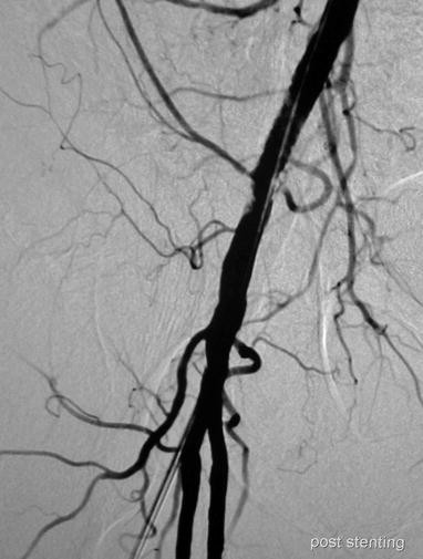
Manual iodinated contrast injection through the femoral sheath confirms successful restoration of lumen patency; there is some residual stenosis at the site of dissection which is not considered haemodynamically significant


Alternative Treatment Options
-
1.
In this case surgical repair would require a major reconstructive procedure which was not desirable due to patient’s history of coronary artery disease [4].
-
2.
Due to the position of the dissection flap very close to the arterial entry site, one could consider in this case a crossover endovascular approach from the left CFA; however, this approach has two limitations: (a) a second puncture should be made and (b) the right femoral sheath should be pulled out and the artery compressed, a manoeuvre which in our experience can potentially cause complete thrombosis of the right CFA, thus making endovascular repair more difficult.
Case 2
A 74-year-old gentleman with a history of stable angina and a past history of right EIA balloon-expandable stenting was referred for percutaneous coronary angiography. The right CFA was chosen as a site of entry and a standard 6-Fr, 11-cm sheath was inserted under local anaesthesia. After sheath insertion, the interventional cardiologist noticed resistance and kinking on guidewire insertion, and a hand contrast injection demonstrated an extensive dissection involving the EIA (Fig. 93.6).

Manual contrast injection through the ipsilateral sheath demonstrates an extensive dissection involving the EIA
After several unsuccessful attempts to redirect the guidewire into the true lumen from ipsilateral approach, the procedure was abandoned, the sheath pulled out and femoral artery haemostasis obtained by manual compression. Following haemostasis, bilateral clinical examination revealed diminished right femoral pulses. At that point, the interventional radiologist was called to assist in the endovascular fixation of the lesion.
Interventional Procedure
-
1.
Following local anaesthesia with lignocaine 1 %, the left CFA was punctured with an 18-G hollow needle under ultrasonographic guidance. A standard 0.035″ J-typed Teflon-coated guidewire was inserted into the arterial lumen, and the reassembled 6-Fr sheath was introduced.
-
2.
Over the 0.035″ guidewire, a catheter was inserted to engage the contralateral CIA. We used a 5-Fr Cobra 1 catheter (Gordis) as we do in the majority of such cases; if the angulation of the aortic bifurcation is very steep, we use instead a Simmons 1 or a SOS Omni (AngioDynamics) catheter.
-
3.
A hand injection of iodinated contrast through the Cobra catheter confirmed the presence of a long EIA flow-limiting dissection.
-
4.
The J-typed Teflon-coated guidewire was exchanged to a 0.035″ angled soft hydrophilic wire (Terumo).
-
5.
The 5-Fr Cobra-hydrophilic wire combination (Fig. 93.7) was used to negotiate the dissection. After several attempts, during which the hydrophilic guidewire was persistently entering the false lumen of the dissection, the true lumen was catheterized and the wire inserted down to the level of the right SFA.
Fig. 93.7 
5-F Cobra diagnostic catheter and 0.035-in. angled soft hydrophilic wire
-
6.
The Cobra catheter was advanced to the right SFA, and the wire was exchanged to a 0.035″180-cm long Amplatz stiff guidewire (Boston Sc). The Cobra catheter was removed and the standard 6-Fr, 11-cm sheath was exchanged for a 6-Fr, 45-cm straight-tip sheath (Destination Terumo).
-
7.
The sheath was used to perform a new angiogram and the diagnosis of right EIA dissection was confirmed (Fig. 93.8).
Fig. 93.8 
Angiography through contralateral 6-F, 45-cm crossover sheath confirms the presence of right EIA dissection
-
8.
An 8 × 150 mm nitinol self-expanding stent (Navalis, Tsunamed) was advanced over the Amplatz guidewire and was deployed so that it covered the dissection flap, from the distal to the proximal EIA. A final angiography confirmed restoration of a fully patent EIA lumen (Figs. 93.9 and 93.10).
Figs. 93.9 and 93.10 
Final angiography confirms restoration of a fully patent EIA lumen


Alternative Treatment Options
-
1.
In this case surgical repair would require a major reconstructive procedure which was not desirable due to patient’s history of coronary artery disease [4].
-
2.
Although the wire passed several times inside the false lumen, a subintimal recanalization was not considered an option because re-entry could be difficult.
-
3.
Also from an endovascular point of view, one could consider first a prolonged inflation of a standard long balloon to fix the arterial dissection; however, in our experience, in the vast majority of EIA dissections, this strategy is inefficient and stent deployment is necessary in order to restore full lumen patency. Furthermore, in our experience, balloon inflation can further extend the dissection or even cause EIA rupture. For all these reasons, we decided to primarily stent the dissection with a nitinol self-expanding stent.
Discussion
These two cases demonstrate the steps for successful endovascular repair of iatrogenic iliofemoral dissections. An ipsilateral approach through the initial femoral access should be considered first; however, as it is shown in the second case, redirection of the guidewire into the true lumen can be unsuccessful; in such cases, the iatrogenic dissection is negotiated from a contralateral crossover approach. If the dissection flap is localized in the CFA, prolonged inflation of a standard angioplasty balloon should be attempted first, as it may result in successful apposition of the intima and underlying media; however, very often a stent covering at least proximal CFA must be deployed in order to restore full lumen patency. Fortunately, with new flexible self-expanding nitinol stents, stenting of the proximal CFA is not prohibitive, as these stents can effectively withstand external compression at the hip flexible point, while the CFA segment which remains uncovered by the stent can be used for surgical or endovascular access [5]. In more extensive dissections involving the EIA, self-expanding stent deployment is essential to restore vessel patency [6].
If the iliofemoral dissection is complicated by acute arterial thrombosis, then catheter-directed thrombolysis (CDT) is the treatment of choice. The most commonly used lytic agents for CDT are rt-PA and urokinase, often with the adjunct of catheter suction. Following successful lysis, percutaneous transluminal angioplasty with prolonged balloon inflation and/or self-expandable stent deployment should be performed as complementary procedures in order to treat the underlying dissection.
If thrombolysis is required, the following protocol is used:
-
Initially, two 5-mg boluses of rt-PA (Actilyse, Boehringer, Germany) are injected into the clot through a 5-Fr end-hole catheter at an interval of 10 min in order to initiate lysis and facilitate subsequent manipulations. Following this, the occlusion is negotiated carefully with a 0.035-in. hydrophilic guidewire, and, after successful passage, the end-hole catheter is exchanged for a multiside-hole catheter (we use a 5-Fr Mewissen/Boston Scientific catheter with 20 side holes/10-cm infusion length) in such a way that the infusion part of the catheter is positioned within the thrombus while its tip is occluded by the guidewire. Two additional 5-mg boluses of rt-PA are given at an interval of 10 min.
-
In cases with thrombus confined to the CFA, only one additional bolus is given through the initial end-hole catheter after this had been advanced further into the clot. Ten minutes after the last bolus, an angiogram is performed, in order to check for the extent of thrombus dissolution, and in case of a significant amount of residual thrombus, a continuous infusion of 2.5 mg/h of rt-PA is started using a calibrated infusion pump. During infusion of the lytic agent, concurrent intravenous heparin at a dose of 500 IU/h is given to prevent rethrombosis. Laboratory monitoring consists of baseline and 2-hourly determinations of plasma fibrinogen and activated partial thromboplastin time (APTT).
References
Samson RH, Sprayregen S, Veith FJ, Scher LA, Gupta SK, Ascer E. Management of angioplasty complications, unsuccessful procedures and early and late failures. Ann Surg. 1984;199:234–40.
Labropoulos N, Giannoukas AD, Volteas SK, Kutoubi AA. Complications of the balloon assisted percutaneous transluminal angioplasty. J Cardiovasc Surg. 1994;35:475–89.
Tsetis DK, Kochiadakis GE, Hatzidakis AA, et al. Transcatheter thrombolysis with high-dose bolus tissue plasminogen activator in iatrogenic arterial occlusion after femoral arterial catheterization. Cardiovasc Intervent Radiol. 2002;25:36–41.
Youkey JR, Clagett GP, Rich NM, et al. Vascular trauma secondary to diagnostic and therapeutic procedures: 1974 through 1982. A comparative review. Am J Surg. 1983;146:788–91.
Tsetis D. Endovascular treatment of complications of femoral arterial access. Cardiovasc Intervent Radiol. 2010;33(3):457–68.
Onal B, Ilgit ET, Akpek S, Erbas G, Akkaya A. Endovascular treatment of obstructive iliac artery dissections. Acta Radiol. 2005;46:359–65.
Author information
Authors and Affiliations
Editor information
Editors and Affiliations
Rights and permissions
Copyright information
© 2014 Springer Science+Business Media New York
About this chapter
Cite this chapter
Tsetis, D., Kehagias, E., Spiliopoulos, S., Siablis, D. (2014). Arterial Dissection. In: Dieter, R., Dieter, Jr., R., Dieter, III, R. (eds) Endovascular Interventions. Springer, New York, NY. https://doi.org/10.1007/978-1-4614-7312-1_93
Download citation
DOI: https://doi.org/10.1007/978-1-4614-7312-1_93
Published:
Publisher Name: Springer, New York, NY
Print ISBN: 978-1-4614-7311-4
Online ISBN: 978-1-4614-7312-1
eBook Packages: MedicineMedicine (R0)