
Abstract
The anatomy of the face is of utmost importance in cosmetic procedures. In this chapter we will review relevant anatomical structures of the face that will be well supported by anatomical drawings.
Access provided by Autonomous University of Puebla. Download chapter PDF
Keywords
- Cosmetic Purposes
- Relevant Anatomical Structures
- Anatomical Drawings
- Cosmetic Procedures
- Superficial Musculoaponeurotic System (SMAS)
These keywords were added by machine and not by the authors. This process is experimental and the keywords may be updated as the learning algorithm improves.
1 Introduction
Boney landmarks permit identification of overlying superficial structures. For example, the zygomatic arch is the upper limit of the parotid gland, the orbital rims mark the path of the infra- and supraorbital nerves, and the mastoid process reveals the emergence of the facial nerve. Similarly, the foramen imaged as boney notches affords a window to the orbital and mental nerves and arteries that run in the subcutaneous tissues.
The superficial musculoaponeurotic system (SMAS) interconnects the facial and neck musculature and is divided into two parts: the superficial SMAS, with the arterial supply and the deep SMAS, wherein lie most of the nerves. While the veins generally parallel the arteries, great variation occurs.
3D imaging is optimal to focus on the facial danger zones to avoid nerve damage. 2D cross-sectional imaging of the small facial nerves is difficult and C-plane reconstruction will confirm the linear and uninterrupted nature of the original picture.
2 Relevant Anatomic Structures and Regions
The following are important anatomical structures and regions for performing cosmetic procedures:
-
1.
Temporal branch of the facial nerve located below a line drawn from approximately 0.5 cm below the tragus to 2 cm above the lateral eyebrow and above the zygoma (Fig. 13.1)
Fig. 13.1 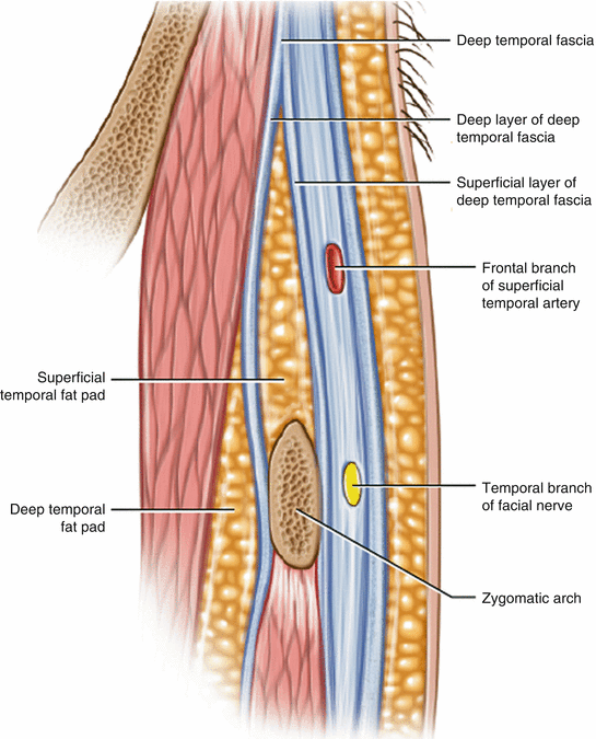

Drawing (sagital axis of the temple and lateral cheek region) shows the location of the temporal branch of the facial nerve, besides other relevant structures
-
2.
Greater auricular nerve located approximately 6.5 cm below the external auditory canal (Fig. 13.2)
Fig. 13.2 
(a) drawing, (b) ultrasound (longitudinal view) of the greater auricular nerve, located approximately 6.5 cm below the external auditory canal
-
3.
Marginal mandibular branch of the facial nerve located mid mandible approximately 2 cm posterior to the oral commissure (Fig. 13.3)
Fig. 13.3 
(a) Drawing, (b) Ultrasound (longitudinal view) of the marginal mandibular branch of the facial nerve located in the mid mandible approximately 2 cm posterior to oral commissure
-
4.
Zygomatic and buccal branches of the facial nerve located in the triangle formed by connecting the dots of the malar eminence, posterior border of the mandibular angle, and oral commissure (Fig. 13.4)
Fig. 13.4 
Drawing of the supraorbital and supratrochlear nerves located at the superior orbital rim above mid pupil
-
5.
Supraorbital and supratrochlear nerves located at the superior orbital rim above the mid pupil (Fig. 13.5)
Fig. 13.5 
Drawing of the zygomatic and buccal branches of facial nerve located in the triangle formed by connecting the dots of the malar eminence, posterior border of mandibular angle and oral commissure
-
6.
Infraorbital nerve located at the inferior orbital rim below the mid pupil (Fig. 13.6)
Fig. 13.6 
(a) drawing (b) ultrasound (transverse view) of the infraorbital nerve (arrow) located at the inferior orbital rim below the mid pupil
-
7.
Mental nerve located midmandible below the second premolar (Fig. 13.7) [1–3]
Fig. 13.7 
Drawing of the mental nerve located midmandible below the second premolar


References
Owsley JQ. SMAS-platysma face lift. Plast Reconstr Surg. 1983;71:573–9.
Hamra ST. The tri-plane face lift dissection. Ann Plast Surg. 1984;12:268–81.
Rudolph R. Depth of the facial nerve in face lift dissections. Plast Reconstr Surg. 1990;85:537–42.
Author information
Authors and Affiliations
Corresponding author
Editor information
Editors and Affiliations
Rights and permissions
Copyright information
© 2013 Springer Science+Business Media New York
About this chapter
Cite this chapter
Bard, R.L. (2013). Anatomy of the Face for Cosmetic Purposes. In: Wortsman, X. (eds) Dermatologic Ultrasound with Clinical and Histologic Correlations. Springer, New York, NY. https://doi.org/10.1007/978-1-4614-7184-4_13
Download citation
DOI: https://doi.org/10.1007/978-1-4614-7184-4_13
Published:
Publisher Name: Springer, New York, NY
Print ISBN: 978-1-4614-7183-7
Online ISBN: 978-1-4614-7184-4
eBook Packages: MedicineMedicine (R0)