
Abstract
A double osteotomy is indicated in the following situations:
-
A situation in which an isolated osteotomy (on the femur or on the tibia) to correct a major angular deformity (>to 10°) in the frontal plane in valgus (Fig. 19.1) or in varus (Figs. 19.2 and 19.3a) would result in an oblique joint line (Fig. 19.3b). This obliquity would create shear forces across the knee joint that can lead to early failure of the intervention. A distal femoral osteotomy combined with a proximal tibial osteotomy is able to correct the axis of the lower limb while maintaining an acceptable obliquity of the joint line (Fig. 19.3c).
-
A situation in which an attempted single site correction with an opening wedge osteotomy results in too much opening, compromising the stability of the osteotomy.
-
A situation in which correction with a single closing wedge osteotomy would be too large and result in poor coaptation of the proximal and distal bone segments, which can cause problems for future total knee arthroplasty.
-
The treatment of osteoarthritis secondary to malunion of the femur. In these cases, the aim of the procedure is to address the frontal or torsional malunion on the femur by a femoral osteotomy and to address the arthritis with a tibial osteotomy (Fig. 19.4). It is of major importance to know that femoral malunions situated close to the knee joint are more significant than those situated at a greater distance from the joint. A femoral osteotomy can only correct a deformation in extension and not in flexion.
Access provided by Autonomous University of Puebla. Download chapter PDF
Keywords
These keywords were added by machine and not by the authors. This process is experimental and the keywords may be updated as the learning algorithm improves.
Introduction
A double osteotomy is indicated in the following situations:
-
A situation in which an isolated osteotomy (on the femur or on the tibia) to correct a major angular deformity (>to 10°) in the frontal plane in valgus (Fig. 19.1) or in varus (Figs. 19.2 and 19.3a) would result in an oblique joint line (Fig. 19.3b). This obliquity would create shear forces across the knee joint that can lead to early failure of the intervention. A distal femoral osteotomy combined with a proximal tibial osteotomy is able to correct the axis of the lower limb while maintaining an acceptable obliquity of the joint line (Fig. 19.3c).
Fig. 19.1 
Major angular deformity in the frontal plane in valgus (right knee)
Fig. 19.2 
Major angular deformity in the frontal plane in varus (both knees)
Fig. 19.3 
(a) Case of major angular deformity in varus. (b) An isolated tibial osteotomy to correct a major angular deformity would create an oblique joint line. (c) A normal or acceptable joint line obliquity after correction of a major angular deformity becomes possible using a distal femoral osteotomy associated with the proximal tibial osteotomy
-
A situation in which an attempted single site correction with an opening wedge osteotomy results in too much opening, compromising the stability of the osteotomy.
-
A situation in which correction with a single closing wedge osteotomy would be too large and result in poor coaptation of the proximal and distal bone segments, which can cause problems for future total knee arthroplasty.
-
The treatment of osteoarthritis secondary to malunion of the femur. In these cases, the aim of the procedure is to address the frontal or torsional malunion on the femur by a femoral osteotomy and to address the arthritis with a tibial osteotomy (Fig. 19.4). It is of major importance to know that femoral malunions situated close to the knee joint are more significant than those situated at a greater distance from the joint. A femoral osteotomy can only correct a deformity in extension and not in flexion.
Fig. 19.4 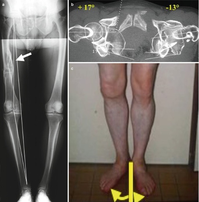
(a) Medial femorotibial osteoarthritis caused by rotational malunion after femoral fracture (external rotation of the right limb). (b) External rotational deformity measured by CT scan. (c) Clinical deformity


Certain difficulties and complications are inherent to a double osteotomy:
-
1.
The risk for a delayed union or malunion is increased compared to an isolated osteotomy.
-
2.
Calculation of the correction remains difficult and complicated. In the case of a femoral malunion, one can perform both interventions separately starting with the femoral derotation osteotomy and then the tibial osteotomy at a later stage. If a computer-assisted navigation is available, both corrections in the frontal and horizontal plane can be combined during the same intervention.
Nevertheless, indications for a double osteotomy remain rare, and in this chapter we will not discuss proximal or diaphyseal femoral osteotomies that are indicated in isolated torsional problems.
The Principles
Varus Knee
In a varus knee with a mechanical axis less than 165°, the combination of a lateral closing wedge distal femoral osteotomy with a lateral closing wedge high tibial osteotomy or medial opening wedge high tibial osteotomy is indicated. The advantage of an opening wedge high tibial osteotomy is preservation of the length of the lower limb. The skin incision is placed laterally on the femur and crosses the midline at the level of the tibial tubercle to continue medially on the tibia. Alternatively, an isolated lateral femoral incision can be combined with an isolated medial tibial incision. In cases of a closing wedge high tibial osteotomy, a laterally based long skin incision is typically used (Fig. 19.5).

Postoperative x-rays (see case Fig. 19.2)
Valgus Knee
In a valgus knee with a mechanical axis greater than 190°, a combination of an opening wedge lateral distal femoral osteotomy with a closing wedge medial high tibial osteotomy is indicated (Fig. 19.6). This combination results in an acceptable orientation of the joint line while lowering the risk of injury to the peroneal nerve.

Postoperative long leg films after correction of a major valgus deformity
Malunion with Torsional Problem
In cases of osteoarthritis secondary to a femoral malunion in combination with a torsional problem greater than 15° and a frontal deviation greater than 10°, we advise the combination of a derotation osteotomy on the femur and a tibial osteotomy to address the frontal plane deformity (Fig. 19.7).

Pre- and postoperative x-rays after correction of an external rotational malunion associated with a medial compartment osteoarthritis (see case Fig. 19.4)
Surgical Technique
On the Femur
The approach has been described in detail in the chapter on femoral osteotomy for varization.
-
1.
Lateral opening wedge osteotomy for valgus knee (see chapter on femoral osteotomy for varization)
-
2.
Closing wedge osteotomy for the varus knee
The area for the osteotomy is prepared. Two additional Kirschner guide pins are introduced in the femur as guide pins for the future osteotomy. One pin is introduced parallel to the joint line approximately 50 mm proximal to the joint line. The second pin is introduced proximally to the first on the lateral cortex but converging with the first medially. This represents the angle and the wedge that will be resected. The quadriceps muscle is retracted at a level proximal to the trochlea with the knee in extension; the posterior side of the knee is cleared. A superficial longitudinal mark on the lateral cortex of the femur with the oscillating saw can serve as a landmark to determine the rotation (Fig. 19.8). The blade plate has to be introduced in the epiphyseal area approximately 30 mm proximal to the joint line. The blade is 5.6 mm thick and 16 mm in width, and the distance between the holes is 16 mm. Its entry point is anterior and proximal to the lateral collateral ligament. The entry angle has been determined by preoperative planning and a specific reamer is used. For a calculated valgus correction of 8°, the guide instrument is set at 93° (85° + 8°; this is the complementary angle to the desired anatomical angle of 95°, plus the angle of correction). The blade is subsequently introduced into the femur. The correct angulations are again checked using the image intensifier.
Fig. 19.8 
Two rotational landmarks are superficially done on the femoral cortex using the saw
-
3.
Derotation osteotomy in the case of femoral malrotation
The area of the osteotomy is prepared in the same manner. Two superficial saw marks are made on the lateral cortex indicating the desired angle of the derotation (Fig. 19.8). By doing this, an isolated derotation osteotomy can be performed as well as a derotation osteotomy in combination with an opening wedge or a closing wedge femoral osteotomy. The derotation osteotomy should not interfere with the patellar tracking or create a step on the anterior cortex.

On the Tibia
For these surgical techniques, please see chapter on tibial osteotomy.
The bone graft obtained in case of a closing wedge femoral osteotomy is used to fill the opening wedge tibial osteotomy.
Postoperative Guidelines
The postoperative guidelines are identical as for a high tibial osteotomy.
Author information
Authors and Affiliations
Corresponding author
Editor information
Editors and Affiliations
Rights and permissions
Copyright information
© 2014 Springer-Verlag London
About this chapter
Cite this chapter
Archbold, P., Verdonk, P., Servien, E. (2014). Surgical Technique for a Double Osteotomy. In: Neyret, P., Demey, G. (eds) Surgery of the Knee. Springer, London. https://doi.org/10.1007/978-1-4471-5631-4_19
Download citation
DOI: https://doi.org/10.1007/978-1-4471-5631-4_19
Published:
Publisher Name: Springer, London
Print ISBN: 978-1-4471-5630-7
Online ISBN: 978-1-4471-5631-4
eBook Packages: MedicineMedicine (R0)