
Abstract
Before considering any surgical intervention a thorough understanding of the patients’ pathology is of paramount importance. In this chapter we will consider how fluoroscopic assessment and MRI arthrography can help those with an interest in hip preserving surgery. We will also consider the contribution of hip injections in the diagnosis and management of hip pain.
Access provided by Autonomous University of Puebla. Download chapter PDF
Keywords
These keywords were added by machine and not by the authors. This process is experimental and the keywords may be updated as the learning algorithm improves.
Introduction
Before considering any surgical intervention a thorough understanding of the patients’ pathology is of paramount importance. In this chapter we will consider how fluoroscopic assessment and MRI arthrography can help those with an interest in hip preserving surgery. We will also consider the contribution of hip injections in the diagnosis and management of hip pain.
Clohisy et al. [1] concluded in their very instructive paper that despite their attempts to define several standard diagnostic criteria to diagnose structural hip abnormalities, there was limited reliability in radiographic diagnosis. They urged caution in basing surgical treatment options on isolated radiographic findings and highlighted the importance of understanding the mechanical pathology of individual patients, particularly in femoro-acetabular impingement (FAI) to avoid relying solely on static radiographic findings.
As emphasized by Bedi et al. [2], impingement is a dynamic problem and therefore is best investigated dynamically. For this reason a dynamic fluoroscopic arthrogram is essential very useful tool.
Dynamic arthrograms can be obtained under fluoroscopic control and ideally should be undertaken by the surgeon who will be performing the definitive surgery. MRI arthrograms can give additional information regarding the integrity of the articular cartilage as well as labral damage and extra-articular pathology. In a static position an arthrogram can be used in conjunction with a CT and MRI to outline the bony structure of the joint surfaces and soft tissues including the labrum and chondral surfaces. With advances in the contrast agents used in MRI arthrograms such as ‘delayed Gadolinium Enhanced MRI of Cartilage’ (dGEMRIC), information regarding the quality of the articular cartilage can also be obtained.
As well as injecting contrast into the hip joint, other topics to be discussed will include the symphysis pubis arthrogram, the psoasogram and injection of the trochanteric bursa, all of which can be useful therapeutic modalities in patients with extra-articular causes for their pain.
Fluoroscopic, Dynamic Hip Arthrograms
An arthrogram is an investigation where a radio-opaque dye is injected into the joint to try to outline the joint surfaces. As mentioned it can be a dynamic investigation which is done preferably by the operating surgeon where the joint is moved under imaging to assess congruence, instability and position of best fit. It can be used for therapeutic and diagnostic intervention.
Technique
It is our practice, having obtained appropriate consent from the patient, to perform the procedure in the operating theatre under a short general anaesthetic. An image intensifier is placed on the contra-lateral side to the hip which allows an unobstructed AP view of the hip of interest to be taken and allows movement of the hip so that lateral views as well as views in rotation and varus/valgus alignment can also be made. Fluoroscopic pictures are taken before injection of contrast as an AP view with the femur in neutral (patella pointing to the ceiling), the Dunn view to assess the anterior aspects of the femoral head and superior impingement as well as the frog lateral. A 22 gauge spinal needle is inserted under fluoroscopic control into the hip joint. In the adult, the authors’ preference is for the anterior approach, aiming the needle at the base of the femoral head directly onto the proximal femoral neck and attempting to insert the needle tip into the sulcus formed as the hip capsule is elevated off the femoral neck. A medial approach is reserved for the paediatric patient group. Once the desired position is reached, the hip can be gently rotated. If the needle is lodged in the periosteum of the femoral neck it will rotate in the same direction as the hip, however, if it is within the capsule but not lodged in soft tissue it will rotate in the opposite direction to the rotation of the hip. A radio-opaque contrast (Omnipaque®, GE Healthcare) is then injected through intravenous tubing into the hip joint. We usually instill approximately 2 ml’s of dye so the joint capsule is not over distended. Further injection of local anaesthetic and steroid (approximately 5 ml’s) 0.5 % chirocaine or marcaine and 40 mg of triamcinolone (Kenalog®-40, Bristol-Myers Squibb) is effected. We try to avoid injecting large volumes that may cause distension and discomfort. It is important to avoid extravasation of the contrast particularly in the supero-lateral aspect of the joint so that the arthrogram yields the best possible information for the surgical plan. The needle is then removed and the hip is circumducted to allow for dispersal of the contrast over the entire femoral head. Having injected the dye the hip is screened dynamically in flexion, then internal and external rotation, abduction and adduction to assess congruence, stability and position of best fit. The hip is then classified into the following five groups:
-
1.
The hip is congruent in all movements. There is no pooling of the dye and there is a round head within the round socket as shown in Figs. 13.1 and 13.2. Shenton’s line is maintained in flexion, abduction, adduction, internal and external rotation.
Fig. 13.1 
AP fluoroscopic image of a normal hip arthrogram
Fig. 13.2 
Dunn view fluoroscopic image demonstarting a normal congruent hip
-
2.
The hip shows reducible subluxation. There is a disrupted Shenton’s line on the AP neutral view shown in Fig. 13.3 which on flexion and internal or abduction of the hip is congruent with no pooling of the dye shown in Fig. 13.4.
Fig. 13.3 
AP fluoroscopic image showing disruption of Shenton’s line in a dysplastic hip
Fig. 13.4 
AP fluoroscopic image of a dysplastic hip in abduction with no pooling and showing good joint congruency
-
3.
The hip shows irreducible subluxation. The Shenton’s line will not restore in any position and therefore a false socket has been created.
-
4.
There is evidence of hinge abduction in extension where the superior aspect of the femoral head impinges on the labrum edge of the acetabulum and the centre of rotation moves out in abduction on dynamic testing and pooling of the dye occurs in abduction shown in Fig. 13.5.
Fig. 13.5 
AP fluoroscopic image of a hip with cam impingement showing impingement of the femoral head on the labrum at the superolateral aspect of the acetabulum
-
5.
There is evidence of impingement anteriorly and superiorly in the Dunn view shown in Fig. 13.6, as the hip is flexed in abduction and internal rotation and again an unstable movement is noted as the hip is flexed and pooling of the dye medially.
Fig. 13.6 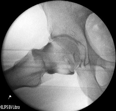
Dunn view fluoroscopic image showing impingement anteriorly and superiorly


While the dynamic movement occurs, a position of best fit, that is, the most congruent is assessed. In a congruent hip this is usually in abduction or internal rotation and in an incongruent hip it may be in adduction of the femur. While the position of best fit is assessed, attention is paid to the position of the greater trochanter in relation to the centre of rotation and acetabular coverage as this will allow for planning of any acetabular surgery. These static images are taken as a record but the dynamic test allows for the feel of the hip and the planning of any intervention.
The injection of local anaesthetic and steroid will give some temporary block to the hip pain and is a useful diagnostic tool in eliminating extra-articular causes of pain that may present as hip pain. The injection can also be used as a therapeutic tool to give some pain relief while a decision is made regarding intervention. The use of CT allows assessment of the bony structures and delineation of the articular cartilage. The use of the MRI allows assessment of the chondral surfaces and of the labrum/chondrolabral junction and soft tissue structures which may also be a source of pain. The dGEMRIC technique allows biochemical assessment of the articular cartilage rather than the standard present methods which better identify anatomical structures. It is important to be aware of the presence of degenerative changes as this would influence the outcome/prognosis of hip preserving surgery.
Examples of Hip Arthrogram Findings
Perthes in the Adult Hip
Figure 13.7 demonstrates an AP fluoroscopic image of a right hip. The typical appearance of a flattened femoral head with a hanging rope sign can be seen and is further illustrated by the Dunn view shown in Fig. 13.8. Figure 13.9 shows an AP fluoroscopic image after injection of contrast into the joint. The intact labrum can be seen as a filling defect sitting on top of the superolateral aspect of the femoral head. Figure 13.10 shows the same view but as an MRI arthrogram confirming that the filling defect is the hypertrophied labrum. Figure 13.11 shows an AP fluoroscopic image with the hip abducted demonstrating medial pooling of the contrast caused by the hinge abduction of the lateral aspect of the femoral head on the lateral aspect of the acetabulum. That is to say, the femoral head does not move concentrically within the acetabulum but levers on the edge of the acetabulum in abduction. Figure 13.12 shows an AP fluoroscopic image with the hip adducted. This shows obliteration of the medial pooling of the contrast and the ‘best fit’ of the femoral head within the acetabulum. The joint is concentric in this position and the outline of the labrum can be seen to be down sloping. The shape of the femoral head roughly represents a broad based cone with the acetabulum a reciprocally similar shape, making concentric movement difficult. This can be demonstrated with live screening of the hip. Once the femoral head hinge abducts on the lateral aspect of the acetabulum, any further abduction is achieved with tilting of the pelvis and not from movement of the femoral head within the acetabulum. The surgical options can be difficult in this situation. In this particular case the patient was very symptomatic and was 15 years old. In this situation a valgus osteotomy can be performed with a head-neck debridement, however the patient should be adequately counselled regarding the realistic expectations of this procedure to succeed. They should be warned that they may require a hip arthroplasty as a young adult.

AP fluoroscopic image of a right hip (Perthes)

Dunn view fluoroscopic image of a right hip (Perthes)

AP fluoroscopic image of a right hip (Perthes) with contrast

Coronal MRI arthrogram right hip (Perthes)

AP fluoroscopic image with right hip (Perthes) abducted with contrast

AP fluoroscopic image with right hip (Perthes) adducted with contrast
Dysplasia in the Adult
Figure 13.13 shows an AP fluoroscopic image of a dysplastic hip with a centre edge angle of 17° and a sourcil angle of 12°. Figure 13.14 shows a fluoroscopic image after the hip has been injected with contrast. With the hip in 20° abduction, the femoral head has good lateral coverage and the labrum which is outlined by the contrast, is horizontal. In this position the joint is congruent and represents the best position of the hip as shown in Fig. 13.15. This is the expected position of the hip following a successful peri-acetabular osteotomy. During the dynamic screening of the hip, careful attention should be paid to the Dunn view ensuring that there is no co-existent cam lesion. A static Dunn view is shown in Fig. 13.16. Patients with dysplasia may not have any symptoms or signs of impingement pre-operatively but following correction of their dysplasia with an osteotomy, a cam lesion may become symptomatic particularly if the socket is retroverted.

AP fluoroscopic image of the left hip (Dysplasia)

AP fluoroscopic image of the left hip (Dysplasia) with contrast

AP fluoroscopic image of the left hip (Dysplasia) abducted with contrast

Dunn view fluoroscopic image of the left hip (Dysplasia) with contrast
MRI Arthrogram
If labral pathology is suspected then MR arthrography is the investigation of choice. Czerny et al. [3] confirmed that the sensitivity and specificity of MR arthrography in the diagnosis of labral tears and detachments was 90 % and 91 % respectively and in MRI’s without contrast these figures dropped to 30 and 36 %. Standard MR arthrography can also show articular cartilage thinning and cartilage defects, but cannot give any information regarding the quality of the articular cartilage. More recent techniques can be used to identify the characteristics of the cartilage itself.
These techniques involve the injection of a contrast such as Gadolinium-DTPA2− (Diethylene Triamine Penta-Acetic Acid) which is an ionic agent that has a negative charge that is able to penetrate cartilage. This contrast agent works since the glycosaminoglycans (GAG’s) found within articular cartilage also have a negative charge so areas within cartilage that have a high GAG content will have low concentration of Gd-DTPA2− and areas with a low GAG content will have a high concentration of Gd- DTPA2−. From the distribution of Gd- DTPA2−, areas of high and low GAG concentration can be determined. This technique is called ‘delayed Gadolinium Enhanced MRI of Cartilage’ (dGEMERIC) and is illustrated in Fig. 13.17. The results of this technique are encouraging when compared to the ‘gold standard’ of estimating GAG content biochemically and histologically [4]. This agent also appears to increase the detection rate of defects as opposed to using an MRI with a non-ionic contrast such as Prohance (Bracco Diagnostics, Princeton, New Jersey) [5]. Using the dGEMERIC technique it is not possible to measure the absolute amount of GAG content, but it is able to provide a baseline with which disease progression and therapeutic measures can be monitored.

This sequence of magnetic resonance images illustrates how dGEMERIC imaging can visualize the glycosaminoglycan composition of articular cartilage. (a) Shows a proton density image of articular cartilage. (b) Following administration of a charged ionic contrast agent (Gd- DTPA2−), the distribution of which is dependent on the concentration of glycoaminoglycans (GAG’s). Areas of high concentration of GAG’s take up less of the contrast due to there negative charge and areas of relatively low GAG content will take up more of the contrast. (c) When the same patient is given a nonionic agent Gd(HPDO3A) the cartilage appears homogenous. This suggests that the selective uptake of the ionic contrast agent seen in 11b is due to charge and hence indicates the GAG distribution [32] (Courtesy of the American Journal of Bone and Joint Surgery)
Some studies have also suggested that the technique can be successfully used in the selection of suitable patients for hip arthroscopy over the more traditional methods of assessment of degenerative disease [6–8]. In an interesting study looking at cam impingement, Pollard et al. [8] reviewed the dGEMRIC images of hips in patients who were asymptomatic and who were subdivided into one of two subgroups depending on the presence of a cam deformity and a positive impingement test. The authors showed that there was evidence of localized cartilage damage in patients who were asymptomatic with a cam deformity and had no evidence of joint space narrowing based on their plain radiographic assessment. In these patients the dGEMRIC technique was able to identify reduced GAG content in the anterosuperior region of the acetabular articular cartilage. The remainder of the joint had a similar GAG content to the hips with a normal head-neck morphology and physical examination. This could suggest that the difference in GAG content is related to the cam impingement. This conclusion is consistent with other studies [6, 7] that dGEMRIC is able to provide objective evidence of disease progression in the absence of any measureable change in the joint space. Additionally, the severity of the cartilage damage has been shown to be proportional to the severity of the cam deformity [8]. However as pointed out in other studies [7], a cam deformity does not inevitably result in progressive osteoarthritis, other factors such as age, activity level, acetabular morphology and the durability of the chondrolabral junction also play a significant role.
In the age of increasing financial pressures where the use of a novel and expensive technique such as dGEMRIC may be limited, patients who are at the upper age limit for hip preservation surgery (≥35 years) may benefit the most from such a technique as age has been shown to be a significant prognostic factor for early failure [9–14].
Symphysis Pubis Arthrogram
Osteitis pubis is a relatively rare pathology in the general orthopaedic clinic but has been quoted to be as high as 7 % in the general athletic population [15–17]. The majority of the reports to date describe the condition affecting athletes who participate in sports involving kicking such as football, although it has also been reported in basketball players and long distance runners [16, 18]. Symptoms can include pain when kicking or during the swing phase of the gait cycle when the hip is flexed. Pain is classically localized over the symphysis pubis and parasymphyseal bone but can also occur within the lower aspect of the abdominal muscles. Clinical evaluation may reveal tenderness over the adductors particularly over the musculotendinous attachments. Painful symptoms can be reproduced with passive and resisted muscle tests of the adductor and abdominal muscles. A Technetium Tc 99 m pubic bone scan has been classically used to detect increased uptake in the pubic symphysis area [19–21]. However, the degree of uptake is poorly correlated with the duration and the severity of symptoms [20] and currently the MRI has become the diagnostic modality of choice shown in Figs. 13.18 and 13.19 [22–25]. Although this condition can be treated non-operatively with anti-inflammatories and rest, resistant cases can be treated with an injection of contrast under fluoroscopic control to first confirm the location of the symphysis pubis joint followed by an injection of steroid and local anaesthetic. There are a number of case series reporting favourable results with patients being able to return to sport after injection of steroid into the symphyseal cleft and surrounding tissues [26–28].

Coronal T2 MRI of the pelvis illustrating oedema adjacent to the symphysis pubis

Axial T2 MRI of the pelvis illustrating oedema adjacent to the symphysis pubis
In O’Connell et al.s [28] description of the technique under fluoroscopic control they introduced a 22 gauge needle into the symphyseal cleft halfway between the upper and lower margins of the symphysis. The needle was advanced into the cleft and 1 ml of non-ionic contrast was injected to confirm the needle position and outlining the fibrocartilaginous disc. In O’Connell’s study a radiographic image was then taken to record the appearance of the disc. Then an aqueous suspension of 20 mg of methylprednisolone acetate and 1 ml of 0.5 % bupivacaine hydrochloride was injected into the cleft as shown in Fig. 13.20. Of the 16 patients who had confirmed osteitis pubis in O’Connell’s study 14 experienced immediate relief of their symptoms and were able to resume athletic activities 48 h after the procedure. One patient had complete resolution of symptoms at their 6 month follow up following a period of rest and one patient had ongoing symptoms. There were no complications reported. The authors concluded that particularly in athletes a symphyseal cleft injection can confirm the diagnosis and give short-term relief enabling return to sport.

AP fluoroscopic image of the pelvis after infiltration of contrast into the symphysis pubis
Injection of the Trochanteric Bursa
Trochanteric bursitis is a relatively common problem in the hip clinic and although it can be successfully treated with conservative interventions in the majority of patients [29] recurrence can be a problem. There are both direct and indirect causes for this condition but in most patients the etiology is multifactorial and can affect patients of all ages. There are a number of non-operative options that can be administered either independently or in combination. Such methods include activity modification, physiotherapy, weight loss non-steroidal anti-inflammatories (NSAIDS) or a corticosteroid injection. Currently the most widely used treatment modality is an injection performed with ultrasound guidance. Furia et al. [30] found from their results following a course of rest, physiotherapy, ultrasound, steroid injection, ice as well as heat that 66 % of patients were able to return to sport and 83 % to jobs that involved a lot of manual labour after 3 months.
In summary, a single steroid injection can be effective in treating patients with trochanteric bursitis but some patients may benefit from a multimodal approach and some recalcitrant cases may require surgery.
Psoasogram
Iliopsoas bursitis and tendinitis are interrelated conditions, in that inflammation of one will inevitably result in inflammation of the other, due to their close proximity. Therefore these conditions can be considered as essentially identical with respect to their presentation, aetiology and treatment. Acute or chronic occupational trauma and sports injuries are thought to be responsible for the majority of iliopsoas bursitis [31] with rheumatoid arthritis being an additional cause.
Initial treatment has classically included rest with targeted physiotherapy consisting of stretching and strengthening exercises along with a course of oral anti-inflammatory medications. However, not all patients respond well to this treatment and as a result may proceed to an injection. Commonly this is performed under ultrasound guidance. In the majority of patients this intervention is able to provide temporary or permanent symptom relief to allow return to activities and may postpone or avoid future surgical intervention.
To conclude, arthrograms and hip injections can be extremely useful to the surgeon with an interest in hip preserving surgery. The dynamic arthrogram should not be underestimated in its usefulness in managing patients with an atypical presentation of hip pain and inconclusive static imaging.
References
Clohisy JC, Carlisle JC, Trousdale RF, Kim YJ, Beaule PE, Morgan PF, et al. Radiographic evaluation of the hip has limited reliability. Clin Orthop Relat Res. 2009;467:666–75.
Bedi AF, Dolan MF, Leunig MF, Kelly BT. Static and dynamic mechanical causes of hip pain. Arthroscopy. 2011;27:235–51.
Czerny CF, Hofmann S, Neuhold AF, Tschauner CF, Engel AF, Recht MP, et al. Lesions of the acetabular labrum: accuracy of MR imaging and MR arthrography in detection and staging. Radiology. 1996;200:225–30.
Bashir A, Gray ML, Hartke J, Burstein D. Nondestructive imaging of human cartilage glycosaminoglycan concentration by MRI. Magn Reson Med. 1999;41:857–65.
Bashir A, Gray ML, Boutin RD, Burstein D. Glycosaminoglycan in articular cartilage: in vivo assessment with delayed Gd(DTPA)(2-)-enhanced MR imaging. Radiology. 1997;205:551–8.
Bittersohl BF, Steppacher S, Haamberg TF, Kim YJ, Werlen S, Beck MF, et al. Cartilage damage in femoroacetabular impingement (FAI): preliminary results on comparison of standard diagnostic vs. delayed gadolinium-enhanced magnetic resonance imaging of cartilage (dGEMRIC). Osteoarthritis Cartilage. 2009;17:1297–306.
Kim YJ, Jaramillo D, Millis MB, Gray ML, Burstein D. Assessment of early osteoarthritis in hip dysplasia with delayed gadolinium-enhanced magnetic resonance imaging of cartilage. J Bone Joint Surg Am. 2003;85:1987–92.
Pollard TC, McNally EG, Wilson DC, Wilson DR, Madler BF, Watson MF, et al. Localized cartilage assessment with three-dimensional dGEMRIC in asymptomatic hips with normal morphology and cam deformity. J Bone Joint Surg Am. 2010;92:2557–69.
Byrd JW, Jones KS. Prospective analysis of hip arthroscopy with 10-year follow up. Clin Orthop Relat Res. 2010;468:741–6.
McCarthy JC, Jarrett BT, Ojeifo OF, Lee JA, Bragdon CR. What factors influence long-term survivorship after hip arthroscopy? Clin Orthop Relat Res. 2011;469:362–71.
Steppacher SD, Tannast MF, Ganz RF, Siebenrock KA. Mean 20-year followup of Bernese periacetabular osteotomy. Clin Orthop Relat Res. 2008;466:1633–44.
Boyer TF, Dorfmann H. Arthroscopy in primary synovial chondromatosis of the hip: description and outcome of treatment. J Bone Joint Surg Br. 2008;90:314–18.
Margheritini FF, Villar RN. The efficacy of arthroscopy in the treatment of hip osteoarthritis. Chir Organi Mov. 1999;84:257–61.
Matheney T, Kim YJ, Zurakowski D, Matero C, Millis M. Intermediate to long-term results following the Bernese periacetabular osteotomy and predictors of clinical outcome. J Bone Joint Surg Am. 2009;91:2113–23.
Dahan R. Rehabilitation of muscle-tendon injuries to the hip, pelvis and groin areas. Sports Med Arthrosc Rev. 1997;5:326–33.
Lovell G. The diagnosis of chronic groin pain in athletes: a review of 189 cases. Aust J Sci Med Sport. 1995;27:76–9.
Westlin N. Groin pain in athletes from southern Sweden. Sports Med Arthrosc Rev. 1997;5:280–4.
McMurtry CT, Avioli LV. Osteitis pubis in an athlete. Calcif Tissue Int. 1986;38:76–7.
Fricker PA, Taunton JE, Ammann W. Osteitis pubis in athletes. Infection, inflammation or injury? Sports Med. 1991;12:266–79.
Verrall GM, Slavotinek JP, Fon GT. Incidence of pubic bone marrow oedema in Australian rules football players: relation to groin pain. Br J Sports Med. 2001;35:28–33.
Briggs RC, Kolbjornsen PH, Southall RC. Osteitis pubis, Tc-99m MDP, and professional hockey players. Clin Nucl Med. 1992;17:861–3.
Johnson R. Osteitis pubis. Curr Sports Med Rep. 2003;2:98–102.
Slavotinek JP, Verrall GM, Fon GT, Sage MR. Groin pain in footballers: the association between preseason clinical and pubic bone magnetic resonance imaging findings and athlete outcome. Am J Sports Med. 2005;33:894–9.
De Paulis FF, Cacchio AF, Michelini OF, Damiani AF, Saggini R. Sports injuries in the pelvis and hip: diagnostic imaging. Eur J Radiol. 1998;27 Suppl 1:S49–59.
Overdeck KH, Palmer WE. Imaging of hip and groin injuries in athletes. Semin Musculoskelet Radiol. 2004;8:41–55.
Batt ME, McShane JM, Dillingham MF. Osteitis pubis in collegiate football players. Med Sci Sports Exerc. 1995;27:629–33.
Holt MA, Keene JS, Graf BK, Helwig DC. Treatment of osteitis pubis in athletes. Results of corticosteroid injections. Am J Sports Med. 1995;23:601–6.
O’Connell MJ, Powell TF, McCaffrey NM, O’Connell DF, Eustace SJ. Symphyseal cleft injection in the diagnosis and treatment of osteitis pubis in athletes. AJR Am J Roentgenol. 2002;179:955–9.
Brooker Jr AF. The surgical approach to refractory trochanteric bursitis. Johns Hopkins Med J. 1979;145:98–100.
Furia JP, Rompe JD, Maffulli N. Low-energy extracorporeal shock wave therapy as a treatment for greater trochanteric pain syndrome. Am J Sports Med. 2009;37:1806–13.
Toohey AK, LaSalle TL, Martinez S, Polisson RP. Iliopsoas bursitis: clinical features, radiographic findings, and disease associations. Semin Arthritis Rheum. 1990;20:41–7.
Burstein D, Gray M. New MRI techniques for imaging cartilage. J Bone Joint Surg Am. 2003;85-A:70–7.
Author information
Authors and Affiliations
Corresponding author
Editor information
Editors and Affiliations
Rights and permissions
Copyright information
© 2014 Springer-Verlag London
About this chapter
Cite this chapter
Gooding, C.R., Hashemi-Nejad, A. (2014). Hip Injections and Arthrography. In: Haddad, F. (eds) The Young Adult Hip in Sport. Springer, London. https://doi.org/10.1007/978-1-4471-5412-9_13
Download citation
DOI: https://doi.org/10.1007/978-1-4471-5412-9_13
Published:
Publisher Name: Springer, London
Print ISBN: 978-1-4471-5411-2
Online ISBN: 978-1-4471-5412-9
eBook Packages: MedicineMedicine (R0)