
Abstract
Pericarditis is an inflammatory lesion of pericardium and parietal layer caused by many factors. According to the etiology, it can be divided into infectious and noninfectious diseases. According to the course of disease, it can be divided into acute (course of disease <6 weeks), subacute (course of disease 6 weeks–3 months), and chronic (course of disease >3 months) diseases [1]. According to the morphology, it can be divided into pericardial effusion, constrictive pericarditis, or both.
Access provided by Autonomous University of Puebla. Download chapter PDF
1 Overview
Pericarditis is an inflammatory lesion of pericardium and parietal layer caused by many factors. According to the etiology, it can be divided into infectious and noninfectious diseases. According to the course of disease, it can be divided into acute (course of disease <6 weeks), subacute (course of disease 6 weeks–3 months), and chronic (course of disease >3 months) diseases [1]. According to the morphology, it can be divided into pericardial effusion, constrictive pericarditis, or both.
Most of pericarditis is caused by infectious causes, and a few are caused by noninfectious causes. Acute pericarditis is often caused by viruses, tuberculosis, and autoimmunity. Chronic pericarditis is mostly caused by prolonged acute pericarditis. The main cause in China is tuberculosis. Acute pericarditis is often accompanied by pericardial effusion, while chronic pericarditis shows pericardial adhesion and constriction. Thickened pericardium surrounds the heart, often accompanied by calcification, which looks like armor and is called “armor heart”, resulting in limited systolic and diastolic activities of the heart.
Acute pericardial effusion causes cardiac tamponade, blocked venous return, and significantly reduced cardiac output due to the surging inner pressure in a short time, which leads to dyspnea and shock. Chronic pericardial effusion increases slowly, and the clinical manifestation of cardiac tamponade will not occur until a large amount of effusion. Patients may have fatigue, fever, precordial pain, and other symptoms, which are aggravated when lying on the back and relieved when sitting or lying on the side. Physical examination shows that the heart boundary expands to both sides, with distant cardiac sound, jugular engorgement, increased venous pressure, and decreased blood pressure and pulse pressure.
The results of laboratory examination are related to the primary diseases, such as the increase of leukocyte count, neutrophil count, C-reactive protein, and erythrocyte sedimentation rate in infectious pericarditis. Autoimmune diseases may have positive immune indexes, and uremic patients may show significant increase in creatinine. Most patients may have abnormal electrocardiogram [1].
2 Pathological Manifestations
Acute pericarditis is usually acute exudative inflammation, which can be divided into four types according to the main components of exudation, namely serous pericarditis, fibrinous pericarditis, suppurative pericarditis, and hemorrhagic pericarditis. Pathologically, chronic pericarditis can be divided into two types: nonspecial type and special type. Nonspecial chronic pericarditis is limited to pericardium, and the common causes are tuberculosis, uremia, allergic diseases, and so on. Special chronic pericarditis includes adhesive mediastinal pericarditis and constrictive pericarditis [2]. The pathological manifestations of tuberculous pericarditis are tuberculous changes of pericardium (caseous necrosis, epithelioid granuloma, multinucleated giant cells, or acid-fast staining positive), and Mycobacterium tuberculosis can be found in pericardial effusion.
In acute pericarditis, if the effusion progresses slowly with relatively small amount, the pressure in pericardium may not rise significantly due to the extension of pericardium, which has no significant influence on hemodynamics. If the effusion increases rapidly, the pressure in pericardial cavity will rise sharply and the ventricular diastolic filling is limited. The body increases the venous pressure to improve the ventricular filling, enhance the myocardial contractility, and accelerate the heart rate to increase the cardiac output. If pericardial exudate continues to increase, the above compensatory mechanism will fail and acute cardiac tamponade will occur [3].
In chronic pericarditis, the adhesion of visceral and parietal pericardium is thickened, which is wrapped in the root of heart and great vessels, so that the ventricular diastolic filling is limited, and the cardiac output is limited, resulting in the obstruction of systemic and pulmonary circulation and congestion [3].
3 Imaging Manifestations
-
1.
Acute Pericarditis
-
(1)
X-ray: X-ray radiography is insensitive to a small amount of pericardial fluid, so X-ray cannot find abnormalities in dry pericarditis or less effusion. If the amount of effusion is large, X-ray can show the cardiac shadow increasing to both sides, forming a “flask” shape (Fig. 38.1), even showing a spherical shape. If pericardial effusion is unevenly distributed, leading to wrapping locally, and the cardiac shadow will increase asymmetrically. X-ray radiographs show atypical manifestations in some cases, with only enlarged cardiac shadow, round, and blunt apex.
Fig. 38.1 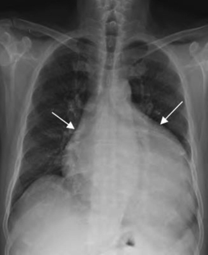
Pericardial effusion (I). The patient, a 55-year-old male. The patient had shortness of breath and fatigue for 3 years, aggravated for half a month. Chest X-ray showed that the cardiac shadow increasing to both sides, forming a “flask” shape (arrow)
-
(2)
CT: Pericardial fluid area with a width >4 mm is considered abnormal. Generally, pericardial effusion is divided into three degrees: Degree I refers to a small amount of effusion, and the amount of effusion is less than 100 mL. Most of the effusion is located outside the posterior wall of left ventricle or outside the lateral wall of right atrium and may be located outside the inferior wall of left ventricle in some patients, especially outside the posterior wall of left ventricle. Degree II refers to a moderate amount of effusion, and the amount of effusion is 100–500 mL. In such case, besides the above sites, effusion also occurs outside the anterior wall of the right ventricle and the apex of the left ventricle. Degree III refers to a large amount of effusion, and the amount of effusion is >500 mL. The effusion involves all parts of pericardial cavity, and the thickness of effusion further increases (Fig. 38.2a, b).
The CT value of effusion with different properties is different. The CT value of transudate is 0–20 HU. The CT value of protein/hemorrhagic effusion is >20 HU. The CT value of effusion in chylous pericarditis is <0 HU [4].
Fig. 38.2 
Pericardial effusion (II). The patient, a 34-year-old male, had chest tightness and shortness of breath for 4 days. (a, b) CT plain scan showed liquid density shadow (arrow) in pericardium. Liquid density shadow (tailless arrow) can also be found on the dorsal side of bilateral thoracic cavity. (c, d) MRI showed pericardial effusion with hypointensities on T1WI and hyperintensities on T2WI (arrow)
-
(3)
MRI: The distribution and morphology of pericardial effusion are the same as those of CT. MRI can show the fluid composition in pericardium, which is helpful for determining the properties of pericardial effusion. On T1WI of SE sequence, serous effusion mostly shows hypointensities, exudative effusion mostly shows hyperintensities, and hemorrhagic effusion shows middle-range intensities or hyperintensities. On T2WI, the effusion mostly shows hyperintensities. Delayed enhancement scan can show pericardial enhancement, which is sensitive to the diagnosis of pericarditis. Besides, other accompanying pathological changes can be shown (Fig. 38.2c, d).
-
(1)
-
2.
Constrictive Pericarditis
-
(1)
X-ray: Constrictive pericarditis is characterized by pericardial calcification, which is manifested as hyperintensities involving the whole cardiac border or wrapping most of the heart. The lesion can also involve local areas, manifested as small patchy opacities, stripe-like opacities, or linear opacities. The predilection sites of calcification are the anterior edge and septum of right ventricle, and a few are located in atrioventricular sulcus (Fig. 38.3).
Fig. 38.3 
Constrictive pericarditis (I). The patient, a 59-year-old female. The patient had intermittent cough for 2 years and shortness of breath for half a month. X-ray chest radiographs showed irregular patchy calcification in the upper part of cardiac shadow (arrow)
-
(2)
CT: Pericardial thickening is manifested as pericardial thickness >4 mm, some cases may be accompanied by a small amount of effusion, and pericardial thickening includes extensive thickening and local thickening. Local thickening is more common in the anterior wall of the heart, atrioventricular sulcus, and the opening of large blood vessels. Constrictive pericarditis is sometimes manifested as different degrees of calcification. Calcification can occur in both visceral and parietal pericardium. Calcification of visceral pericardium is mostly located in atrioventricular sulcus, interventricular sulcus, and cross sulcus, while calcification of parietal pericardium is mostly located in ventral side and septum of right ventricle, which cannot be distinguished on CT. In the case of less effusion in the pericardial cavity, thickened or calcified visceral and parietal pericardium can be distinguished (Fig. 38.4).
The compressed right ventricle become stiff and deformed, and in severe cases, the ventricular cavity is deformed into tubular stenosis. Ventricular constriction hinders cardiac output and causes atrial enlargement, which is more common in right atrium. The normal ventricular septum shows a straight or shallow arcuate structure, while the ventricular septum is stiff and twisted into angles in constrictive pericarditis. Thickening and calcification of pericardium in atrioventricular sulcus can cause cardiac output disorder, resulting in significant atrial enlargement. Limitation of cardiac filling can cause widening of superior and inferior vena cava.
Fig. 38.4 
Constrictive pericarditis (II). The patient, a 79-year-old male, had fever with fatigue for 4 days. CT plain scan (a), CT-enhanced scan (c) in the arterial phase (b) and the venous phase (c) showed pericardium thickening with extensive calcification (arrow) in right ventricular anterior wall and left ventricular anterior wall and cardiac apex, compressed bilateral ventricular shrank, especially in right ventricle, bilateral atrial enlargement, and bilateral pleural effusion
-
(3)
MRI: For the diagnosis of constrictive pericarditis, MRI is inferior to CT because it is insensitive to calcification. Calcification is manifested as linear or patchy hypointensities or even no signal. MRI is superior to CT in differentiating small amount of pericardial effusion from pericardial thickening. MRI has high value in evaluating atrioventricular morphology, and systolic and diastolic function of the heart (Fig. 38.5). MRI can also show the hemodynamic characteristics of constrictive pericarditis. Constrictive pericarditis can be manifested as abnormal movement of ventricular septum in early diastole, that is, moving to the left ventricle first and then moving away from the left ventricle, which is called ventricular septal jitter. This sign is more significant during deep inspiration [5]. Gadolinium contrast-enhanced scan show constrictive pericarditis manifested as delayed enhancement of thickened pericardium, indicating fibroblast proliferation and neovascularization.
Fig. 38.5 
Constrictive pericarditis (III). The patient, a 14-year-old male, suffered from recurrent syncope for half a year. (a, b) MRI T1WI black-blood sequence showed hypointensities of pericardial effusion (arrow) and widened inferior vena cava (arrow) and (c, d) end-systolic and end-diastolic images. MR cardiac cine showed uneven thickening of the pericardium and localized fluid signal in the pericardial cavity. Bilateral atria were enlarged, and each segment of the left ventricle was thinner (ventricular septum 7–8 mm, left ventricular lateral wall 5–6 mm). The systolic and diastolic motions of left ventricle were limited, the motion of ventricular septum was uncoordinated, and ventricular septal jitter was found in diastolic phase
-
(1)


4 Diagnostic Key Points
Imaging examination has some limitations in determining the etiology and properties of acute pericarditis and pericardial effusion, which should be combined with clinical data and laboratory examination results.
Among the imaging manifestations of constrictive pericarditis, pericardial thickening, ventricular septum distortion, and inferior vena cava dilatation are the most characteristic. If three signs occur at the same time, or pericardial thickening is accompanied by inferior vena cava dilatation or ventricular septum distortion, the diagnosis of constrictive pericarditis can be confirmed.
5 Differential Diagnosis
Constrictive pericarditis should be distinguished from chronic pericardial effusion, adhesive pericarditis, and restrictive cardiomyopathy.
-
1.
Chronic pericardial effusion: Constrictive pericarditis without calcification is difficult to be distinguished from chronic pericardial effusion on X-ray chest radiograph and CT. However, the effusion is manifested as long T1 and long T2 signal on MRI, while the thickened pericardium is manifested as long T1 and short T2 signal on MRI.
-
2.
Adhesive pericarditis: This disease may be manifested as pericardial thickening and pericardial calcification, but without hemodynamic change of constrictive pericarditis, not accompanied by dilation of inferior vena cava.
-
3.
Restrictive cardiomyopathy: This disease has no pericardial thickening and calcification. In MRI cine sequences, the decrease of myocardial compliance can be manifested as the shortening of the left-right movement distance of ventricular septum. Contrast-enhanced MR scan can show abnormal myocardial enhancement.
6 Research Status and Progress
-
1.
Characteristic tracing technique of cardiac magnetic resonance imaging: It can be used to distinguish constrictive pericarditis from restrictive cardiomyopathy. The parameter of global longitudinal strain (GLS) can be measured by MRI. The parameter of GLS in constrictive pericarditis is significantly higher than those in restrictive cardiomyopathy (P < 0.001). Combined with conventional cardiac function parameters, this parameter is helpful to improve the differential diagnosis efficiency of cardiac MRI in the above diseases [6].
-
2.
PET/CT: 18F-FDG can be used as an inflammatory imaging agent with high sensitivity. In patients with acute pericarditis accompanied by pericardial effusion, increased uptake of 18F-FDG in pericardium is related to increased risk of recurrence [7]. In addition, pericardial uptake of 18F-FDG at baseline can predict the response of patients with constrictive pericarditis to steroid therapy [8].
References
Ge J, Xu Y, Wang C. Internal medicine. 9th ed. Beijing: People’s Medical Publishing House; 2018.
Bu H, Li Y. Pathology. 9th ed. Beijing: People’s Medical Publishing House; 2018.
Hoit BD. Pathophysiology of the pericardium. Prog Cardiovasc Dis. 2017;59(4):341–8.
Adler Y, Charron P, Imazio M, et al. 2015 ESC guidelines for the diagnosis and management of pericardial diseases: the Task Force for the Diagnosis and Management of Pericardial Diseases of the European Society of Cardiology (ESC) endorsed by: the European Association for Cardio-Thoracic Surgery (EACTS). Eur Heart J. 2015;36(42):2921–64.
Groves R, Chan D, Zagurovskaya M, et al. MR imaging evaluation of pericardial constriction. Magn Reson Imaging Clin N Am. 2015;23(1):81–7.
Amaki M, Savino J, Ain DL, et al. Diagnostic concordance of echocardiography and cardiac magnetic resonance-based tissue tracking for differentiating constrictive pericarditis from restrictive cardiomyopathy. Circ Cardiovasc Imaging. 2014;7(5):819–27.
Gerardin C, Mageau A, Benali K, et al. Increased FDG-PET/CT pericardial uptake identifies acute pericarditis patients at high risk for relapse. Int J Cardiol. 2018;271:192–4.
Chang SA, Choi JY, Kim EK, et al. 18F-Fluorodeoxyglucose PET/CT predicts response to steroid therapy in constrictive pericarditis. J Am Coll Cardiol. 2017;69(6):750–2.
Author information
Authors and Affiliations
Editor information
Editors and Affiliations
Rights and permissions
Copyright information
© 2023 Science Press
About this chapter

Cite this chapter
Hou, Y., Sui, S. (2023). Pericarditis. In: Li, H., Liu, J., Li, L. (eds) Radiology of Infectious and Inflammatory Diseases - Volume 3. Springer, Singapore. https://doi.org/10.1007/978-981-99-4614-3_38
Download citation
DOI: https://doi.org/10.1007/978-981-99-4614-3_38
Published:
Publisher Name: Springer, Singapore
Print ISBN: 978-981-99-4613-6
Online ISBN: 978-981-99-4614-3
eBook Packages: MedicineMedicine (R0)