
Abstract
The evolution of lacrimal surgeries and the understanding of lacrimal disorders have been an amazing journey! From time immemorial, lacrimal disorders have continued to intrigue mankind and pose significant challenges. The chapter summarizes the historical perspectives of major landmark developments in the world of lacrimal sciences right from the ancient times to the very recent past.
Access provided by CONRICYT-eBooks. Download chapter PDF
Introduction
The evolution of lacrimal surgeries and the understanding of lacrimal disorders have been an amazing journey! From times immemorial, lacrimal disorders have continued to intrigue mankind and pose significant challenges. Tough problems have fortunately met tougher wise men at the right time, and this science continued to evolve at a rapid pace. The spectrum of events in this journey can be captured in two wise quotations, one of Sir Isaac Newton (1642–1727) who lauded the culture of each contributor building a higher platform for the subsequent one to fly higher and of Sir Rudolph Virchow (1821–1905) who not long ago expressed his pain at the diminishing number of students who learn from history in subsequent generations:
If I have seen further, it is by standing on the shoulders of ye giants. (Sir Isaac Newton)
It is one of the worst aspects of our present development that historical knowledge diminishes with each generation of students. (Sir Rudolph Virchow)
Ancient Dacryology
The earliest documented reference to any ophthalmic plastics surgery is that of an incision to an infected lacrimal sac in the Code of Hammurabi (2250 BC) (Fig. 1.1). The ancient Egyptians document lacrimal sac infections in the Ebers Papyrus (1500 BC) (Fig. 1.2) and recommended a mixture of antimony, wood powder, myrrh, and dried honey rubbed into the eyes for 4 days! [1]. Hippocrates (460 BC–377 BC) (Fig. 1.3) believed that watery eyes set in an old age and if it turns thicker (discharge), a dried juice of white grapes mixed with copper sulfate is recommended [1].

Code of Hammurabi (2250 BC)

Ebers Papyrus (1500 BC)

Hippocrates (460–377 BC)
The Greeks made significant contributions in the early days. Most diseases of the lacrimal system were referred to as “fistules.” Celsus (25 BC–50 AD) in his landmark text “Da Medicina” advocated cautery and burning of the lacrimal abscess to cure “fistules” [2]. Claude Galen (129–200 AD), a century after Celsus, advocated the use of hot iron to achieve charring of the “fistules” and hence a cure! He believed that puncta evacuate and secrete into the eye! [2]. However, the most remarkable contribution of Galen (Fig. 1.4) has been his description of the causes of epiphora. He documented as follows [2]:
A canal goes from the eyes to the palate and empties there the secretion formed in the eye. Watering may have three causes; either this canal is blocked, or the secretion is excessive or a scar at the nasal canthus. The latter most is incurable.

Claude Galen (129–200 AD)
Medieval Times and Renaissance
The medieval times as well as the renaissance were unfortunately a bit laid-back as far as the scientific progress related to lacrimal system was concerned. The Arabians chipped in with Rhazes (854–925 AD) evaluating the lacrimal passage further down into the nose and later Avicenna (980–1037 AD) (Fig. 1.5) advocating application of mongo bean pastes for lacrimal fistulas. Andreas Vesalius (1514–1564) described lacrimal drainage anatomy with reasonable details [3, 4], and his pupil, Gabriele Falloppio (1523–1562), documented regurgitation of purulent material from the punctum on compression of the lacrimal sac [4, 5]. Leonardo da Vinci’s (1453–1519) and later William Harvey’s (1578–1657) embryologic works were notable during these times.

Avicenna (980–1037 AD)
Modern Dacryology
Major Contributors of Early Days
-
(a)
George Ernst Stahl (1660–1734): Stahl was a German physician (Fig. 1.6), who established nasolacrimal duct obstruction as a cause of dacryocystitis. He also suggested probing using a violin thread!
Fig. 1.6 
George Ernst Stahl (1660–1734)
-
(b)
Dominique Anel (1679–1730): Anel was a French surgeon and among the earliest to device a probe and a syringe (Anel’s Probes and Syringes) and became famous in 1713 after he treated the Duchess of Savoy for lacrimal fistulae in a period of 10 days! [6, 7].
-
(c)
Giovanni Battista Morgagni (1682–1771): Morgagni was an Italian anatomist (Fig. 1.7) and among the earliest to give a description of lacrimal drainage system. He concluded that there were no valves in this system and the flow was bidirectional! He published his account in the treatise “Adversaria Anatomica Omnia” in 1718.
Fig. 1.7 
Giovanni Battista Morgagni (1682–1771)
-
(d)
Lorenz Heister (1683–1758): Heister was the first to classify lacrimal disorders in 1753. He divided the disorders into four chapters, namely, a tearing eye, a tumefaction of the lacrimal system, an ulcer of the lacrimal system, and lacrimal fistulae. The treatise published in 1753 was named “Chirurgische Wahrnehmungen” [8].
-
(e)
John Louis Petit (1664–1741): Petit explained the flow of tears in the lacrimal system and devised a grooved probe for exploration [9].
-
(f)
Sir William Bowman (1816–1892): Sir Bowman was an English anatomist and surgeon (Fig. 1.8), and his contributions to lacrimal surgery are many. He described Bowman’s probes in 1851, punctoplasty in 1853, and canaliculotomy in 1857 [10].
Fig. 1.8 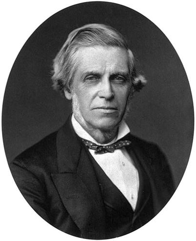
Sir William Bowman (1816–1892)
-
(g)
Joseph Hasner (1819–1892): Hasner was an Austrian ophthalmologist who contributed immensely toward lacrimal physiology and mechanics of the flow of tears and devised surgical procedures for the treatment of lacrimal fistules. The distal most valve of the lacrimal drainage pathway is named after him.


Influential Treatise that Paved the Way Early on
-
1.
Descriptio Anatomica Oculi Humani: This treatise was published in Gottingen in 1755 by the famous German anatomist Johann Gottfried Zinn (1727–1759) (Fig. 1.9). He was among the earliest to describe complete anatomical course of the lacrimal drainage pathway.
Fig. 1.9 
Johann Gottfried Zinn (1727–1759)
-
2.
Observations on That Disorder of Corner of the Eye Commonly Called Fistula Lacrimalis: Published by Percival Pott (1714–1788), an English surgeon (Fig. 1.10) and one of the founders of orthopedics, this work of his was one of the earliest texts on lacrimal disorders.
Fig. 1.10 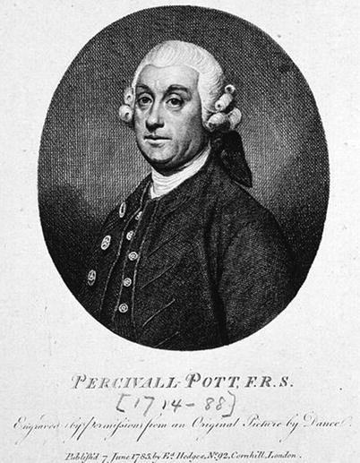
Percival Pott (1714–1788)
-
3.
Chirurgische Wahrnehmungen: This treatise was published in 1753 by Lorenz Heister (1683–1758) and was the first to classify lacrimal disorders into four separate subdivisions. Some of the surgical instruments and their design he published are legendary (Fig. 1.11).
Fig. 1.11 
Surgical instruments of Lorenz Heister (1683–1758)
-
4.
Organic Lacrimalis Pretiumque Externum Oculi Humanos Description Anatomica: This treatise was published in 1797 in Leipzig by Johann Christian Rosenmuller (1771–1820) (Fig. 1.12). In comparison to Zinn’s work, this was very specific treatise only on lacrimal system with advance anatomical details.
Fig. 1.12 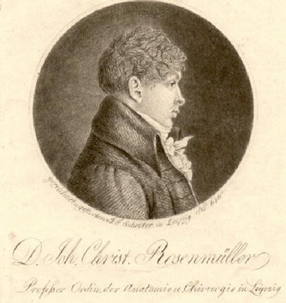
Johann Christian Rosenmuller (1771–1820)
-
5.
Comprehensive Text on Lacrimal Disorders: Johann Adam Schmidt (1759–1809) was the first to bring out an influential treatise on lacrimal system in German and was published on copper plates!


History of Dacryocystectomy (DCT)
The earliest ways of dealing with lacrimal sac infections have been to burn or char it down with the help of molten lead or iron [1, 2], which is practically destroying the lacrimal sac. The first refined way of surgical dacryocystectomy can be traced back to John Thomas Woolhouse in 1724 [11]. Johannes Platner (1694–1747) practiced Woolhouse’s technique and described DCT with trephination of the lacrimal sac and cautery [11]. Most of these surgeries were incomplete and obviously unintentional because of incomplete knowledge of anatomical details. The modern DCT was described by Rudolph Berlin (1833–1897) (Fig. 1.13) in 1868, and he documented [11, 12] as follows:
Dacryocystectomy is the principal operation against incurable epiphora. It is the main protection against corneal abscess and purulent infections against cataract.

Rudolph Berlin (1833–1897)
Although not much progress has been made in surgical advancement of dacryocystectomy, its indications have become limited but much more refined today [13].
History of Dacryocystorhinostomy (DCR)
Dacryocystorhinostomy is among those surgeries whose fascinating history is paralleled by only a few in medicine. It has tremendously evolved in techniques, instrumentation, and, above all, the approaches! The earliest attempts to create communications can be traced back to John Thomas Woolhouse (1650–1734), who described extirpation of sac, perforation of lacrimal bone, and insertion of drains made of gold, silver, or lead. Antonio Scarpa (1752–1832), an Italian anatomist (Fig. 1.14), designed a lead nail, slit the lacrimal sac, and introduced it in a 50-year-old woman, who died in 4 days following surgery [14], possibly because of tetanus or septicemia! Around the same time, Dupuytren (1777–1835) designed a gold tubule for similar purpose, but the patient had a palatal perforation and suffocation [14]. Laguier attempted to drain the sac into the maxillary antrum in 1830 [15, 16].

Antonio Scarpa (1752–1832)
Endonasal DCR was first conceptualized by Caldwell in 1893 [16, 17]. John West in 1914 modified this technique by creating a bony window within the lacrimal and maxillary bone to clear the area of lacrimal sac and nasolacrimal duct into the middle meatus [16, 18]. Rice first introduced the concept of endoscopic endonasal DCR in cadavers in 1988 and showed its feasibility as a good alternative to an external DCR [19]. Mc Donogh and Meiring in 1989 introduced endoscopic endonasal DCR in patients [20], and since then there was no looking back for the endoscopic approach! The techniques have refined, and newer adjunctive technologies have evolved since 1989. Powered and mechanical endoscopic DCR was described by Peter-John Wormald in 2002 [21].
External DCR was described by Italian rhinologist Addeo Toti in 1904 with a 35 mm incision where both the medial wall of the sac and nasal mucosa were excised [22]. Significant change to this procedure happened soon in 1920 when Dupuy-Dutemps and Bourguet introduced the creation of lacrimal sac and nasal mucosal flaps with suturing to create an epithelium-lined fistula [23]. Very few modifications have happened since then, for example, by Viers in 1969 [24] and Iliff in 1971 [25].
Other approaches for a DCR mostly evolved much later [26]. Bruce Massaro in 1990 introduced endoscopic laser-assisted DCR using argon-blue laser in cadavers [27]. Shortly thereafter in 1992, Levin and Stormogipson introduced endocanalicular laser-assisted DCR in cadavers [28], and later Silkiss introduced it in patients in 1992 [29]. Subsequently, various different types of lasers have been used for the bone removal [30, 31]. Endoscopic radiofrequency-assisted DCR as a different technique was introduced by Reynaldo Javate in 1995 [32]. Ultrasonic DCR was first performed by Krasnov in 1971 [33] and reintroduced in 2005 by Sivak-Callcott [34] and has subsequently generated some interest. Nine millimeter balloon DCR was pioneered by David Silbert [35].
Conjunctivodacryocystorhinostomy (CDCR) was introduced by Von Hoffman in 1904 by opening the lacrimal sac and suturing it to conjunctiva without a stent [36]. This procedure was subsequently revisited by Goar [37], Stallard [38], and Bangerter [39]. Lester Jones in 1962 described slitting of the canaliculus to overcome the frustrating problem of proximal lacrimal drainage obstruction [40]. Later on, in collaboration with Gunther Weiss Scientific Glass Blowing Company from Portland, Oregon, he subsequently developed the famous Pyrex Jones Tubes and published his techniques in 1965 with the use of this new stent [41,42,43]. Subsequently, various stents and their modifications as well as buccal mucosa and vein grafts were used [44].
History of Other Lacrimal Surgeries
Interventional radiological procedures for nasolacrimal duct dilatation were described by Hanafee and Dayton in 1978 using the sialography canulas and fluoroscopic guidance [45]. Dacryoendoscopy was introduced by Junemann in 1975 [46]. Becker and Berry introduced balloon dacryoplasty in 1989 [47], and in the same year, Busse conceptualized microdrill dacryoplasty [48]. Canalicular obstructions beyond proximal canaliculi are usually managed by trephination, and modern canalicular trephines were introduced by Hampson Sisler in 1990 [49].
Ophthalmology with Otolaryngology: Historical Perspectives
Ophthalmology and otolaryngology are used to be practiced together by the “EENT” specialists for most of the nineteenth and twentieth centuries [50,51,52]. Major institutes where this was practiced include the London Infirmary for Curing Diseases of the Eye and Ear (inaugurated in 1805) which later became the Moorfields Eye Hospital and the Massachusetts Eye and Ear Infirmary (inaugurated in 1824 as the Boston Eye Infirmary) [50]. Two major global societies were formed, namely, “American Ophthalmological and Otological Society (established in 1864)” and “Western Ophthalmological and Otological Society (established in 1896)” [50]. These EENT specialists used to meet and publish together in common journals. With exponential increase in knowledge and evolution of subspecialties within each branch, there was an increase felt need for separation and this led to the formation of separate American societies in 1979 [51]. Nonetheless, it is important to realize that there are many overlapping areas and lacrimal drainage system happens to be on the forefront. A surgeon dealing with lacrimal disorders, whether ophthalmologists or otolaryngologists, needs to understand that this system traverses both the areas and it is essential to gain sound understanding of the anatomy, physiology, and pathologies of both the areas. A healthy collaboration, knowledge transfer, learning from each other’s experiences, and teamwork can enhance patient care and help achieve the goal of optimal management of lacrimal disorders.
Conclusion
It is very important to know the depths of history, at least in the area of one’s expertise, and this helps greatly in innovating further and advancing medicine. The take-home message can be summarized in the words of Dr. Paul Lichter, the president of the American Academy of Ophthalmology, in its centennial year, who said:
Ophthalmic History must be taught in our residency programs and a must read for all Ophthalmologists as too few of our students revere history and the lessons it can provide.
Note: The photographs used are courtesy of Wikipedia.
References
Hirschberg J. The renaissance of ophthalmology in the 18th century. In:The history of ophthalmology, vol. 1. Amsterdam: Wayenborg Publications; 1984. p. 11.
Hirschberg J. The renaissance of ophthalmology in the 18th century. In:The history of ophthalmology, vol. 3. Amsterdam: Wayenborg Publications; 1984. p. 250–5.
Vesalius AWRJC. On the fabrik of the human body: a translation of De Humani Corporis Fabrika Libri Septem. Novato: Norman Publications; 2007.
Harish V, Benger RS. Origins of lacrimal surgery, and evolution of dacryocystorhinostomy to the present. Clin Experiment Ophthalmol. 2014;52:284–7.
Falloppio G. Observationes Anatomicae. Venice, 1561.
Anel D. New method to cure a lacrimal fistule and a report on different arguments, for and against, and in favour of the newly invented method. Torino, 1713.
Hirschberg J. The renaissance of ophthalmology in the 18th century. In:The history of ophthalmology, vol. 3. Amsterdam: Wayenborg Publications; 1984. p. 246–9.
Hirschberg J. The renaissance of ophthalmology in the 18th century. In:The history of ophthalmology, vol. 3. Amsterdam: Wayenborg Publications; 1984. p. 259–60.
Petit JL. Memoire sur la fistule lacrimale. Mem Acad R Sci. 1734;135
Bowman W. Ann d’Ocul. XXXIX. p. 79–83.
Hirschberg J. The renaissance of ophthalmology in the 18th century. In:The history of ophthalmology, vol. 3. Amsterdam: Wayenborg Publications; 1984. p. 264.
Berlin R. Report of the Heidelberg Ophthalmologische Gesell schaft. 1868. p. 355.
Ali MJ. Dacryocystectomy: goals, indications, techniques and complications. Ophthal Plast Reconstr Surg. 2014;30:512–6.
Hirschberg J. The renaissance of ophthalmology in the 18th century. In:The history of ophthalmology, vol. 3. Amsterdam: Wayenborg Publications; 1984. p. 262.
Laguier. Arch Gen de Med. 1830. p. Xxiii.
Chandler PA. Dacryocystorhinostomy. Trans Am Ophthalmol Soc. 1936;34:240–63.
Caldwell GW. Two new operations for the obstruction of nasolacrimal duct with preservation of the canaliculi. Am J Ophthalmol. 1893;10:189.
Watkins LM, Janfaza P, Rubin P. Evolution of endonasal dacryocystorhinostomy. Surv Ophthalmol. 2003;48:73–84.
Rice DH. Endoscopic intranasal dacryocystorhinostomy. A cadaver study. Am J Rhinol. 1988;2:127.
Mc Donogh M, Meiring JH. Endoscopic transnasal dacryocystorhinostomy. J Laryngol Otol. 1989;103:585–7.
Wormald PJ. Powered endonasal DCR. Laryngoscope. 2002;112:69–71.
Toti A. La dacriocistorhinostomia. Ann d’Ocul. 1910;CXIiii:417.
Dupuy-Dutemps MM, Bourguet ET. Note preliminaire sur un proded de dacryocystorhinostomie. Ann d’Ocul. 1920;157:1445–7.
Viers ER. The use of cautery in external dacryocystorhinostomy. Arch Ophthalmol. 1969;82:489–90.
Iliff CE. A simplified dacryocystorhinostomy. Arch Ophthalmol. 1971;85:586–91.
Ali MJ. Lacrimal surgery: glorious past, exciting present era and the audacity of hope for a brilliant future. Saudi J Ophthalmol. 2014;28:1–2.
Massaro BM, Gonnering RS, Harris GJ. Endonasal dacryocystorhinostomy. A new approach to nasolacrimal duct obstruction. Arch Ophthalmol. 1990;108:1172–6.
Levin PS, Stormogipson DJ. Endocanalicular laser-assisted dacryocystorhinostomy. An anatomic study. Arch Ophthalmol. 1992;110:1488–90.
Silkiss RZ, Axelrod RN, Iwach AG, et al. Transcanalicular THC:YAG dacryocystorhinostomy. Ophthalmic Surg. 1992;23:351–3.
Metson R, Woog JJ, Puliafito CA. Endoscopic laser dacryocystorhinostomy. Laryngoscope. 1994;104:269–74.
Pearlman SJ, Michalos P, Leib ML, et al. Translacrimal transnasal laser assisted dacryocystorhinostomy. Laryngoscope. 1997;107:1362–5.
Javate RM, Campomanes BS, Co ND, et al. The endoscope and the radiofrequency unit in DCR surgery. Ophthal Plast Reconstr Surg. 1995;11:54–8.
Krasnov MM. Ultrasonic dacryocystorhinostomy. Am J Ophthalmol. 1971;72:200–1.
Sivak-Callcott JA, Linberg JV, et al. Ultrasonic bone removal with Sonopet OMNI: a new instrument for orbital and lacrimal surgery. Arch Ophthalmol. 2005;123:1595–7.
Silbert DI. Outcomes of 9mm balloon-assisted endoscopic DCR: retrospective review of 97 cases. Orbit. 2010;29:131–4.
Athanasiov PA, Madge S, Kakizaki H, et al. A review of bypass tubes for proximal drainage obstruction. Surv Ophthalmol. 2011;56:252–66.
Goar EL. Congenital absence of the lacrimal puncta and canaliculi. Trans Am Ophthalmol Soc. 1931;29:91–9.
Stallard HB. An operation for epiphora. Lancet. 1940;2:743–4.
Bangerter A. Behandlung bei Tränenröhrchenstenose. Ophthalmologica. 1947;114:195–202.
Jones LT. The cure of epiphora due to canalicular disorders, trauma, and surgical failures on the lacrimal passage. Trans Am Acad Ophthalmol Otolaryngol. 1962;66:506–24.
Jones LT. Conjunctivodacryocystorhinostomy. Am J Ophthalmol. 1965;59:773–83.
Jones LT, Wobig JL. Surgery of the eyelid and lacrimal system. Birmingham, UK: Aesculapius; 1976.
Steele EA. Conjunctivodacryocystorhinostomy with Jones tube: a history and update. Curr Opin Ophthalmol. 2016;27:439–42.
Paufique L, Durand L. Surgical treatment of eye watering: repair of the canaliculus with a venous graft. Ann Ocul. 1969;202:337–44.
Hanafee WN, Dayton GO. Dilatation of the nasolacrimal duct under radiographic control. Radiology. 1978;127:813–5.
Junemann G, Shulte D. Moderne Probleme der Erkrankungen der Lider und des Tränenapparates. I ed. Stuttgart: Enke; 1995. p. 243–64.
Becker BB, Berry FD. Balloon catheter dilatation in lacrimal surgery. Ophthalmic Surg. 1989;20:193–8.
Busse H. Microsurgery in lacrimal disorders. Dev Ophthalmol. 1989;18:50–2.
Sisler HA, Allarakhia L. A new ophthalmic microtrephine. Ophthalmic Surg. 1990;21:656–7.
Thawley SE. The otolaryngologist-ophthalmologist relationship: an historical perspective. Otolaryngol Clin N Am. 2006;39:845–53.
Truhlsen SM. Whatever happened to the EENT specialists? Surv Ophthalmol. 2013;58:92–4.
Patel BC, Anderson RL. History of oculoplastic surgery (1896–1996). Ophthalmology. 1996;103:S74–95.
Author information
Authors and Affiliations
Corresponding author
Editor information
Editors and Affiliations
Rights and permissions
Copyright information
© 2018 Springer Nature Singapore Pte Ltd.
About this chapter
Cite this chapter
Ali, M.J. (2018). Lacrimal Disorders and Surgery: Historical Perspectives. In: Ali, M. (eds) Principles and Practice of Lacrimal Surgery. Springer, Singapore. https://doi.org/10.1007/978-981-10-5442-6_1
Download citation
DOI: https://doi.org/10.1007/978-981-10-5442-6_1
Published:
Publisher Name: Springer, Singapore
Print ISBN: 978-981-10-5441-9
Online ISBN: 978-981-10-5442-6
eBook Packages: MedicineMedicine (R0)