
Abstract
The maxilla is the cornerstone of the appearance and function of the midface and has important functions such as supporting the skull base, eyeballs, and midface, bearing the chewing force, and separating the oral and nasal cavities. The main causes of maxillary defects include acquired defects caused by tumor-reductive surgery and severe trauma and the congenital developmental malformations. The effect of the maxillary defect on the appearance and function of the patient counts for much, the maxillary defect is often accompanied by the destruction or deletion of surrounding vital structures, and therefore it will cause facial deformities and severe loss of oral functions, bring catastrophic blow to the patient’s physiology and psychology, and seriously affect the quality of life of patients.
Access provided by CONRICYT-eBooks. Download chapter PDF
1 Overview
The maxilla is the cornerstone of the appearance and function of the midface and has important functions such as supporting the skull base, eyeballs, and midface, bearing the chewing force, and separating the oral and nasal cavities. The main causes of maxillary defects include acquired defects caused by tumor-reductive surgery and severe trauma and the congenital developmental malformations. The effect of the maxillary defect on the appearance and function of the patient counts for much, the maxillary defect is often accompanied by the destruction or deletion of surrounding vital structures, and therefore it will cause facial deformities and severe loss of oral functions, bring catastrophic blow to the patient’s physiology and psychology, and seriously affect the quality of life of patients.
Because the maxilla is connected to adjacent various bones in the middle and upper parts of the face, the maxillary defect is often accompanied by defects in the ethmoid bone, nasal bone, cheekbones, palate bone, orbital bone, and skull base bone. The resection range for the maxillectomy performed due to tumor often includes part of cheekbone, and sometimes it still needs to include the nasal bone, ethmoid bone, and orbital bone depending on the specific situation; the subtotal or total maxillary resection often leads to palatomaxillary defects; for example, when carrying out the cranio-maxillofacial combined resection, it is often needed to remove the skull base bone, including the anterior cranial fossa and middle cranial fossa, even occasionally also involving backward the posterior cranial fossa; the extended radical resection of the malignant advanced tumor in the middle part of the face will lead to defects which are usually perforating defects, while the defects caused by trauma or war injury are combined with varying degrees of soft and hard tissue defects according to different injury causes, state of injury, and wound tract, which increase the difficulty of maxillary defect reconstruction to some extent. Therefore, the repair and reconstruction of the maxillary defect, especially the functional reconstruction, have been a challenging task faced by surgeons in departments of maxillofacial and oral head and neck surgery, plastic surgery, and prosthodontics.
So far, domestic and foreign scholars still have controversies on the reconstruction of maxillary defect, and its treatment still remains in the level of maxillofacial prosthetic treatment and the repair with free composite tissue flap [1,2,3,4,5,6,7,8,9,10,11,12,13,14,15,16,17,18,19,20]. In recent years, along with the improved consensus of both doctors and patients on increasing the survival rate and survival quality, coupled with increasingly sophisticated microsurgical techniques, widely used medical biological materials, and the introduction of digital medicine, especially the development of rapid prototyping technology, the ideal or proper functional maxillary reconstruction proposed and implemented by domestic and foreign scholars is made possible. For this purpose, on the basis of obtaining a lot of successful experiences after years of unremitting efforts, they gradually promote the in-depth researches in this area. On that account, this chapter is intended to take the surgical reconstruction as the fundamental key, give consideration to both appearance and function combined with the authors’ experience and knowledge, emphatically introduce the reconstruction and repair of defects after maxillary tumor surgery, and discuss issues of common concern.
1.1 Maxillary Defect Classification
Since the sites and contents of maxillary defect caused by tumor resection or severe trauma are often not the same, the methods for repair of maxillary defect reconstruction selected by the surgeons as well as the appearance and functional effect after reconstruction are also somewhat different. Meanwhile, there are a wide variety of methods for maxillary reconstruction, and currently, there is no uniform standard to follow; thus, it is necessary to classify the maxillary defects, in order to seek methods which are not only beneficial to clinical diagnosis, treatment plan development, and postoperative functional evaluation but also are helpful in carrying out discussions on the reconstructive method for maxillary defects on the same platform and setting up selection criteria for a variety of methods for maxillary reconstruction while can also effectively evaluate and compare the efficacies. According to domestic and foreign reports, the methods for maxillary defect classification mainly include HS classification, Fan Sen classification, Zhao Yimin classification, Brown classification, Cordeiro and Santamaria classification, Okay classification, Triana classification, and Yamamoto classification. In these classifications, the first three classifications are based on the perspective of prosthodontic rehabilitation and thus have relatively limited application; the later several classifications are proposed by surgeons from the perspective of surgical reconstruction. Given the limited space available, this chapter describes only the more popular and widely used Brown classification (2000) and the modified classification by Brown (2010).
Brown classification (2000) is a classification system proposed by British scholar Brown et al. [21] according to maxillary defects, respectively, in the vertical and horizontal planes (Fig. 8.1).
-
Vertical defects (the English word of 垂直缺陷)
-
Horizontal defects (the English word of 水平缺陷)

Brown classification of maxillary defects (2000). (a, e) Class 1, (b) Class 2, (c) Class 3, (d) Class 4, (f) Subclass a, (g) Subclass b, (h) Subclass c
1.1.1 Classification of Vertical Defects
The vertical defects are divided into four classes according to the status of unilateral maxillary defect, of which they are divided into Class 1 and Class 2 according to whether there exists an area of oronasal fistula and they are divided into Class 3 and Class 4 according to the degree of orbital invasion. Specific classification is as follows:
-
1.
Class 1 defects – Maxillectomy without involvement of the sinus cavity.
-
2.
Class 2 defects – Low maxillectomy, including resection of walls of maxillary sinus and alveolar processes with preservation of the orbital floor and part of orbital tissue.
-
3.
Class 3 defects – High maxillectomy, including orbital floor or part of orbital tissues; the skull base may be involved, but the eye ball is preserved.
-
4.
Class 4 defects – Radical maxillectomy includes orbital exenteration, and the anterior skull base resection may or may not be included.
1.1.2 Classification of Horizontal Defects
The horizontal defects are divided into three subclasses according to the extent of resection of alveolar bone and palate:
-
1.
Subclass a – Unilateral resection of alveolar bone and palate without exceeding the midline and involving the nasal septum
-
2.
Subclass b – Resection of alveolar bone and palate exceeding the midline and involving the nasal septum
-
3.
Subclass c – Resection of total alveolar bone and palate
The vertical defects in the maxilla will make a huge impact on the appearance of the midface, while the horizontal defects will cause more functional disorders in chewing, swallowing, and pronunciation.
Brown classification covers the two aspects such as deformity and dysfunction (teeth occlusion, chewing, and pronunciation) in the midface (nose and paranasal sinuses, eyeballs) caused by the maxillary defects.
After Brown classification had been applied in clinic for years, Brown et al. (2010) also proposed a modified classification on this basis (Fig. 8.2). In addition to the original four classes, the vertical defects additionally include Class 5 defects that there exist the orbital and maxillary defects, but the alveolar process and palate are intact, and Class 6 defects that there exist nasal and surrounding maxillary defects; the horizontal defects additionally include a subclass of transverse defects that are less than or reach half of the hard palate. Since this modified classification is latestly proposed and has not yet been acknowledged by scholars worldwide, this chapter still uses Brown classification (2000).
-
Vertical defects
-
Horizontal defects

Brown modified classification of maxillary defect (2010). (a) Class 1, (b) Class 2, (c) Class 3, (d) Class 4, (e) Class 5, (f) Class 6, (g) Subclass a, (h) Subclass b, (i) Subclass c, (j) Subclass d
1.2 The Principles for Repair and Reconstruction of the Maxilla
1.2.1 The Open Repair Should be Applied in Patients with Primary Maxillary Sinus Cancer and Highly Malignant Tumor
Because of the importance and complexity of the invasion range and the adjacent area of primary maxillary sinus cancer, and its poor biological behavior and survival rate, there is a certain difficulty in controlling the radical resection and safety margin. In view of such principles for facilitating the timely detection and treatment of postoperative recurrence of maxillary malignant tumor, the authors suggest that, for some maxillary tumors with higher malignant degree such as primary maxillary sinus cancer and osteosarcoma and combined with sinus wall damage, at the same time of performing radical tumor resection, the open repair methods of using the man-made support such as titanium mesh to maintain the appearance of the midface and wearing a prosthesis can be applied firstly, and then the surgical reconstruction can be implemented at 2 years after surgery when there is no local recurrence and distant metastasis.
1.2.2 The Patients with Tumors Which Can Be Radically Resected Can Be Treated with Immediate Closed Repair and Reconstruction
After radical maxillary tumor resection, due to simultaneous defects in bone tissues such as the orbital bone, cheekbone, and nasal bone, the patient’s appearance can be affected in different degrees. With the development of CT, MRI, and endoscopic techniques, the concerns in the past that the immediate use of autologous tissue to repair and reconstruct the maxilla may affect the examination of tumor recurrence are gradually eliminated, and there is no data to support that the prognoses of patients with surgical reconstruction are poorer than those of patients without surgical reconstruction. Therefore, for patients with tumors of higher malignant degree that primarily occurred in the palate and gingiva, the lesions are relatively limited and do not invade the maxillary sinus; for patients with tumors which can be completely removed or some patients with tumors of low malignant degree, the lesions invade the maxilla but do not invade through the sinus wall; the authors advocate that the closed repair and functional reconstruction are performed at one stage, according to the defect statuses in maxilla and adjacent bones; the artificial prosthesis (biological material or titanium mesh) can be used as scaffolds to reconstruct the anatomical shape, and then the oral surface or nasal surface is covered with the free composite tissue flap to restore the alveolar crest and palate and reconstruct the nasal passage and separate the oral and nasal cavities. The implants can be transplanted immediately or delayedly to restore the patient’s chewing function finally.
1.3 The Objectives and Requirements for Repair and Reconstruction of Maxillary Defects
The repair and reconstruction of maxillary defects should also take into account the recovery of function and appearance, and the effective targeted measures should be taken in accordance with the cause, location, extent, and type of the defect. The ideal reconstruction method must meet the following objectives and requirements [22]:
-
1.
Fill the defects caused by tumor surgery or trauma.
-
2.
Separate the oral and nasal cavities.
-
3.
Restore the support structure of the maxilla.
-
4.
Recover the function of tissues and organs of midface such as chewing, pronunciation, and swallowing.
-
5.
Reconstruct the position of the eye or fill and beautify the orbit after eye enucleation.
-
6.
Maintain a specific nasal airway.
-
7.
Provide necessary bony support for midface tissues such as the upper lip, nose, and cheek, thus avoiding the lower eyelid ectropion.
-
8.
Repair and reconstruct the appearance of the midface.
However, to date, there is no any kind of reconstruction method which can reach the objectives of all these maxillary reconstruction. For this purpose, the scholars from various countries continue to explore the ideal method of reconstruction.
1.4 The Basis for Repair and Reconstruction of Maxillary Defects and Its Meaning
At present, the method and timing of repairing the defects after resection of maxillary tumor remain controversial. For a very long time in the past, the traditional prosthesis occupied a dominant position in the repair of maxillary defects, and a surgery on donor site can be omitted. The prosthesis can fill the dead space, separate the nasal and oral cavities, and restore some chewing function and can be removed and put on at will, which is very favorable for observing whether there is early tumor recurrence. But its disadvantages are also obvious. Due to the fact that its retention condition is poor and it is not closed with the surrounding tissue, its adhesive force and attachment force are reduced accordingly; it tends to produce leakage and tilting, thereby affecting the functions such as sucking, chewing, and speech; and it is also not conducive to cleaning the oral environment; the long-term compression of prosthesis can cause secondary trauma and form the traumatic ulcers, so the postoperative recovery of the patient is not satisfactory.
In recent years, the vascularized tissue is transplanted to repair the maxillary defect, and this has been accepted by a growing number of physicians and patients; these techniques also make up the flaws of repair of maxillary defects with prosthesis fundamentally [5, 7,8,9,10,11,12, 17, 18, 22]. Therefore, the immediate repair with free composite tissue flap combined with the endosseous implant appears; thereby, not only the oronasal fistulas in the patients can be immediately closed, but also the chewing function, phonetic function, and nasal ventilation function of the patients after surgery can be recovered to varying degrees because of the repair with bone graft and the implant of better quality. In the past, the main concerns against the immediate repair are the worries that due to the covering of the dead space after maxillectomy by tissue flap, if the tumor recurs in future, the recurrent tumor foci cannot be directly observed with the naked eye, which may delay the diagnosis and treatment of the patient. With the development and popularization of modern medical imaging such as nasal endoscopy, CT, and MRI, it is possible to find the recurrent tumor foci earlier and earlier, which is conducive to the monitoring of early disease recurrence. Meanwhile, there is no literature suggesting that the survival rate of patients with immediate reconstruction is lower than that of the patients without immediate reconstruction; on the contrary, the survival rate of patients with immediate reconstruction is greater than that of the patients without immediate reconstruction.
For the ideal timing for repair and reconstruction of maxillary defects, the author thinks it should be performed immediately as soon as possible after surgery, because the immediate repair after surgery is conducive to early functional recovery and prevention of scar contracture; otherwise, the surgical reconstruction at the second stage will be harmfully affected, and the scar contracture will be more serious during postoperative radiotherapy. The long-term scar contracture and no hard tissue support in the infraorbital region after surgery often result in collapse and deformity of the midface of the patient, and it also brings some difficulties to the second-stage reconstruction. For the patients with defects in the intraoral mucosa, especially at the rear of the soft palate, if the tissues are not fixed and the muscle bundles are not accurately aligned during surgery, the obvious postoperative contracture can also occur. Therefore, the soft palate function will gradually decline and even is lost, and this leads to secondary velopharyngeal insufficiency and hypernasality in the patients after surgery, even if the second-stage surgery is performed in such patients, and it is quite difficult to improve the soft palate function after surgery. Therefore, the author believes that it should be advocated that the maxillary defects are immediately repaired with vascularized composite tissue flaps on the basis of strictly following the surgical indications and ensuring the safety margin.
2 Method for Repair and Reconstruction of Maxillary Defects
Because the pathological patterns and size ranges of various tumors involving the maxilla are different, and the anatomical structure of the maxilla itself is complex, the types and respective contents of the maxillectomy are different; thus, the resulted maxillary defects are not confined to a single defect and are complex series of diverse areas including small communication between the mouth and nose, and even the larger cranio-maxillofacial defect and the defects of different types and in different sites require different methods for repair and reconstruction; the surgeons engaged in the repair and reconstruction should select the most appropriate method for maxillary reconstruction according to the respective needs of each type of defects and each patient and reach the doctor-patient consensus as far as possible. So far, a lot of methods for repair and reconstruction have been used in repair and reconstruction of maxillary defects by various scholars worldwide, and they have gone through the test of time and practice, and especially the long-term effects are evaluated. Under the premise of strictly selecting indications, all the properly selected methods for repair and reconstruction will play their respective roles. These methods include skin graft transplantation and prosthesis repair, local tissue flap repair, regional tissue flap repair, artificial implant material, free bone (autograft, allograft, xenograft bones) transplantation, and vascularized tissue flap (fascia skin flap, myocutaneous flap, osseous myocutaneous flap, sandwich tissue flap, perforator skin flap, prefabricated or pre-formed tissue flaps, etc.). Of course, some of the abovementioned methods have gradually been eliminated, while other methods are being vigorously promoted by various scholars worldwide.
2.1 The Traditional Methods for Repair and Reconstruction of Maxillary Defects
The prosthodontic repair is mainly used for limited maxillary defects such as Class 1 defects of Brown classification, and the patients who are not suitable for repair with vascularized tissue flap and whose remaining teeth have enough support strength. The local tissue flaps such as palate island flap and buccal fat pad flap allow the surgeons to exchange for repair of smaller maxillary defects with minimal damage; some regional tissue flaps such as temporalis muscle flap and submental island flap have all been successfully used to reconstruct relatively larger defects in midface and maxilla. But because the regional tissue flaps often lack sufficient amount of tissue to fill the defect, as well as the length of the vascular pedicle is insufficient to reach the defect area, it is subjected to certain restrictions in application in repair and reconstruction of larger maxillary defects.
The artificial implant materials used in the reconstruction of the maxilla include titanium mesh, titanium, and biomaterials, of which the titanium mesh is most widely used and reliable. The safety of maxillary reconstruction with titanium mesh has long been recognized by the majority of scholars, because the titanium and titanium alloys have stable physical, chemical, and biological properties, good biocompatibility, light weight, high strength, corrosion resistance, and low conductivity and have the advantage of being transmitted by X-ray which other metals do not have. The application of titanium mesh in repair and reconstruction is dated back to its application in repair of defects in the skull, skull base, and orbital floor caused by tumors or trauma, and some scholars use it for reconstruction of maxillary defects, and the titanium mesh can be used alone and can also be used in combination with soft tissue flaps or free bone transplantation. Because the titanium mesh has a better image quality on CT and MRI, therefore, the use of the titanium mesh to reconstruct the mandible does not have an effect on the monitoring of tumor recurrence. Another advantage of the titanium mesh is that it has sufficient strength to support the midface and orbital contents. Tideman (1993) [3] first performed repair of maxillary defects by means of casting titanium mesh scaffold, filling it with autologous iliac bone, and wrapping it with temporalis myofascial flap; its recent application in four cases has obtained satisfactory results. But the shaping precision and flexibility of the casted titanium mesh are poor; there is a certain difficulty in making relatively great adjustment during surgery, coupled with reasons such as inadequate tissue volume in single temporalis myofascial flap; and it may result in exposure of titanium mesh after surgery; therefore, the technology has not been promoted. In addition, although the postoperative appearance of titanium mesh is generally satisfactory and the operation is relatively simple, however, if the surgeon is inexperienced and local blood supply and decreasing tension are ineffective, this can cause postoperative wound infection, fistula formation, and increase the probability of exposure of titanium mesh; it should be used with caution especially in patients with Class 3–4 defects of Brown classification or in patients who have received radiotherapy or need to have postoperative radiotherapy.
2.2 Reconstruction of the Maxillary Defect with Vascularized Free Tissue Flap
The vascularized free tissue flap can be used to reconstruct simultaneously the compound and complex defects in maxilla and midface and is not affected by the location of the donor site, and the vascularized free tissue flap includes two types such as soft tissue flap and hard tissue flap. The soft tissue flap mainly plays roles in covering or filling the defects and eliminating the dead space, but the soft tissue flap cannot be used for bone reconstruction of the maxillary defect and therefore cannot achieve the purpose of implanting artificial tooth for repair. The new alveolar crest repaired by soft tissue flap is relatively blunt, and at the same time, it is more difficult to restore the shapes of the buccal gingival sulcus and palatal arch, showing a trampoline-like form, and most patients are unable to wear partial or half mouth denture. In addition, although the recent results are often unsatisfactory, because of factors such as muscle atrophy and gravity action, the long-term effect of soft tissue flap reconstruction, especially the recovery of appearance, will be far less than expected. Since the 1990s, along with the application of vascularized composite bone muscle (skin) flaps in reconstruction of maxillary defects by various scholars worldwide, the vascularized composite bone muscle (skin) flap combined with the planting techniques are extensively used, which opens up a new era for repair and reconstruction of maxillary defects. The advantage of vascularized composite bone muscle (skin) flap is that it can reconstruct the osseous pillar and appearance of the midface and make up the shortcoming that the soft tissue flap cannot serve as the support due to the long-term atrophy; the chewing function can be reconstructed combined with the implant denture technique, so as to achieve the true meaning of the functional reconstruction of maxilla.
2.2.1 The Indications for Maxillary Reconstruction with the Vascularized Free Tissue Flap
The related indications for maxillary reconstruction with the vascularized free tissue flap are a problem worthy to be discussed. Although a variety of pedicle or free soft tissue flaps were used to fill or cover the dead space caused by maxillary defects, the author thinks it can only be called as repair; contemporarily, the application of composite bone muscle (skin) flap combined with implant placement recovers the occlusal relationship and the chewing function of the maxilla. Since both the function and appearance are recovered, it can only be called as reconstruction.
In the following circumstances, it may be considered that the vascularized free flaps are used for maxillary reconstruction:
-
1.
The patients have concomitant oral mucosa (skin) defects in the adjacent site and the defects are larger, for example, the concomitant larger tissue defects in sites such as the buccal mucosa (skin), soft palate, and lateral pharyngeal wall can be repaired with the latissimus dorsi muscle, pectoralis major muscle, transverse rectus abdominis myocutaneous flap, or anterolateral thigh skin flap.
-
2.
For the patients with more limited range of tumor, such as Class 2 defects of Brown classification and with younger age, the authors advocate that the fibula composite flap is used for repair; if the bilateral defects exist, it is also considered that the iliac bone muscle (skin) muscle flap or the shoulder blade myocutaneous flap is used for repair, and the implant denture repair can be performed immediately or at the second stage.
-
3.
The resection range is larger; for the patients with Class 3 defect of Brown classification, the authors recommend that the titanium mesh is taken as the support frames of the anterior wall of maxillary sinus and the inferior wall of orbit; the fibula composite flap is used to close the communication between mouth and nose simultaneously in the tooth socket area.
-
4.
The follow-up has been performed for 2 years after maxillectomy, and there are no patients with recurrent tumors who ask for autologous tissue repair.
2.2.2 The Common Methods for Maxillary Reconstruction with Vascularized Free Tissue Flap
As mentioned earlier, currently, the common methods for maxillary reconstruction with vascularized free tissue flap are divided into two types, soft tissue flap and hard tissue flap, and the soft tissue flaps include the radial forearm skin flap, anterolateral thigh skin flap, pectoralis major myocutaneous flap, transverse rectus abdominis myocutaneous flap, and the transverse rectus abdominis myocutaneous flap; the hard tissue flaps include the fibula myocutaneous flap, iliac osteo-myocutaneous flap, shoulder blade myocutaneous flap, and radial forearm osteocutaneous flap. Various methods have their own indications and advantages and disadvantages. Given the limited space, this section will focus on the reconstruction of the maxilla with more commonly used radial forearm osteocutaneous flap and the fibula myocutaneous flap.
2.3 Application of the Computer-Aided Design/Computer-Aided Manufacturing Technology in the Maxillary Reconstruction
Although the vascularized composite bone flap has played a dominant role in the reconstruction of maxillary defects, for bone graft shaping and facial appearance reconstruction, except on the basis of skull specimens, the previous operators always carried out estimation according to individual clinical experience, whose subjectivities can be imagined, so that the repeatability was poor, and it was difficult to recover the ideal appearance of the midface, which had a certain distance from the requirements for reconstruction of maxillofacial appearance and function. Therefore, how to achieve the optimum combination and have a good construction and prediction before surgery? The appearances and the application of technologies such as the rapid prototyping and computer-aided design (CAD)/computer-aided manufacturing (CAM) provide reliable guarantees for reconstructing new ideal morphology of the maxilla to restore its original appearance and function and realize individualized reconstruction in the true sense. The rapid prototyping is a high-tech manufacturing technology which started to be commercialized at the end of the 1980s. Since the CAD/CAM system had characteristics such as precision, visualization, and strong operability, this technology was introduced into surgery soon and gradually showed its advantages in the field of oral and maxillofacial surgery. For example, Tideman et al. (1993) performed immediate repair for the defect after maxillectomy using combined methods such as pure titanium mesh scaffold casted by CAD/CAM technology, filling with autogenous iliac bone block and the transfer of temporalis myofascial flap to cover the inner and outer layers of the scaffold, which recently received satisfactory results. Since 2000, the author [10, 11] had been applying the CAD/CAM technology as the means of model surgery for reconstruction of large maxillary defects (Class 2–3 defects of Brown classification), had been using the preoperative prefabricated titanium mesh to recover the appearance of the maxilla, and thus had achieved a good reconstruction effect through reconstructing the anatomical shape of the maxilla. The specific surgical approach will be detailed later.
The application of CAD/CAM technology in the reconstruction of maxillary defects has the following advantages compared with the traditional method: First, it can more accurately recover the appearance of the maxilla and thus effectively carry out anatomical reconstruction. Secondly, the osteotomy guide is designed preoperatively according to the model to determine the fixed locations of osteotomy line, titanium mesh, and titanium plate, which is conducive to guiding the accurate placement of the bone graft and the axial direction of implant placement during surgery to prevent postoperative deviation, in addition, which can effectively save time and attain the result with half effort. But it still has disadvantages such as longer production cycle, weakened bone area which is not conducive to stent bending, and slightly higher costs.
2.4 Reconstruct of the Maxilla Using the Radial Forearm Skin Flap Combined with CAD/CAM Prefabricated Titanium Mesh
The radial forearm skin flap was formed initially by a Chinese scholar Professor Yang Guofan (1978). Because it has advantages such as long vascular pedicle, thick diameter, larger area for harvesting the skin and fascia, and simpler preparation, it is considered to be a good choice for the reconstruction of oral mucosa and has been widely used. In the reconstruction of maxilla, the radial forearm skin flap is used previously to mainly reconstruct the soft palate defects and the limited Class 1 defects of Brown classification. Since 2000, the author had been applying the CAD/CAM technology as the means of model surgery for reconstruction of large maxillary defects – Class 2–3 defects of Brown classification – and had been using the preoperative prefabricated titanium mesh to recover the appearance of the maxilla. The anatomic structure of the maxilla at one stage was reconstructed with the method of folding the free radial forearm skin flap to repair the oral and nasal wounds, and thus the functions such as chewing, speaking, and ventilation were restored. To 2002, it had been clinically applied in 19 cases, and the satisfactory results had been obtained. This method makes the three-dimensional reconstruction of large maxillary defects more accurate and more individualized. This method will be introduced in the following paragraph through taking the reconstruction of Class 3 defects of Brown classification for an example.
2.4.1 Preoperative Preparation
-
1.
In addition to routine preoperative examination to exclude systemic disease and other surgical contraindications, the patients with the maxillary cancer should undergo three-dimensional CT examination to determine the lesion range and the invasion status in the adjacent tissues; the patients with malignant tumors should be examined to determine whether the cervical lymph node metastasis exists and to exclude the possibility of distant metastasis.
-
2.
Allen’s test is performed in the forearm, the refluxes of the superficial palmar arch and deep palmar arch are inspected, and the patency of the cephalic vein is also inspected. One side with better reflux and no history of previous surgery, trauma, and intravenous chemotherapy is selected as the donor site and is protected.
-
3.
Three-dimensional CT data of the maxilla are recorded on a CD. The engineering technicians will read data via CAD/CAM software and then simulate the maxillary resection on the computer and form the maxillary defect images on the affected side. The maxilla at the healthy side will be copied to the affected side using the principle of mirror symmetry to produce a mirror image of the affected side after reconstruction, namely, the rehabilitation image. According to computer images, the rapid prototyping technology is used to produce the corresponding rehabilitation model. Three-dimensional titanium mesh stents of the orbital floor, the inner side of the nasal cavity, the maxillary anterior wall, and the bottom wall are prefabricated on the rehabilitation model of the maxilla before surgery.
-
4.
Before surgery, the oral cavity clean governance is performed, and the skin in surgical area is shaved and cleaned, and the 400–600 ml of blood is prepared.
2.4.2 Surgical Procedures
-
1.
The general anesthesia with tracheal intubation is carried out, and then the routine disinfection and draping are performed.
-
2.
Incision design. The extraoral incision starts from the vermilion border of the lower lip at 1 cm from the medial side of the corner of the mouth, along the vermilion border, outward along the vermilion border to the corner of the mouth, then from the corner of the mouth and parallel to the nasolabial groove (correspond to the body surface projection of the posterior margin of deltoid muscle) and travels obliquely downward to cross the lower margin of the mandible and reach an area at 2 cm below the lower margin, and then a parallel incision is performed in the neck at 2 cm below the lower margin of the mandible, bypass the underneath of the mandibular angle. The cervical incision may be extended to a site below the mastoid tip, or an additional longitudinal cervical incision is performed for cervical lymph node dissection. The starting point of the intraoral incision is continued to the starting point of extraoral incision in vermilion border of lower li and travels obliquely downward to the upward side of the mandibular buccal gingival sulcus and then parallel to the mandibular buccal gingival sulcus and along pterygomandibular folds to bypass the maxillary tuberosity and then along the maxillary gingival sulcus to cross the midline (Fig. 8.3).
-
3.
Firstly, incise the cervical and facial incisions along the deep surface of the platysma muscle and turn over the flap to the lower margin of the mandible. Dissect out the marginal mandibular branch of facial nerve in the range of at about 1 cm, respectively, above and below mandibular angle. Protect the marginal mandibular branch of facial nerve and trace it to the site where it enters into the lower lip, incise the orbicularis oris muscle on its top, and ligate the superior labial artery.
-
4.
Incise the full-thickness intraoral lower lip incision, and incise the mandibular buccal gingival sulcus and pterygomandibular fold incisions as well as the maxillary buccal gingival sulcus incision, turn over upward the entire buccal flap along the superficial surface of the parotid masseter muscle fascia and under the periosteum of the anterolateral wall of the maxilla to reach the infraorbital margin, ligate the infraorbital neurovascular bundle, and expose the anterolateral wall of the maxilla.
-
5.
Separate the nasal mucosa and the periosteum above the infraorbital margin, incise the mucoperiosteum in the midline of the palate, bypass the maxillary tuberosity along the posterior margin of the hard palate, and continue to the incision on the buccal-labial side. Use the electric motor saw to saw off, respectively, from the midpalatal suture, sutura nasomaxillaris, and sutura zygomaticomaxillaris, chisel off the root of the alar plate with a bone chisel, and perform en bloc resection of the lesion-affected maxilla. Ligate the internal maxillary artery, and pack the pterygoid plexus for compression hemostasis.
-
6.
Place the prefabricated titanium mesh in the defect area at the affected side; select the root of nose, cheekbone, and contralateral alveolar bone as fixation sites; and perform fixation with titanium nails after drilling and restore the three-dimensional structure of the midface. It is appropriate to use two to three titanium nails for fixation in each site.
-
7.
Prepare the forearm skin flap in another group, and design the skin flap according to defect size. Mark out the body surface projections of the radial artery and cephalic vein and the midpoint of the cubital fossa, and design the skin flap taking the midline of radial artery and cephalic vein as the axis; the distal end does not exceed the first transverse wrist crease; a S-shaped incision of about 10 cm is designed in the proximal end to expose the vascular pedicle, and the midpoint of the cubital fossa and its extended line.
-
8.
Use the exsanguination band or pneumatic tourniquet to temporarily block the blood flow in the forearm. Firstly incise the proximal incision and S-shaped incision of skin flap, and bluntly dissect out the cephalic vein. Incise full-thickness S-shaped incision to expose the cephalic vein, and turn over the skin flap toward the both sides, so as to facilitate the exposure of the radial artery in later time.
-
9.
Design the incision and incise along both sides of the skin flap, first at the inner side and then at the lateral side. Incise the skin and subcutaneous tissue to reach between the deep fascia and the myolemma; the inner side reaches the radial wrist flexor tendons, and the lateral side reaches the brachioradialis tendon. It should be avoided that the fine branches given off from the radial artery are damaged during surgery. Incise the skin and subcutaneous tissue in the distal skin flap; ligate and cut off, respectively, the radial artery, accompanying veins, and cephalic vein in the distal skin flap. Dissociate and protect the radial cutaneous nerve, and separate it from the skin flap; if it is needed to carry the radial cutaneous nerve, this nerve can be included in the skin flap.
-
10.
Lift up the skin flap along the superficial surface of the myolemma, and prevent the detachments of the vascular pedicle of radial artery and vein from the skin flap. Isolate the vascular pedicle firstly, and ligate the radial artery perforators one by one. After being completely dissociated, the vascular pedicle is wrapped with warm saline gauze along with skin flap, and the vascular pedicle is not cut off temporarily. The exsanguination band is removed, and the blood supply situation of the skin flap is observed.
-
11.
Prepare the blood vessels of the receptor site; separate the facial artery and vein from anastomosing with the blood vessels of the donor site.
-
12.
The required length of the vascular pedicle is examined before being cut off, the artery is ligated at first, and then the vein is ligated. The time for cutting off the pedicle and implanting the skin flap into the receptor site should be shortened as far as possible. And then thorough hemostasis is carried out for the wound after cutting off the pedicle, the full-thickness skin graft is harvested from the abdomen for transplantation, and the pressure dressing is performed.
-
13.
The forearm skin flap is transferred to the receptor site, is covered with titanium mesh after placement according to the defect shape, and is sutured, respectively, with mucosas around the defect to repair the soft tissue defect at the oral side of the palate. The vascular pedicle of radial artery and cephalic vein is passed out from intraoral area to the submaxillary area, the vascular torsion is prevented, and the artery and vein are anastomosed successively under the microscope; the good venous reflux is confirmed by blood vessel patency test.
-
14.
The complete hemostasis is carried out for the cervical wound; after placement of drainage tube, the buccal flap is put back into place, and the intraoral and extraoral incisions are sutured, respectively.

Incision design. (a) Facial incision. (b) Intraoral incision
2.4.3 Postoperative Care
The patient can usually wear the removable partial denture at 6–12 months after reconstruction.
From 2000 to 2002, the author reconstructed 19 cases of Class 2–3 maxillary defects of Brown classification according to the abovementioned method, including nine cases of Class 2 defect and ten cases of Class 3. All free radial forearm skin flap survive, and the facial appearances of the patients are satisfactory. The pronunciation is clear, and the degree of mouth opening ranged from 2.5 to 4.0 cm. Of all patients, 16 patients undergoing repair with removable partial denture can have a full diet or soft diet. Ten patients underwent detection of bite force and occlusal function before and after surgery. The results indicated that the recovery rate of the bite force of full mouth was between 27.05% and 74.06% after the occlusion of dentures was restored. The speech intelligibility tests showed that the value of speech intelligibility of patients was between 92.5% and 99.5%, compared with the control group consisting of normal person (99.0% ± 0.71%), and there was no significant difference.
2.5 Reconstruction of the Maxilla with Fibular Myocutaneous Flap Combined the Titanium Mesh Produced by CAD/CAM Technology
Since 2001, the author [20, 22, 23] has designed the method for the new individualized closed three-dimensional reconstruction of Class 2–3 defects of Brown classification using the fibular myocutaneous flap combined with titanium mesh and has performed implant denture repair at the first or second stage, now which are briefly described as follows:
2.5.1 The Method of Producing the Titanium Mesh to Reconstruct the Maxilla
-
1.
Production of maxillary model and prefabrication of titanium mesh. The operations for using CAD/CAM technology to produce the maxillary model and prefabricate the titanium mesh are the same with the former, which are not repeatedly described here.
-
2.
Preparation of fibular myocutaneous flap. Generally the fibular myocutaneous flap in the ipsilateral lower limb is selectively used to facilitate the placement of the vascular pedicle. The length of the harvested fibula refers to the length determined on the maxillary model before surgery; the skin island is usually designed in the lower one third of the lower limb and is harvested along the deep surface of the deep fascia of the lower limb, and the perforator of skin island is carefully protected. The skin defect is closed by transplantation of the abdominal full-thickness skin graft.
-
3.
Shaping and fixation of fibula. The biteplate made before surgery is used to guide the osteotomy and placement of the fibula and determine the implantation site of the implant. For Class 2a and 3a defects of Brown classification, the fibula is cut into two sections, which are, respectively, used to reconstruct the infrazygomatic crest and zygomaticomaxillary pillar at the affected side; and for Class 2b–2c and Class 3b–3c defects of Brown classification, the fibula is cut into two to three sections, which are, respectively, used to reconstruct the bilateral infrazygomatic crest and the pterygomaxillary pillar at the affected side (Fig. 8.4). The fibula is fixed with the contralateral alveolar crest or cheekbone and the ipsilateral alveolar crest using mini titanium plates. For Class 3 defects of Brown classification, the titanium mesh is additionally used and fixed onto the fibula and the remaining pillar in midface to reconstruct the outer wall of the maxillary sinus and the orbital floor.
-
4.
Reconstructions of soft palate and nasal passage. The skin island fibular myocutaneous flap is cut into two parts, and each part carries a separate perforating branch and is used to reconstruct the soft palate and nasal airway, respectively. If there is a large amount of soft tissue defects, the free radial forearm skin flap and fibular myocutaneous flap can be harvested for repair by series connection. Among the blood vessels in receptor site, the external maxillary artery and the anterior facial vein are usually used for anastomosis; when the external maxillary artery and the anterior facial vein cannot be used, it can also be considered that other blood vessels such as the superior thyroid artery, lingual artery or superficial temporal artery, common facial vein, and external jugular vein can be selectively used.
-
5.
Reconstructions of chewing function. After the completion of the reconstruction of the maxilla, in order to restore the full chewing function, the implant can be implanted at the first or second stage according to the need. If the implantation is performed in the same period, the implanting direction and angle of the implant will be determined by referring to the implanting direction reserved during making the biteplate before surgery and the direction and angle of antagonistic teeth. The implantation at the second stage can be performed at 6 months after reconstruction of 6 months, and it is needed to trim the thick soft tissue before implantation. The patients without conditions for implant denture repair can undergo repair with removable partial denture at 6 months after surgery.

Shaping of fibula and titanium mesh in reconstruction of Class 2–3 defects of Brown classification using fibular myocutaneous flap combined with titanium mesh. (a) Class 2 defects, (b) Class 3 defects, (c) Subclass a defects, (d) Subclass b defects, (e) Subclass c defects
2.5.2 Typical Case
-
1.
Case I Immediate reconstruction of maxillary defects of Class 3 of Brown classification.
-
1.
Incision design: Consistent with the foregoing, the admission passage was still split in lateral lip in this case; the extraoral incision started from the vermilion border of the lower lip at 1 cm on the medial side of the corner of the mouth and outward along the vermilion border to the corner of the mouth and then started from the corner of the mouth and parallel to the nasolabial groove (corresponding to the body surface projection of the posterior margin of the deltoid muscle) and diagonally downward across the lower mandible to 2 cm below the lower margin, and then a parallel incision was made in the neck at 2 cm below the lower margin of the mandible (Fig. 8.5a).
-
2.
Incised and exposed the maxilla: Firstly incised the cervical incision, and then turned over the skin flap along the deep surface of platysma muscle to the lower margin of the mandible; dissected out the marginal mandibular branch of the facial nerve in the range of about 1 cm above and under the mandibular angle, and protected the marginal mandibular branch of the facial nerve and traced it. Incised the skin incision in the facial buccal area, and traced the marginal mandibular branch of the facial nerve to the point where it entered into the lower lip and protected the marginal mandibular branch of the facial nerve. Incised the orbicularis oris muscle, and ligated the superior labial artery. Incised the full-thickness intraoral incision in the lower lip, and made an incision, respectively, in the mandibular buccal gingival sulcus and pterygomandibular fold. Turned over the whole buccal flap upward along the superficial surface of the parotid masseter muscle fascia to the level of the maxillary buccal gingival sulcus, and attentions should also be paid to the protection of the buccal branch of the facial nerve if encountered in this process. Separated and dissected the facial artery and facial vein for being anastomosed with the blood vessels in donor site. Designed and incised the incisions in maxillary buccal gingival sulcus and palatal side, separated and exposed the anterolateral wall of the maxilla, and ligated the infraorbital neurovascular bundle (Fig. 8.5b).
-
3.
Total maxillectomy: Separated the nasal mucosa and the periosteum above the inferior orbital rim; made an incision in the midpalatal suture to reach the level of the junction of the hard and soft palate and then turn outward, which is continuous with the incision on the buccal side. Used the electric motor saw to saw off, respectively, from the midpalatal suture, sutura nasomaxillaris, and sutura zygomaticomaxillaris, chiseled off the root of the alar plate with a bone chisel, and performed en bloc resection of the lesion-affected maxilla. Ligated the internal maxillary artery, and packed the pterygoid plexus for compression hemostasis. The status of the right maxillary defect was revealed after the wound was washed (Fig. 8.6).
-
4.
Harvesting of fibular myocutaneous flap: The incision in the lower limb was made (Fig. 8.7a); the skin island was designed along the position of the skin perforator marked by the ultrasound before surgery. The incision in the lower limb was incised by layers; the separation was performed along the clearance between the long peroneal muscle, the short peroneal muscle, and the soleus muscle; the long peroneal muscle and the short peroneal muscle were stripped off; the fibula was exposed; and attentions were paid to protecting the perforator. After the fibular myocutaneous flap was made, the vascular pedicle was cut off when the receptor site was readily prepared, and the fibular myocutaneous flap was harvested for transplantation (Fig. 8.7b).
-
5.
Fixation: The harvested fibula was cut into two segments, which were fixed, respectively, with the contralateral maxilla and the ipsilateral cheekbone to reconstruct the right maxillary alveolar crest and infrazygomatic crest, and the two bone segments needed to be fixed between each other (Fig. 8.8a). The titanium mesh prefabricated on the model was used to reconstruct the anterolateral wall of the maxilla; the titanium mesh was fixed, respectively, with the fibula for reconstructing the alveolar crest, ipsilateral maxillary process in frontal bone, and cheekbone; it was appropriate to use two to threetitanium nails for fixation in each site (Fig. 8.8b). The vascular pedicle of peroneal artery and vein was passed out from intraoral area to the right submaxillary area; the vascular torsion was prevented. Under the microscope, the facial artery was anastomosed with the peroneal artery, and then the facial vein was anastomosed with the peroneal vein in turn. Good venous reflux was confirmed by blood vessel patency tests for three times. The skin island of the fibula myocutaneous flap was placed in the palate and was sutured with the surrounding mucosa to repair the soft tissue defect. The complete hemostasis for the cervical wound was performed, and the layered suture was performed after being put back original place; a rubber drainage sheet was placed in submaxillary area.
-
6.
Postoperative treatment: The head was immobilized for 7 days after surgery; the routine anti-infection, anticoagulation, and support treatment were carried out; and the patient received the nasal feeding of liquid food. The submandibular rubber drainage sheet was removed at 3–5 days after surgery, and the stitches in wound were taken out at 7–10 days after surgery.
-
7.
The reexamination was carried at a year and a half after surgery, and the positive profile of the patient showed that the bilateral zygomaticofacial region was symmetrical; the three-dimensional CT showed that the appearance of the right maxilla after reconstruction was satisfactory, and bilateral sides were symmetrical (Fig. 8.9).
Fig. 8.5 
Designed the incision, incised the skin flap, and separated and exposed the anterolateral wall of the maxilla. (a) Designed the incision. (b) Exposed the anterolateral wall of the maxilla
Fig. 8.6 
The defect status after maxillectomy
Fig. 8.7 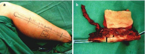
Harvesting of fibular myocutaneous flap. (a) Design of the incision in the lower limb. (b) The prepared fibular myocutaneous flap
Fig. 8.8 
Reconstruction of the right maxillary alveolar crest, infrazygomatic crest, and maxilla. (a) Reconstruction of the maxillary alveolar crest and infrazygomatic crest with the fibula. (b) Reconstruction of the anterior wall of the maxilla with the titanium mesh
Fig. 8.9 
The positive profile at a year and a half after surgery and the appearances of the right maxilla on three-dimensional CT after reconstruction. (a) Positive profile. (b) Lateral profile. (c) The appearance of positive side of the right maxilla on three-dimensional CT after reconstruction. (d) The appearance of the lateral side of the right maxilla on three-dimensional CT after reconstruction
-
1.
-
2.
Case II Second-stage reconstruction of Class 3 maxillary defect of Brown classification.
-
1.
Incision design: because the patient had a history of left total maxillectomy, it was proposed that the subtotal resection of the right maxilla was performed at this time; therefore, the admission passage in the right lateral lip was split selectively combining the incision scar in the original left nasal side; the right submaxillary area was taken as the receptor site (Fig. 8.10a). Incised the right extraoral incision at first, and turned over the skin flap along the deep surface of the platysma muscle to the lower margin of the mandible. Dissected out the marginal mandibular branch of facial nerve within the range of about 1 cm, respectively, above and below the mandibular angle, and protected the marginal mandibular branch of facial nerve and traced it to the site where it entered into the lower lip (Fig. 8.10b).
-
2.
Then the intraoral incision was incised, the detailed surgical procedure is the same as in Case I, and the subtotal resection of right maxilla was performed. Attentions should be paid to protecting the nasal cannula, loosening scar, and lifting up the collapsed upper lip and nosewing (Fig. 8.10c); removed the residual palatine bone (Fig. 8.10d).
-
3.
Harvesting of fibular myocutaneous flap: the skin island was designed along the position of the skin perforator marked by the ultrasound before surgery. The procedures for designing the incision (Fig. 8.11a) and harvesting the skin flap were the same as those in Case I, the vascular pedicle was cut off when the receptor site was readily prepared, and the fibular myocutaneous flap was harvested for transplantation (Fig. 8.11b).
-
4.
At the same time, incised the original surgical scar along the left face, and turned over the skin flap to expose the left maxillary defect. The harvested fibula was cut into four segments, which were fixed, respectively, with the bilateral cheekbones to reconstruct the bilateral alveolar crests and infrazygomatic crests, and the four bone segments were fixed between each other. The prefabricated titanium mesh was used to reconstruct the anterolateral wall of the maxilla, and the titanium mesh was fixed, respectively, with the fibula for reconstructing the alveolar crest and the ipsilateral cheekbone; it was appropriate to use two to three titanium nails for fixation in each site (Fig. 8.11c). The vascular pedicle of peroneal artery and vein was passed out from intraoral area to the right submaxillary area, the vascular torsion was prevented. The facial artery was anastomosed with the peroneal artery and the facial vein was anastomosed with the peroneal vein in turn under the microscope; the good venous reflux was confirmed by three blood vessel patency tests.
-
5.
The skin island of the fibula myocutaneous flap was placed in the palate and was sutured with the surrounding mucosa to repair the soft tissue defect. The complete hemostasis for the cervical wound was performed, and the layered suture was performed after being put back to its original place (Fig. 8.12), and a rubber drainage sheet was placed in submaxillary area.
-
6.
The positive profile at 3 months after surgery showed that the bilateral zygomaticofacial regions were symmetrical; the three-dimensional CT showed that the appearance of bilateral maxilla after reconstruction was satisfactory (Fig. 8.13).
Fig. 8.10 
Designed the incision, and carried out subtotal resection of the right maxilla. (a) Incision design. (b) Incised the right extraoral incision; turned over the skin flap along the deep surface of platysma muscle to the lower margin of the mandible. (c) Lifted up the collapsed upper lip and nosewing. (d) Resected the residual palate bone. (e) The maxillary defect status
Fig. 8.11 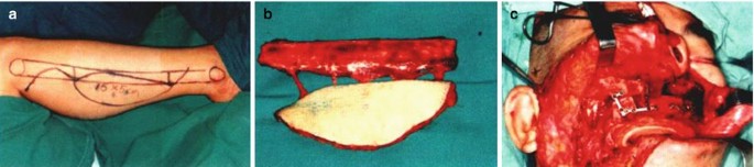
Harvesting of fibular myocutaneous flap and reconstruction of bilateral maxilla. (a) Incision design. (b) The fibular myocutaneous flap after the pedicle was cut off. (c) The fibula after shaping was fixed to reconstruct the maxilla
Fig. 8.12 
After suture. (a) Front appearance after the suture. (b) Intraoral appearance after the suture
Fig. 8.13 
The positive profile at 3 months after surgery and the appearances of the bilateral maxilla on three-dimensional CT after reconstruction. (a) Positive profile. (b) Lateral profile. (c) The appearance of positive side of the maxilla on three-dimensional CT after reconstruction. (d) The appearance of the maxilla from the perspective of looking up on three-dimensional CT after reconstruction
-
1.
-
3.
Case III Second-stage reconstruction of Class 2 defect of Brown classification under the assistance of virtual operation plan.
-
1.
Preoperative computerized surgery simulation and surgical program design.
-
1.
The maxillofacial CT scan data were read by and maxillofacial CT scan data by SurgiCase 5.0 medical image processing software (Materialise Company, Belgium); the three-dimensional reconstruction was carried out using the scanned sequential tomographic images. Three-dimensional reconstructed images of the jawbone were as shown in Fig. 8.14a–c.
-
2.
The CT scan data of the lower limbs were read with SurgiCase 5.0 software, and the three-dimensional reconstruction was carried out on the scanned sequential tomographic images. Harvesting of the fibula was simulated on three-dimensional reconstructed images, and the length of the fibula to be harvested was determined. The fibula bone segment harvested by the computer was transferred onto the maxilla at the affected side, the adjustment was carried out according to the appearance of maxillary alveolar crest after image restoration, the shaping curve for harvesting the fibula was determined, and the position and direction of the implant were designed with reference to the tooth along axial of maxillary teeth after image restoration. The front, lateral, and bottom images after fibula reconstruction and implantation were shown in Fig. 8.14d–f. Finally, the fibular graft after shaping was compared with the original model, the final effect images of computerized surgery simulation were obtained after adjustment, and the rapid prototyping was made in accordance with the final image.
Fig. 8.14 
Images of patients after three-dimensional reconstruction of jawbones, fibula reconstruction, and implant implantation. (a) Front image. (b) Lateral image. (c) Bottom image. (d) Front image after reconstruction. (e) Lateral image after reconstruction. (f) Bottom image after reconstruction
-
1.
-
2.
The surgery was performed in accordance with preoperative computerized surgical program design.
-
1.
Defect exposure: designed the incision along the original surgical scar (Fig. 8.15a), made a full-thickness incision flap to expose the bilateral maxillary defect, loosened the scar (Fig. 8.15b, c), and lifted up the collapsed upper lip and nosewing.
-
2.
Used a chainsaw to trim both broken ends until the blood oozed out, and avoided punching through the maxillary sinus. Designed the incision along the front of left antilobium, incised and turned over the skin flap, and dissected and protected the superficial temporal artery and vein as the blood vessels in receptor site (Fig. 8.15d).
-
3.
The titanium plate was prefabricated on the reconstruction model of maxilla produced with rapid prototyping technology, and the fixation position of the titanium plate was identified (Fig. 8.16a). According to the fixation position of the titanium plate determined on the model, the maxillary buccal cortical bone was drilled and fixed with the prefabricated titanium plate, and both broken ends of the maxilla were connected (Fig. 8.16b).
-
4.
The fibular myocutaneous flap was prepared in another group, and the fibula was harvested according to the required length by preoperative computer simulation. The steps for designing the incision (Fig. 8.17a) and harvesting the skin flap (Fig. 8.17b) were shown in the above article.
-
5.
The vascular pedicle was cut off when the receptor site was readily prepared, and the fibular myocutaneous flap was harvested for transplantation (Fig. 8.17c). According to the shaping guide plate, the prepared and shaped fibular myocutaneous flap was cut into three segments. The fibular myocutaneous flap after shaping was placed in the maxillary defect area and was drilled and fixed (Fig. 8.17d).
-
6.
The superficial temporal artery and vein were anastomosed with the peroneal artery and vein in turn under the microscope, and the good venous reflux was confirmed by three blood vessel patency tests (Fig. 8.17e). The skin island was used to repair the intraoral mucosa defect (Fig. 8.17f).
-
7.
The complete hemostases for bilateral upper lip wounds were performed, and the layered suture was performed after being put back to its original place (Fig. 8.18); a rubber drainage sheet was placed in front of the left ear.
-
8.
At 3 months after surgery, the positive and lateral profiles showed that bilateral upper lips had no collapse and the intraoral wound healed well. The three-dimensional CT and panoramic radiograph showed that the appearance of the maxilla after reconstruction was satisfactory and the bilateral sides were symmetrical (Fig. 8.19).
Fig. 8.15 
Designed the incision along the original surgical scar, loosened the scar, exposed the defect area, used a chainsaw to trim both broken ends until the blood oozed out, designed the incision along the front of left antilobium, incised and turned over the skin flap, and dissected out and protected the superficial temporal artery and vein as the blood vessels in receptor site. (a) Designed the incision along the original surgical scar. (b) Loosened the scar. (c) The defect area after complete loosening of the scar. (d) Dissected out the blood vessels in receptor site
Fig. 8.16 
The reconstruction model of maxilla was used to prefabricate the titanium plate and determine the fixation position of the titanium plate. (a) Determined the fixation position of the titanium plate. (b) The maxillary buccal cortical bone was drilled and fixed with the prefabricated titanium plate, and both broken ends of the maxilla were connected
Fig. 8.17 
The fibular myocutaneous flap was prepared; the fibular myocutaneous flap after shaping was placed in the maxillary defect area and was drilled and fixed. (a) Incision design. (b) The prepared fibular myocutaneous flap. (c) The fibular myocutaneous flap after the vascular pedicle was cut off. (d) The fibular myocutaneous flap was shaped and fixed. (e) The vascular anastomosis was performed and good venous reflux was confirmed. (f) The appearance after repair of the intraoral mucosa defect with the skin island
Fig. 8.18 
After suture. (a) Positive profile after suture. (b) Lateral profile after the suture
Fig. 8.19 
Positive profile at 3 months after surgery, three-dimensional CT, and panoramic radiograph. (a) Positive profile. (b) Lateral profile. (c) The intraoral wound healing well. (d) Three-dimensional CT. (e) Oral panoramic radiograph
-
1.
-
1.


From 2001 to 2008, the authors had reconstructed a total of 28 cases of maxillary defects of Class 2–3 of Brown classification according to abovementioned methods, of which there were 9 cases of Class 2 defects and 19 cases of Class 3 defects; 6 patients with extensive soft tissue defects were repaired using series connected radial forearm free skin flap. In addition to that, one patient had skin island necrosis due to the compression of the perforator of the skin flap; all the remaining 27 fibular myocutaneous flaps and 6 radial forearm free skin flaps survived. The imageological examination showed that the bony fusions between the bone segments of fibula and the adjacent bones were good. After a follow-up of 9–72 months, the appearance of the midface of the patient was satisfactory, and the bilateral sides were basically symmetrical, the oral and nasal cavities were completely separated, and the pronunciation was clear. The speech intelligibility test showed that the speech intelligibility values of the patients had no significant difference compared with the people in normal control group, all patients could have a full diet or soft diet, three patients underwent implant-supported denture restoration, and 15 patients underwent removable partial denture restoration. The detections of bite force and occlusal function were performed before and after surgery; the results indicated that, after restoring the occlusion of dentures, the recovery rate of the bite force of full mouth was between 42.50% and 79.28% with an average of 61.35%, which was greater than that in the patients undergoing reconstruction using the titanium mesh scaffold combined with the radial forearm free skin flap (50.15% ± 14.59%). Among the 15 patients with Class 3 maxillary defect of Brown classification in whom the titanium mesh was used to reconstruct the lateral wall of maxilla and support the eyeballs, two patients undergoing second-stage reconstruction had exposure to part of the titanium mesh under the inner canthus and in buccal gingival sulcus, respectively, at 4 months and 36 months after surgery and thus needed to undergo the second surgery to eliminate the exposure of the titanium mesh, and the remaining 13 patients had no exposure of the titanium mesh; the probability of titanium mesh exposure was 13.3% (2/15), which was significantly lower than that in patients undergoing reconstruction with the aforementioned titanium mesh combined with the transverse rectus abdominis myocutaneous flap or the anterolateral femoral skin flap (27.8%, 5/18) and was also lower than that in patients undergoing reconstruction with the titanium mesh combined with radial forearm free skin flap (21.1%, 4/19) [22].
The authors’ experiences show that the fibular myocutaneous flap combined with the titanium mesh can effectively reconstruct the Class 3 maxillary defect of Brown classification. The fibula can be used to reconstruct the alveolar crest and the zygomaticomaxillary pillar, and the titanium mesh can be used to reconstruct the lateral wall of the maxillary sinus and inferior orbital rim and orbital floor, of which the alveolar crest, the inferior orbital rim, and orbital floor are the horizontal pillars of the midface, and the zygomaticomaxillary pillar is the vertical pillar of midface. In addition to the nasomaxillary pillar, a few major pillars which maintain the appearance and function of the midface have been effectively restored. Upon completion of implanting dentures or partial dentures, the chewing stress is not only distributed onto the new alveolar crest and the zygomaticomaxillary pillar, and the lateral wall of the maxillary sinus reconstructed by the titanium mesh can also play a role in conducting part of the stress. The stress distribution is very similar to the normal maxilla which is extremely similar to the stress distribution of the normal maxilla. Thus, it can be seen that the fibular myocutaneous flap combined with titanium mesh is a relatively simple and reasonable method for reconstruction of Class 3 maxillary defects of Brown classification. But for the patients with Class 4 defects of Brown classification, whether this method is applicable or whether it is needed to be in combination with other methods still requires further study and discussion.
3 Related Problems and Prospects for Maxillary Reconstruction
3.1 Application of Functional Surgical Concepts in the Maxillary Reconstruction
Oral and maxillofacial functional surgery refers to a new surgical connotation and category which carries out immediate or deferred reconstruction of tissue defects and organ loss in oral and maxillofacial area caused by tumors or trauma to achieve the purpose of restoring the function and appearance. The oral and maxillofacial functional surgery mainly includes the following three aspects: (1) The diseased tissues are removed and the normal tissues are preserved under the premise that the principles of surgical oncology are not violated. (2) The immediate repair and reconstruction should be performed after removal of defects caused by the diseased tissues. (3) The functional repair should be promoted on the basis of tissue repair and anatomical reconstruction, including sensory or dynamic reconstruction. The authors believe that the application of functional surgical concepts in the reconstruction of the maxilla should follow the following aspects and should be comprehensively considered combined with the demand of patients:
3.1.1 Facial Appearance
After preoperative CT information of the patients is input into the CAD system, the rapid prototyping technology is used to make the maxillary virtual model with defects. According to the principle of facial symmetry, maxillary model for rehabilitation is designed, which thus accurately guide the individualized shaping and placement of the maxilla. The upper and anterior walls of the maxillary sinus can be supported with the prefabricated titanium mesh, and the retentive force can be strengthened, and thus the patient’s appearance can be effectively restored.
3.1.2 Chewing Function
The functional reconstruction of maxilla is performed with free composite osseous myocutaneous flap combined with the CAD/CAM technology, the accurate three-dimensional bony structure of the midface is reconstructed anatomically, and the original appearance of the maxillary alveolar crest is restored, so that the reconstructed tissues can withstand a certain chewing pressure. For patients with defects equal to or greater than Class 3 according to Brown classification, the zygomatic implant can also be implanted to strengthen and effectively conduct the biting force. The CAD/CAM technology can also guide the surgeon to design the osteotomy line and the fixation sites according to the model before surgery, which is conducive to guiding the accurate placement of the transplanted bone and thus maximizes the possibility of appearance of the stress concentration regions when performing function after surgery; the planting technology can provide reliable retention, stabilization, and support for the dentures, so that the optimal chewing efficiency can be achieved in the maxilla after reconstruction.
3.1.3 Voice Function
The free radial forearm skin flap or composite osseous myocutaneous flap can be used to repair the bottom wall of the maxilla completely and tightly and close the oral surface and nasal surface at the same time, thus preventing the occurrence of the oronasal fistula. The radial forearm skin flap sometimes overcomes the limitation that the tissue volume is insufficient when the larger soft tissue wound is repaired simply with the fibular myocutaneous flap. Its sufficient length ensures a good adhesion of the soft palate and also ensures the accuracy of the tongue-palatal contact in the process of pronunciation; therefore, the soft palate will not shrink back at the same time, and the occurrence of velopharyngeal incompetence can be maximally prevented and the incidence rate of hypernasality can be greatly reduced in patients after maxilla resection.
3.1.4 Ventilation Function
Because the free radial forearm skin flap can be prepared according to required sufficient length, and its placement also has considerable flexibility, the use of the radial forearm skin flap can not only close the wound at the oral side but also reconstruct the nasal air passage of the patient through folding the skin flap to recover the nasal ventilation function of the patient after surgery.
3.2 The Factors to be Considered in Maxillary Reconstruction
The maxillary reconstruction is a very challenging and complex issue; the reconstruction surgeon should fully consider the various factors when making a surgical plan, such as the site and volume of the maxillary defect, whether combined with the surrounding tissue defect, the structural condition of the remaining bone, the general condition of the patient, the need for adjuvant radiotherapy, the technological level of the operator, the location of the tissue flap to be harvested, the scope available for harvesting, and the situation of the vascular pedicle. The relevant parameters needed to be taken into full account when the maxilla is reconstructed are listed in Table 8.1, which are available for consideration by the surgeons engaged in repair and reconstruction when making a surgical plan, in order to select the most effective and appropriate reconstruction method to obtain the best aesthetic and functional results.
3.3 The Prospect of the Functional Reconstruction of the Maxilla
At present, the maxillary defect reconstruction has achieved considerable progress, the postoperative problems on basic oral functions and aesthetic requirement in patients with maxillary defects have also been better resolved, and the composite osseous myocutaneous flap combined with planting technology has established its dominance in functional reconstruction of maxilla and will continue to be improved in the future. Of course, the combined use of multiple repair methods such as composite osseous myocutaneous flap, local tissue flap, and prostheses to reconstruct the maxillary defect can get better results than the reconstruction with single technology, but the current functional maxillary reconstruction still has the following deficiencies to be improved: how to further restore the sinus cavity structure within the maxilla, how to solve the problem of taking the inner mucosa to replace the current skin tissue to repair the intraoral defect, and how to reconstruct more accurately the osseous pillar in the midface, the soft tissue coverage in the surface, and related dentures. Solving these problems not only depends on the continuous improvement and upgrading of the existing biological materials and technology but also places the hope on that some subjects which are still at the experimental stage are solved one by one, such as vascularized transplantation of ligamental tissues and application of in situ tissue-forming techniques. The functional reconstruction of the maxilla still requires careful planning and close cooperation among surgeons in department of surgical oncology, department of reparative and reconstructive surgery, department of prosthodontics, and department of radiotherapy to achieve the satisfactory long-term outcome in function and appearance. To this end, scholars from various countries are constantly working hard to achieve the same goal, that is, to reconstruct the appearance and function of the maxilla and reproduce the original structure and stomatognathic system in the midface of the patient.
References
Gullane PJ, Arena S. Palatal island flap for reconstruction of oral defects [J]. Arch Otolaryngol. 1977;103(10):598–9.
Martin D, Pascal JF, Baudet J, et al. The submental island flap:a new donor site: anatomy and clinical applications as a free or pedicled flap [J]. Plast Reconstr Surg. 1993;92(5):867–73.
Tideman H, Samman N, Cheung LK, et al. Immediate reconstruction following maxillectomy: a new method [J]. Int J Oral Maxillofac Surg. 1993;22(4):221–5.
Muzaffar AR, Adams WP, Hartog MJ, et al. Maxillary reconstruction:functional and aesthetic consideration [J]. Plast Reconstr Surg. 1999;104(7):2172–83.
Triana RJ, Uglesic V, Virag M, et al. Microvascular free flap reconstructive options in patients with partial and total maxillectomy defects [J]. Arch Facial Plast Surg. 2000;2(2):91–101.
Brown JS, Roger SN, McNally DN, et al. A modified classification for the maxillectomy defect [J]. Head Neck. 2000;22(1):17–26.
Uglesi V, Virag M, Varga S, et al. Reconstruction following radical maxillectomy with flaps supplied by the subscapular artery [J]. J Craniomaxillofac Surg. 2000;28(3):153–60.
Futran ND, Wadsworth JT, Villaret D, et al. Midface reconstruction with the fibula free flap [J]. Arch Otolaryngol Head Neck Surg. 2002;128(2):161–6.
Brown JS, Jones DC, Summerwill A, et al. Vascularized iliac crest with internal oblique muscle for immediate reconstruction after maxillectomy [J]. Br J Oral Maxillofac Surg. 2002;40(3):183–90.
Jian S, Jun L, Zhiyuan Z, et al. Closed and three-dimensional reconstruction of maxillary defects with titanium mesh and free forearm flap [J]. Journal of Pract Stomatol. 2002;18(4):291–3.
Jun L, Jian S, Ma H. Application of individual titanium bracket in construction of three-dimensional shape of the mandible [J]. Oral Maxillofac Surg. 2003;13(1):17–20.
Jian S, Jun L, Zhiyuan Z, et al. Closed three-dimensional reconstruction of large maxillary defects with titanium mesh and myocutanous fibular flap and Chinese flap [J]. China J Oral Maxillofac Surg. 2003;1(1):3–8.
Hong S, Jian S. Functional maxillofacial surgery [M]. Shanghai: The Second Military Medical University Press; 2003. p. 366–90.
Ma H, Jian S, Jun L, et al. Evaluation of mastication function after three-dimensional maxillary reconstruction [J]. West China J Stomatol. 2005;23(1):29–31.
Yanqiu W, Jian S, Yang C, et al. Articulatory function after maxillectomy: a comparison between surgical rehabilitation and prosthodontic rehabilitation [J]. Chinese J Oral Maxillofac Surg. 2005;3(1):43–7.
Jian S. Repair and reconstruction of mandibular defects [J]. J Oral Maxillofac Surg. 2005;15(1):5–7.
Bidros RS, Metzinger SE, Guerra AB. The thoracodorsal artery perforator-scapular osteocutaneous (TDAP-SOC) flap for reconstruction of palatal and maxillary defects [J]. Ann Plast Surg. 2005;54(1):59–65.
Yazar S, Cheng MH, Wei FC, et al. Osteomyocutaneous peroneal artery perforator flap for reconstruction of composite maxillary defects [J]. Head Neck. 2006;28(4):297–304.
Arce K. Buccal fat pad in maxillary reconstruction [J]. Atlas Oral Maxillofac Surg Clin North Am. 2007;15(1):23–32.
Sun J, Shen Y, Weng YQ, et al. Lateral lip-splitting approach for total and subtotal maxillectomy [J]. J Oral Maxillofac Surg. 2009;67(6):1197–205.
Brown JS, Shaw RJ. Reconstruction of the maxilla and midface:introducing a new classification [J]. Lancet Oncol. 2010;11(10):1001–8.
Sun J, Shen Y, Li J, et al. Reconstruction of high maxillectomy defects with the fibula osteomyOCUtaneous flap in combination with titanium mesh or a zygomatic implant [J]. Plast Reconstr Surg. 2011;127(1):150–60.
Shen Y, Sun J, Li J, et al. Special considerations in virtual surgical planning for secondary accurate maxillary reconstruction with vascularized fibula osteomyoeutaneous flap [J]. J Plast Reconstr Aesthet Surg. 2012;65(7):893–902.
Author information
Authors and Affiliations
Corresponding author
Editor information
Editors and Affiliations
Rights and permissions
Copyright information
© 2018 Springer Nature Singapore Pte Ltd. and Zhejiang Science and Technology Publishing House
About this chapter
Cite this chapter
Sun, J., Shen, Y. (2018). Repair and Reconstruction of Maxillary Defects. In: Zhou, X., Cao, Y., Wang, W. (eds) Oncoplastic surgery. Plastic and Reconstructive Surgery. Springer, Singapore. https://doi.org/10.1007/978-981-10-3400-8_8
Download citation
DOI: https://doi.org/10.1007/978-981-10-3400-8_8
Published:
Publisher Name: Springer, Singapore
Print ISBN: 978-981-10-3399-5
Online ISBN: 978-981-10-3400-8
eBook Packages: MedicineMedicine (R0)