
Abstract
Trauma is the second leading cause of mortality in all ages and the leading cause of mortality under age 44 years.
Access provided by Autonomous University of Puebla. Download chapter PDF
Keywords
These keywords were added by machine and not by the authors. This process is experimental and the keywords may be updated as the learning algorithm improves.
FormalPara Key Points-
Trauma is the second leading cause of mortality in all ages and the leading cause of mortality under age 44 years.
-
Undertake evaluation and resuscitation in a systematic and methodical manner – to identify and treat according to priority.
-
Treat injuries that are life-threatening as they are found and not at the end of the evaluation.
-
Newer resuscitation strategies are associated with better outcome.
-
Surgical intervention may take priority over completing the entire assessment – intervene and return to the assessment later.
Introduction
Trauma remains the major killer of persons in the age groups 15–44 years of age in the majority of lower- and middle-income countries, with many factors influencing the causes and consequences, and the second biggest killer, after infective diseases, in children. Systems of prehospital care, early adequate resuscitation and timely definitive care result in improved survival, with the need for rehabilitation to re-integrate the survivors into gainful societal activity [1–3].
Pathophysiology and Mechanism of Injury [4]
There are basically two mechanisms of injury in any trauma situation, although they may co-exist, with the actual clinical features primarily governed by the organ injury, be it solid organ, hollow viscus or soft tissue and bone:
Blunt trauma such as:
-
Motor vehicle and other transport-related incidents
-
Assault with a blunt object
-
Fall onto a blunt object
-
Fall from a height
Penetrating trauma:
-
Broken glass penetration
-
Knife wound
-
Low-energy bullet wound (velocity has become less important)
-
High-energy bullet wound
Combined:
-
Bomb blast
-
Some motor vehicle accidents with impalement of body parts
The pathophysiology of injury is twofold, namely, that caused by the injurious event and that resulting from the inflammatory reaction mounted by the body in response to the injury. Both of these processes may make the patient prone to sepsis or multiple organ dysfunctions [5].
Injury patterns may be predictable from knowing the site of impact and the forces generated. A good history from emergency medical service (EMS) staff is helpful here.
Approach to Trauma Evaluation and Resuscitation
Initial Approach
Severe trauma does not allow for the opportunity to follow the normal pattern of medical evaluation, namely, that of history, followed by examination, side room tests and followed by imaging and laboratory investigations. One must focus on saving the patient from death.
Trauma is best managed by a team of committed providers, with no proof that smaller teams fair worse than larger teams, as long as all members of the team understand and perform their role [6] (Table 36.1).
Standard precautions should be followed by all members of the trauma team including wearing: mask and visor, gloves, apron or scrub gown and overshoes or boots.
It is essential that any medical equipment in the emergency department, drugs and fluids are checked regularly for functional and expiry issues. This should be performed at least daily by both nurses and doctors to avoid delays to care when the patient arrives. If pre-notification is received prior to the patient’s arrival, equipment should be made ready and blood bank placed on standby for urgent transfusion supplies if the history is suggestive (Fig. 36.1).

The correct environment and personal protective equipment is essential
There are a number of systematic trauma evaluation programmes, such as the International Trauma Life Support® or Prehospital Trauma Life Support® (for EMS personnel), National Trauma Management Course (IATSIC) or Primary Trauma Care (Oxford University), and the traditional gold standard has been the Advanced Trauma Life Support (ATLS®) for doctors (or the Trauma Nurse Core Course) approach as it is simple and ONE safe way: the ATLS® has become progressively more evidence-based over time [8–14].
Handover
The use of an acronym type handover system such as the De-MIST (demographics, mechanism, injuries, signs, treatment) is useful to impart essential knowledge before patient assessment without undue delay.
-
Demographics (age, gender, where from, etc.)
-
Mechanism of injury (what happened)
-
Injuries identified prehospital/at referring facility
-
Signs and symptoms (and any results from another facility)
-
Treatment given till the patient arrived at your facility
Patient Care Sequence
The aim of the approach to the treatment of the injured is to attempt to identify life-threatening injuries, intervene to treat these as they are found and then progress to the next part of the resuscitation. The traditional approach is based on so-called primary survey, followed by secondary survey and eventually a tertiary survey in the admitting service ward or ICU.
-
Primary survey – A, B, C, D and E – is the resuscitation phase.
-
History (AMPLE) and baseline special exams are performed after life-saving intervention.
-
Secondary survey follows: the head-to-toe review of all injuries – aim to avoid missed injury.
-
Definitive care traditionally follows after stabilisation with disposition to a trauma surgeon to coordinate care.
What has become clear over the past 10 years is that this approach works well for the patient that does not need urgent surgical intervention; however, the newer literature espouses the need to undertake life-saving surgery during the primary survey if needed and to continue thereafter with further assessment. An example of this would be immediate sternotomy or thoracotomy to relieve a pericardial tamponade or laparotomy for control of intra-abdominal bleeding. An even more recent addition is urgent extraperitoneal pelvic packing for exsanguinating pelvic fracture haemorrhage [15].
A: Airway and C-Spinal Control [16]
There is always the assumption with blunt trauma that there is an injury to the C-spine and, therefore, to the cord, so absolute care is required. This is much less true for penetrating trauma, where airway control must not be delayed or impeded by spinal motion restriction.
Assess the airway for:
-
Patency
-
Maintenance: Is it maintained or is intervention and support needed?
-
Identification of potentially threatened airway – intervene early to reduce complications.
-
Airway-specific injuries.
DO NOT use head-tilt chin lift: JAW THRUST is the basic airway manoeuvre of choice (Tables 36.2, 36.3, and 36.4).
The main aim of airway management is a DEFINITIVE airway, which is usually defined as a cuffed endotracheal tube below the vocal cords with the cuff inflated, placed either transoral, transnasal or surgical (cricothyroidotomy in emergency/tracheostomy in elective) (Fig. 36.2).

Equipment for trauma airway management
If two attempts by two operators are unsuccessful, then proceed to RESCUE OPTION, currently using a laryngeal mask airway (LMA) is the procedure of choice as this allows time to prepare for elective tracheostomy, while not a definitive airway, it offers more protection than an oropharyngeal tube. Alternatively if there is an inadequate seal, one can proceed to needle cricothyroidotomy as a temporary solution – using jet-ventilation will allow about 20 min to obtain further assistance.
Should one face the dreaded “cannot intubate-cannot ventilate” scenario, then one is forced to move towards the surgical definitive airway, namely, cricothyroidotomy. So-called slash tracheostomy is not recommended. Only once the airway is secured, proceed to breathing.
B: Breathing and Ventilation
Assessment of breathing must be independent of airway. If the patient is breathing, the assessment of rate, mechanism (e.g. flail chest/diaphragmatic breathing) and oxygenation will guide the need for either chest tubes or ventilation. The apnoeic patient requires not only airway management but full ventilation support.
The primary survey goal of breathing assessment serves to exclude life-threatening chest injuries: treatment is determined by clinical findings (Table 36.5).
Indications for ventilation:
-
The patient with apnoea
-
Adult RR >30 or <10/min
-
Poor mechanism with severe flail chest and contusion >25 % lung volume on CXR/CT
-
Blood gas: pO2 <8 kPa/60 mmHg or pCO2 >6,5 kPa/50 mmHg
-
GCS <9/15
Placement of a chest tube:
-
Fourth to fifth ICS, anterior to mid-axillary line (“safe triangle”).
-
Clean and drape. Give plenty of local anaesthetic – give it time to work!
-
Incise skin and underlying fat with the scalpel only.
-
Take a blunt-tipped spreadable forceps and run it over the underling fifth rib until you feel the pleural “pop” and then open the clamp and pull back while open, spreading the intercostal muscles off the underlying bone – make a hole big enough for a finger to check for intrapleural adhesions – cause no further harm!
-
DO NOT USE ANY SHARP-POINTED TROCHARS – remove these devices if they are in the drain tube; preferably use a clear PVC-type Argyle catheter with at least two drain holes, less chance of blockage.
-
Place the tip of the drain on the blunt spreadable forceps and direct it into the chest aiming always apico-posterior, so that blood can drain from the supine patient and air will collect at the apex.
-
Fix the drain to the chest wall with sutures and tape, and connect it to a valve-based [17] or water-seal chest drain set (the advantage of the former: allows for autotransfusion). Use saline in the drain to allow for autotransfuse the blood that drains (Fig. 36.3).
Fig. 36.3 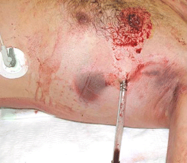
Well-placed chest tube after gunshot chest

C: Circulation and Haemorrhage Control
The best indicator that a patient is in hypovolaemic shock is the pulse rate: the patient with a tachycardia (adult >120/min) [18] is shocked till proven otherwise. Importantly it is more about perfusion than pressure!
The treatment of bleeding is to STOP the bleeding. It is pointless to resuscitate the patient with fluids, and they bleed onto the floor. Control the bleeding with DIRECT pressure. It may be necessary in C to stop what you are doing, take the patient to the operation room and surgically control the bleeding if it is in the chest, abdomen, pelvis or long bones!!! FAST (focussed assessment sonar in trauma) is a recommended extension of the clinician’s armamentarium to identify intra-abdominal haemorrhage in the acutely unstable trauma patient. Laparotomy is the default operation unless the chest is the clear bleeding source [15].
The routine use of procoagulants (rVIIa and tranexamic acid) is currently still controversial outside of specific time frames and guidance from coagulation tests such as ROTEM/TEG.
Apart from major bleeding, one must identify end-organ ischaemia (usually a mark of a peripheral or cervical vascular injury), cardiac tamponade (mainly with penetrating trauma) and possible blunt aortic arch rupture (usually acceleration-deceleration injury) (Fig. 36.4, Table 36.6).

Life-threatening cardiovascular injury: CXR blunt trauma – wide mediastinum and knife wound causing pericardial tamponade
Fluid in resuscitation: traditionally crystalloid fluid in high volumes has been used in fluid resuscitation; however, it is now recognised [19] that it is far better to use blood and blood products in a balanced product ratio giving the patient what they have lost. An initial bolus of up to 2 l of Ringer’s lactate (20 ml/kg in children) is acceptable to differentiate responders from non-responders. Consider permissive hypotension, which means keeping the SBP around 90 mmHg until surgical control of bleeding and then resuscitate fully for obvious noncompressible bleeding in the context of rapid access to surgical facilities. Also consider autotransfusion if capabilities exist. Blood should be given in the 1:1:1 ratio of red cells, to plasma, to platelets – this is a goal, not an absolute target; about 1:2:1 is probably optimal [20, 21]. While restrictive bolus therapy is appropriate for non-responders who can access surgery, large-bore central access (such as dialysis or “sheath introducer” large-bore catheters) and the use of high-capacity giving sets are advised intraoperatively (Fig. 36.5).

Autotransfusion with chest drain device [17] and large-bore central catheter
The best gauge of whether or not you are gaining the upper hand in the resuscitation is the perfusion of vital organs, such as the heart, brain and kidneys: so urine output is an objective assessment of the degree of your success. Place a urine catheter early to assess this, after doing a rectal exam on the male patient with pelvic trauma to exclude a urethral rupture. Blood gas lactate is the best marker! [22] Aim for lactate <2,5 mmol/l. Base deficit of less than 4 is a good surrogate.
Exclude the life-threatening circulation injuries:
-
Cardiac tamponade (Beck’s triad): muffled heart sounds and pulsus paradoxus with hypotension and distended neck veins. Treatment is OPEN the chest in the operation room.
-
Thoracic aorta rupture with a pseudoaneurysm – good upper limb pulses, weaker lower limb pulses and radio-femoral delay but may have no signs and only be suspected on CXR with a widened mediastinum (>8 cm at T4 in adults). Most die on scene!
Also quickly check the limbs for distal pulses and reduce long bone and pelvic fractures to decrease both pain and fluid loss. Exclude compartment syndromes! Once the obvious bleeding is controlled, look for hidden losses such as the chest, abdomen and pelvis.
Vascular injures to the peripheral circulation may result in limb-threatening situations, and this must be detected early. Early involvement of the SURGEON is essential.
D: Disability and Neurological Impairment
During the primary survey there are only THREE things of importance:
-
Pupil reactions
-
Any obvious neurological deficit: Localising signs
-
Glasgow coma score: Eye opening, speech and motor function/15
Any abnormality detected here requires that the patient be supported and further harm prevented by avoiding hypoxia, hypotension and hypothermia. Good A, B and C impact outcome of abnormal D. The additional requirement is for neuroimaging, either a CT scan of ideally the head and spine (Fig. 36.6) [23].

CT head – intracerebral haemorrhage
E: Expose the Patient, but Maintain Environmental Control
-
Undress them completely – ideally cut off the clothing!
-
Keep warm with light blanket, warm fluids and warm room (recommended at least 24 °C) [24].
-
Log-roll now and examine the back – get them off the spine-board onto a padded but firm stretcher (5 cm padding adequate).
After addressing the life-threatening injuries, proceed to detailed history and other baseline special examinations.
History
-
A = Allergies
-
M = Medications used
-
P = Previous medical/surgical history
-
L = Time of the last meal before the time of the trauma
-
E = Events surrounding the current admission
The patient, if conscious, family, onlookers or the EMS staff may provide information. Ask pertinent questions related to the type of incident.
Baseline investigations:
-
AP chest and pelvis X-ray are ESSENTIAL (neck X-ray less important if spinal motion restriction maintained and is unnecessary for most penetrating trauma).
-
Blood gas, FBC, U&E, crossmatch and urine dipstick, toxicology screen and pregnancy tests are routine as appropriate. TEG/ROTEM coagulation studies are gaining importance (Figs. 36.7 and 36.8).
Fig. 36.7 
CXR after gunshot, NXR after gunshot, PXR – open book with lateral shear injury
Fig. 36.8 
Point-of-care blood gas and coagulation analysers should be part of a well-equipped resuscitation area


While this is being processed, one can now begin the SECONDARY SURVEY: This is head-to-toe examination of the entire patient with “fingers or probes into every orifice”, so as to not miss any injury, including a “log-roll” if not yet performed (Table 36.7).
References
Soriede K. Editorial: strengthening the trauma chain of survival. Br J Surg. 2012;99 Suppl 1:1–3.
Hardcastle TC, Brysiewicz P. Trauma in South Africa: from humble beginnings to an Afrocentric outreach. Int Emerg Nurs. 2013;21:118–22.
Gosselin RA. The increasing burden of injuries in developing countries. Tech Orthop. 2009;24:230–2.
Brochnner AC, Toft P. Pathophysiology of systemic inflammatory response after major accidental trauma. Scand J Trauma Emerg Med. 2009;17:43.
Bianchi ME. DAMPs, PAMPs and alarmins: all we need to know about danger. J Leukoc Biol. 2007;81:1–5.
Deo SD, Knottenbelt JD, Peden MM. Evaluation of a small trauma team for major resuscitation. Injury. 1997;28(9-10):633–7.
American College of Surgeons, Committee on Trauma. Resources for the optimal care of the injured patient. Chicago: Committee on Trauma American College of Surgeons; 2006.
International Association of Trauma and Surgical Intensive Care/Academy of Traumatology (India). NTMC. National Trauma Management Course™. Available from www.indiatrauma.org. Accessed Jun 2014.
American College of Surgeons. ATLS® student course. 9th ed. Chicago: ACS; 2012.
International Society of Surgery/International Association of Trauma and Surgical Intensive Care. DSTC Course™. Available from www.iatsic.org/dstc.html. Accessed Jun 2014.
National Association of Emergency Medical Technicians. PreHospital trauma life support. 7th ed. St. Louis: Mosby-JEMS; 2011.
Campbell J, editor. International trauma life support. Alabama: Alabama Chapter – American College of Emergency Physicians; 2011.
Primary trauma care. www.primarytraumacare.org (updated 2013). Accessed Jun 2014.
Emergency Nurses Association. Trauma Nurse Core Course, 7th ed, www.ena.org. Accessed Jun 2014.
Boffard KD, editor. Manual of Definitive Surgical Trauma Care (DSTC). 3rd ed. London: Hodder; 2011.
Kovacs G, Law JA. Airway management in emergencies. Connecticut: Peoples Publishing House; 2011.
Cooper C, Hardcastle TC. Prospective randomised trial of a new chest drain device. S Afr J Surg. 2006;44(4):132–5.
Jacobsen J, Secher NH. Heart rate during haemorrhagic shock. Clin Physiol. 1992;12(6):659–66.
Jansen JO, Thomas R, Loudon MA, Brooks A. Damage control resuscitation for patients with major trauma. Br Med J. 2009;338:b1778.
Miller TE. New evidence in trauma resuscitation – Is 1:1:1 the answer? Perioper Med. 2013;2:13.
Hardcastle TC. Complications of massive transfusion in trauma patients. ISBT Sci Ser. 2006;1(1):180–4.
Heinonen E, Hardcastle TC, Barle H, Muckart DJJ. Lactate clearance predicts outcome after major trauma: audit of a trauma ICU population. Afr J Emerg Med. 2014;4:61–5.
Brain Trauma Foundation. Guidelines for the management of severe traumatic brain injury, 3rd ed, 2007. Available from www.braintrauma.org/coma-guidelines. Accessed Jun 2014.
Hardcastle TC, Stander M, Kalafatis N, Hodgson E, Gopalan D. External patient temperature control in emergency centres, trauma centres, intensive care units and operating theatres: a multi-society literature review. S Afr Med J. 2013;103:609–11.
Author information
Authors and Affiliations
Corresponding author
Editor information
Editors and Affiliations
Additional information
All photographs and X-ray images are the property of the author and were personally captured using a digital camera; permission is granted for publication, and copyright is held by the author
Rights and permissions
Copyright information
© 2016 Springer India
About this chapter
Cite this chapter
Hardcastle, T.C. (2016). Early Management of Trauma. In: David, S. (eds) Clinical Pathways in Emergency Medicine. Springer, New Delhi. https://doi.org/10.1007/978-81-322-2713-7_36
Download citation
DOI: https://doi.org/10.1007/978-81-322-2713-7_36
Published:
Publisher Name: Springer, New Delhi
Print ISBN: 978-81-322-2711-3
Online ISBN: 978-81-322-2713-7
eBook Packages: MedicineMedicine (R0)