
Abstract
The modern ribosomal machinery is very complex, and its core subsystems and many of its individual components are universally found in all three domains of life. This indicates that much of the story of ribosome origins and its subsequent evolution predates the last universal common ancestor (LUCA). Thus, ribosome history relates to other early life issues such as the possibility and nature of an RNA World, the early history of chirality, and always most hopefully the origins of the genetic code. However, this is not the end of the story. As discussed elsewhere in this volume, important events have also occurred since the LUCA, especially in eukaryotic ribosomes that have served to integrate the machinery with other cellular systems. Ribosome origins and subsequent evolution are in reality somewhat separate problems. In addressing the former, this chapter initially examines the source and nature of the peptidyl transferase center (PTC), including where and how the peptide bond is made. This is followed by efforts to understand the subsequent evolution of the ribosome, which led to the addition and refinement of various other functional centers including the decoding center. This is being accomplished using what is in essence a reverse engineering approach to develop a timeline of major events in the ribosome history. Finally, significant events on the timeline are discussed in detail.
Access provided by Autonomous University of Puebla. Download chapter PDF
Keywords
These keywords were added by machine and not by the authors. This process is experimental and the keywords may be updated as the learning algorithm improves.
1 The Beginnings: Origins of the PTC and Possible Existence of an RNA World
1.1 Background
A central question in the origin of the modern translation machinery is the source of the extremely conserved PTC where the peptide bond is formed. Indeed, the formation of the PTC is by definition at the beginning of ribosome history even if some of the components actually predate it. For many years, investigators sought to discover which ribosomal protein or combinations thereof were responsible for catalyzing peptide bond formation. Failure to solve this riddle gradually led to the hypothesis that the ribosomal RNA (rRNA) had a significant role as reviewed by Noller [1]. When a high-resolution crystal structure of the 50S ribosomal subunit was obtained [2], it quickly became clear that since no protein appeared to be in proximity to the PTC region, the RNA itself must be catalytic [3]. Later it was found, in the case of the Thermus thermophilus ribosome, that ribosomal protein L27 did in fact closely approach the PTC [4, 5]. However, elimination of multiple residues that most closely approach the PTC does not completely prevent peptide bond formation [6]. Most recently it was shown that the rate of peptide bond formation was independent of the presence or absence of L27 [7]. Thus, the ribosome is widely and likely correctly regarded as an RNA machine.
1.2 RNA World
This brings the question of the RNA World to the fore. The evidence that is most indicative of the existence of a primitive RNA World that predated protein synthesis is the potential involvement of nucleotide coenzymes in prebiotic reactions as originally proposed by White in 1976 [8] and recently reviewed and pursued further by Yarus [9]. In many cases, the core structure of the coenzyme is an adenine diphosphate with a side chain that sometimes contains amide bonds attached to the 5′ diphosphate. This is seen, for example, in nicotinamide adenine dinucleotide and coenzyme A. This same feature is also seen in modern aminoacyl tRNA synthetases that initially attach the cognate amino in the same position creating the aminoacyl adenylate.
However, to obtain an extensive RNA World in which RNA catalyzes biochemical pathways that many have envisioned, it is thought necessary to obtain an RNA-based RNA replicase. This would need to be accomplished before peptide synthesis became established as the latter could quickly terminate the RNA World in favor of an RNA/peptide World. Proteins that can replicate RNA clearly exist, whereas effective RNA-catalyzed RNA replication of RNAs has proven difficult to demonstrate. Although template-directed synthesis by ribozymes is feasible [10], enzymes that synthesize RNAs approaching their own size had been difficult to obtain [11]. Using selection in ice, this hurdle was recently overcome in a specialized laboratory setting [12].
More recent efforts have highlighted the value of freeze-thaw cycles in the possible generation of self-replicating RNAs [13]. This progress notwithstanding, the complexity of these synthetic self-replicating RNAs is not substantially less than that of the peptidyl transferase center (PTC) found in extant ribosomes. So perhaps the quest to find a self-replicating RNA should be augmented/replaced with a quest to find a minimal peptide that can replicate an RNA. At the least, such a peptide RNA polymerase must emerge at some early stage as protein polymerases, and other complex proteins such as aminoacyl tRNA synthetases are likely present at the time of the LUCA [14]. Depending on its size, evidence of such a peptide could strongly support an abbreviated RNA World where the discovery of peptide synthesis predated the discovery of an RNA replicase. Indeed, RNAs that can synthesize peptides have been obtained in selection experiments [15]. As noted by Lilley [16], “Ultimately the finest achievement of the RNA World was probably the creation of proteins. These then took over most of the catalytic functions, leaving the ribosome as the most permanent monument to a heroic era.” However, even this may be an overstatement, as the RNA World may simply have never existed, as is suggested by the primitive nature of the mechanism of peptide synthesis in ribosomes and limited evidence, if any, of proteins replacing ribozymes [17].
A second problem with even an abbreviated RNA World, which is shared with ribosomal origins, is of course the source of the RNA. Although recent progress has been made [18, 19], the prebiotic synthesis of chiral RNA or a precursor nucleic acid remains uncertain. One alternative view is a prebiotic world in which peptides of perhaps 3–8 residues could be produced without ribosomes [20]. The synthesis of even longer polymers of amino acids on illite and hydroxyapatite has been demonstrated [21]. Non-coded peptide synthesis in fact occurs in extant organisms using coded synthetases [22]. In the absence of the ribosomal machinery, various atypical features can be utilized including the incorporation of D-amino acids and/or non-standard amino acids [23]. Irrespective of how or when RNA or a precursor nucleic acid first became available, it would clearly be an important step in origins. This is because complexity is readily increased even in the absence of a replicase by hybridization and/or ligation. These processes are obtainable in random RNA pools [10]. It has been shown, for example, that a ligation of a simple RNA stem loop structure to itself could in principal produce a tRNA with the full complexity of the modern molecule [24].
1.3 Peptidyl Transferase Center
With regard to the modern PTC, Ilana Agmon in Yonath’s research group found evidence of structural similarity between the A-site and P-site portions of the modern PTC [25]. This suggests that the modern PTC actually arose from an ancient duplication or hybridization event [26, 27]. Consistent with this, in the LSU rRNAs of Chlamydomonas reinhaardtii mitochondria [28] and the Euglena gracilis cytoplasm [29], the PTC is actually formed by noncovalent base pairing between two separate RNA fragments. These fragments are, however, very large and likely more indicative of possibility rather than actual history.
Regardless of its origin, a popular question regarding the PTC is the mechanism of the two reactions it is involved in [30]. The more studied of these is the entropic peptidyl transferase reaction in which the ester bond that links the nascent peptide to the 3′ hydroxyl of the 3′ terminal ribose of the P-site tRNA is subject to aminolysis by the alpha-amino group of the incoming tRNA. The reaction is fast with 15–50 peptide bonds produced per second [31]. The PTC is thought to be an entropy trap, which means it is the positioning of the substrates that is central rather than conventional chemical catalysis [32, 33]. The chemistry has been the subject of considerable discussion [30, 34–39] with no consensus resolution. Unlike a typical enzyme, the reaction is also not specific. The PTC can synthesize other products including esters and thioesters [40–44] and utilize non-standard amino acids [45–47]. The second reaction is the subsequent release of the peptide from the P-site tRNA by ester bond hydrolysis, which occurs when a stop codon is reached. This reaction is much slower [48].
However, a focus on the specifics of the chemistry obscures the larger picture of the utility of a proto-ribosome in a prebiotic world. It is not just the ability to synthesize peptide or perhaps ester bonds that matters, but rather the ability of the process to be done over and over again in order to create a polymer [49]. The chemistry itself is inherently favorable in that the synthesis of a dipeptide is relatively easy, but its subsequent release is slow. Thus, further extension is possible if additional activated amino acids approach the growing chain before it is released. The modern ribosome exploits this by holding the growing chain in place in the P site while the next charged tRNA is recruited to the A site. When the PTC region of the ribosome is closely examined, one sees that the PTC actually forms a nanopore (Fig. 1).

Space filling model of the PTC region highlighting the residues that surround the exit pore. Backbone atoms are colored in brown, and the bases are in green. The black line traces the RNA backbone throughout the region. Unlike other known RNA pores that are lined almost exclusively with backbone atoms, the PTC pore provides access to the bases
The PTC pore serves as the entrance to the exit tunnel that provides a path for the growing peptide to pass through the ribosome and ultimately emerge as a mature protein. To accomplish this, various bases in the PTC are accessible at the pore surface. In the modern ribosome, any blockage of the exit tunnel rapidly inhibits protein synthesis, in essence because the active site becomes clogged. This is the mechanism employed by many macrolide antibiotics [50–56]. Many other antibiotics simply prevent the correct positioning of acceptor or donor substrates at the PTC [30].
With these facts in mind, it was previously proposed [49] that the entrance to the exit tunnel was part of the PTC from the very beginning. In fact, in combination with the favorable chemistry, it was likely this feature that provided a critical advantage over alternative peptide synthesis systems by facilitating the polymerization process as well as the synthesis of individual peptide bonds. For example, if pairs of activated amino acids bound to very small RNAs [57] are brought to the surface of a proto-ribosome they could, if positioned properly, form a peptide bond such that one of the RNAs now carries a dipeptide while the other is vacant. This is then the critical moment. If complexity is to continue to increase, the small RNA that contains the dipeptide must now be more likely to stay associated with the proto-ribosome. In contrast, the now naked small RNA must be more likely to leave the reaction center thereby making the creation of a trimer possible when a new RNA carrying an amino acid reaches the surface of the proto-ribosome. It is argued that the presence of the exit pore will facilitate precisely this by providing a means to slow the departure of the small RNA carrying the dipeptide and preventing it from interfering with the next synthesis step.
What amino acid would the small RNAs carry? This question has been addressed not from the PTC perspective, but rather from the origins of coding. The basic idea is that the proposed small aminoacylated RNAs would be the ancestors of modern tRNAs. These RNAs may have delivered different amino acid to the proto-PTC depending on their individual structure and sequence. For example, the CCA stem of early proto-tRNAs may have favored some amino acids over others. Thus, as recently revisited [58], it has been speculated that there was originally an operational genetic code [59, 60] that would have predated the usual genetic code. In support of this idea, some modern aminoacyl tRNA synthetases do not rely on the anticodon to determine tRNA identity. For example, an alanine minihelix RNA and an even smaller microhelix consisting of only the acceptor stem are readily charged [61, 62].
What peptides would be made? It is not envisioned that any magic peptide would have emerged. In fact, as stated explicitly by Noller [1], “it is proposed that translation originally arose not to synthesize functional proteins, but to provide simple (perhaps random) peptides that bound to RNA, increasing its available structure space, and therefore its functional capabilities.” Being random, noncoded, and likely lacking complex structures, the earliest peptides would provide one obvious benefit: stabilization of the growing proto-ribosome [49]. For example, simply increasing the lifetime of the machinery would facilitate further developments in the pre-LUCA World. However, stabilization of the very core of the emerging PTC was likely initially facilitated by metal ion interactions, not peptides. The modern PTC region is largely devoid of protein interactions. Instead, inner sphere interactions of magnesium with phosphate oxygens play a key role [63, 64]. However, at the time the ribosome was first developing, the Earth was anaerobic. As a result, ferrous ions may have been used rather than magnesium [65]. If a ribosome structure from an anaerobic organism becomes available, it will be of interest to see if ferrous iron is associated with the ribosomes.
Given the proposed importance of the PTC in terminating the RNA World one would expect that a suitable PTC analog would be stable, able to bind the substrates, and easy to form. Although it remains to be proven experimentally, initial quantum kernel energy studies indicate that Yonath’s proto-ribosome would in fact form a stable structure to which small RNA substrates could stably attach [66]. But, how easily could a PTC-like structure be formed in a prebiotic world? A search of known RNA structures was performed, and in fact 11 additional nanometer size pores were found in the large rRNAs, and more examples were found in other RNAs [67]. These additional pores are made by folding of the RNA and in several cases encompass less than 100 nucleotides. However, unlike the PTC case, all of these additional pores are formed primarily by the backbone with the bases facing away from the pore surface. One might argue that a second pore in the rRNAs with a structure akin to the PTC pore would be detrimental because of possible competition. However, pores were also found in other RNAs, and although smaller, these too typically hide the bases from the pore surface. A second issue is conservation. The residues that comprise the modern PTC region are found to be extremely conserved when a representative group of organisms is examined [68]. This reflects on one hand the importance of the PTC, but if only a few sequences can perform the function, then its discovery in the prebiotic world would be unlikely. Of course, the modern PTC is central to all extant organisms, and it likely was highly optimized by selective forces well before the emergence of the LUCA. It would be helpful to further study whether an artificial RNA capable of performing the PTC reaction is available. However, to date such an RNA-alone system has not yet been obtained.
2 Toward a Timeline for the Subsequent Evolution of the Translation Machinery
2.1 Initial Models of Ribosomal RNA Age
Compared to the PTC, the modern ribosome is incredibly complex. The large subunit RNA in bacteria is alone approximately 2900 residues in length, and clearly some parts of it must be older than other parts [69]. This raises the obvious challenge to develop the history and function of individual sections of the RNA and possibly ribosomal proteins, (r-proteins). Thus, structural insights have been used to deduce the relative age of various ribosomal components. The large subunit RNA in particular has been targeted by multiple approaches. An initial attempt examined sequence and secondary structure conservation to identify functionally important regions of the RNA [68]. One might also infer relative age from this comparison as the most conserved regions might be expected to be the oldest.
Further progress was spawned by the availability of atomic resolution structures. Initially, Hury et al. [70] examined tertiary interactions that provide connectivity between distant regions of the 23S rRNA. It was argued that connectivity likely increased over time with the most connected regions being the oldest. It was concluded that the oldest regions were likely domain 5, e.g. the PTC, followed essentially simultaneously by a portion of domain 2 (helices 31–35), which encompasses part of the exit tunnel, and domain IV, which is a major site of bridges to the 30S subunit. Somewhat later by this criterion would be the addition of parts of domain I and domain VI. Domain 3 and the GTPase center would be the most recent additions. A comparison with the LSU secondary structure analysis revealed that with the exception of the GTPase Center, essentially these same regions were universally conserved [70]. The GTPase exception reminds us that universality of structure is primarily about maintaining function.
An alternative approach was developed by Hsiao et al. [64]. They aligned three-dimensional structures of the 23S rRNAHM (23S rRNA of Haloarcula marismortui) and 23S rRNATT (23S rRNA of Thermus thermophilus) and obtained objective local and global superimpositions of the two LSU rRNAs. They then sectioned the superimposed LSUs into concentric shells, like an onion, using the site of peptidyl transfer as the origin. Next they approximated ribosomal evolution by accretion of spherical layers. Thus, RNA regions near the PTC are regarded as the oldest and increasingly recent as one approaches the surface.
This approximation appears to capture significant information along the evolutionary timeline revealing, for example, that the sequence and conformational similarity of these 23S rRNAs are greatest near the PTC origin and diverge smoothly with distance from it (i.e., with increasing spherical shell radius). Unlike the Hury model [70], the onion model [64] provided a relative age for individual helices in the RNA rather than just local regions. It was found that characteristics such as (1) rRNA conformation, (2) rRNA base pairing interactions, (3) rRNA interactions with Mg2+ ions, and (4) ribosomal protein conformation and interactions vary with distance from the PTC origin. The results suggest that the conformation, environment, and interactions of both RNA and protein can be described as changing in an observable manner over evolutionary time. An examination of the exit tunnel similarly provides insight into the age of ribosome regions as it was present from the beginning and necessarily maintained as the ribosome grew larger. Thus, it progresses from its beginning at the PTC in domain V to domains IV, II, I, and III respectively.
Subsequently, the Hury approach [70] was substantially refined [71]. In particular, it was recognized that A-minor tertiary interactions [72, 73] were potentially directional in time. These interactions connect adenosine stacks with helical regions that are distant in the primary sequence. The adenosine stack is not stable by itself, whereas the helical stack is thereby implying the latter is older. This idea coupled with a dismantling of the ribosome by systematically eliminating regions whose absence does not compromise the integrity of the remaining structure allowed construction of the first helix-by-helix model of rRNA history [71]. It was found that essentially all of the A-minor interactions associated with the PTC region of domain 5 involved a helix that interacted with an A-stack elsewhere in the RNA. Thus, the PTC was concluded to be the oldest portion of the LSU rRNA. Other old regions again included helices H31–H35 of domain II and helices H61 and H64–H67 of domain IV. In contrast, regions identified as very recent included helices H42–H44, which comprise much of the modern GTPase center, and helix H38, the A-site finger.
2.2 Accretion Model for Ribosomal RNA History
All of these initial studies focused on the bacterial and/or archaeal ribosomes. Eukaryotic ribosomes typically have larger RNAs. However, there is a common structural core shared by archaea, bacteria, and eukaryotes. The increased size of the eukaryotic rRNAs results from local blocks of additional residues, which are often referred to as expansion segments [74, 75]. In actuality, the first expansion segment that was observed was found in the 5S rRNA of Halococcus morrhuae (Fig. 2) [76].

The secondary structure of H. morrhuae 5S rRNA is shown with the location of the 108-nucleotide expansion sequence indicated [76]. The four primary helical regions in the 5S rRNA secondary structure are labeled by bold Roman numerals. In the presence of the insert, the backbone connection between residues 108 and 109 is broken, but the base pairs needed to make the usual fifth helical region appear to be present
In this case, the 108-nucleotide insertion nearly doubles the size of the 5S rRNA. The insertion emerges from what would otherwise be a helical segment of the RNA. By simply removing the insertion, one can reconnect the bases at the beginning and end of the insert and create a perfectly normal 5S rRNA. Because the insert is only present in one genus, one can readily infer that it was a recent addition. The crucial insight that had previously been overlooked is that the same processes were likely also in effect before the LUCA. With this in mind, one can basically work backwards looking for examples in the common core structure where a helix can be removed without disrupting the rest of the core. These frequently appear to have arisen as the result of insertion of a branch helix into a preexisting trunk helix as in the case of H. morrhuae 5S rRNA [77]. Such a helix, which is now universal, is then envisioned as having been inserted at a pre-LUCA time. A second example of insertion fingerprints arises by elongation of an existing helix [77]. In addition, local rearrangements of a stem may occur [78]. In multiple cases, one can see that additional structure has have been added over time while preserving more widely found inserts that presumably occurred at earlier times. Thus, by looking for different types of insertion fingerprints in combination with A-minor interactions and continuity, Petrov sought to infer the relative age of local regions in the LSU [77]. A total of 58 accretion elements were identified and numbered (not shown) according to their possible order of addition to the growing LSU. The various insertion elements were then grouped into one of six phases (Fig. 3). Eukaryotes frequently have post-LUCA insertions and are envisioned to have two additional phases.

The resulting grouping is in many respects essentially a phylogenetic tree of the accretion events. As in a phylogenetic tree, timing along different branches can differ once they have diverged. Thus, in particular the exact temporal order of many peripheral additions is uncertain as they were acquired independently of one another [77]. It should also be appreciated that individual events in ribosome evolution such as the introduction of coding are likely occurring both over extended time periods and in parallel with other events. Thus, a linear timeline, although useful, is not realistic. Instead, the idea of phases is introduced to group events that are likely occurring in a similar time period. The accretion model addresses the order of additions along various branches in the structure, but phase assignment must take into account other information. In sectors where additions have occurred in all the proposed phases, one can correctly infer their likely order, for example, from dark blue to light blue to green, to yellow, to orange, to red. However, if accretion was very fast in some periods, from a larger perspective two accretions may actually have occurred in the same phase. In many instances, not all phases are represented in a lineage that progresses, for example, directly from green to red. In this case one does not immediately know from the accretion data alone whether the final accretion actually occurred in phase 4, 5, or 6. The phase assignment usually takes into account other information, too. Thus, phase assignment is sometimes subjective, so future refinement of the model can be expected.
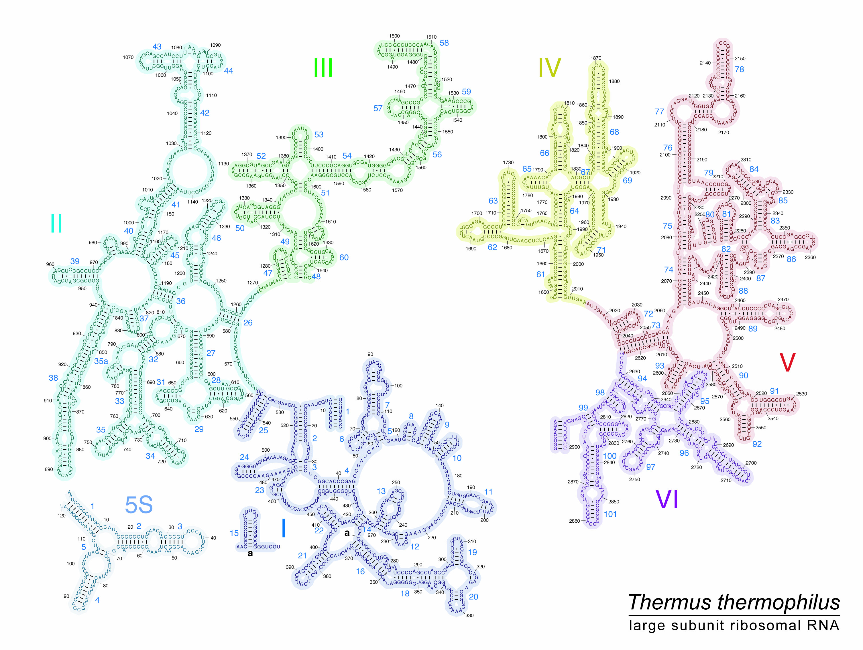
Another complication is relating traditional helix numbering and definitions to the accretion model and various ribosome studies. This reflects the fact that the traditional helix numbering scheme is rather random. Diagrams indicating the numbering system are available at (http://rna.ucsc.edu/rnacenter/images/figs/thermus_23s_2ndry.jpg) and for the SSU at (http://rna.ucsc.edu/rnacenter/images/figs/thermus_16s_2ndry.jpg). For example, both helix H34 and helix H42 actually consist of two stems separated by an interior loop. Using the recently proposed naming system for ribosomal proteins [80], it is seen for the case of H34 that ribosomal protein uL4 interacts in part with one of these helices but not the other. So saying uL4 interacts with helix H34 is misleading. In the accretion model, this numbering system is discarded in favor of a system in which each accretion element is given a number. So now the portion of helix H34 that interacts with uL4 is seen to be part of accretion element 10. However, accretion element 10 also contains what in the traditional helix numbering system is helix H35. Because the traditional numbering system has been widely used in numerous papers, the best solution might be to break offending helices in the traditional nomenclature up such that each stem has a specific name that relates to the original naming system. Then we would see that uL4 binds to H34a but not H34b.
Most recently, the accretion analysis was extended to the small subunit [81]. Again, it was possible to assign a relative age to various helices, and phases could be assigned. The crucial issue at this point is the time of initial collaboration between the two subunits. The small subunit might be envisioned to initially start as a small growing RNA with its own history that begins to interact with the emerging large subunit. In the most extreme case, the small subunit RNA is envisioned to have absolutely no prior history and thus is associated perhaps by hybridization with the large subunit as a small RNA. The key to correlating the small and large subunit timelines is the bridge elements and the extended two-domain tRNAs, which ultimately connect the two subunits. The bridges can only form when both partners exist. The timing relationship between the two subunits was based on bridge elements with directionality supported by A-minor elements in which the younger A-minor donors were found to be in the SSU. In the accretion model, the earliest bridge elements are B3 and B2a. Once the bridge elements were in place, it was possible to correlate subsequent events in the two subunits. However, prior to the amalgamation of the two subunits, each would have a separate timeline. The combined model provides an approximate time of appearance for each helical region and thereby provides an approximate timeline (Fig. 4) against which various ribosomal features and activities can be mapped. This timeline is organized into six phases for bacterial ribosomes, and events associated with each phase are described in detail [81].

A summary timeline representing major events in the evolution of the full ribosome is reprinted with permission [81]. In this version, the LSU and SSU are envisioned to have initially evolved independently and subsequently are joined together in phase 3
2.3 Is the PTC the Oldest Portion of the Ribosome?
Separately an alternative model of ribosome origins that putatively relies primarily on phylogenetic methods applied to the ribosomal RNA structure has been proposed [82, 83]. This model and Petrov’s accretion model [80] have generated recent controversy [84–86]. A primary issue is whether or not the origin of protein synthesis began with the creation of the PTC region of the large subunit. One strong indicator of this is that the PTC can be made from a single self-folding RNA, whereas the decoding center is not [87]. From a purely semantic point of view, it would appear the origin should be the structure that was first able to make peptide bonds. However, there is a possible complication in that it has been suggested on several occasions that the translocation process that moves the mRNA may have originated in the RNA World, possibly as the original replicase [88–90].
In fact, both Petrov and Caetano-Anollés recognize that a portion of the ancestral small subunit rRNA may have existed as an independent body prior to any association with a precursor large subunit. Harish and Caetano-Anollés [83] clearly state, “Intersubunit bridge history indicates early independent evolution of subunits.” In this view, both subunits already consist of multiple helices at the time they first associate. In the case of the accretion model, the large subunit precursor is envisioned to contain the major components of the PTC and thought to already be making non-coded peptides at the time it acquires (or is acquired by) the proto-SSU in phase 3. Indeed, the modern 50S particle can synthesize peptides by itself. It therefore is appropriate to simply consider the addition of the small subunit as another step toward the modern ribosome thereby indicating that translation evolved from the PTC even if a portion of the 30S subunit had an earlier prior history. However, this argument does not apply to the Caetano-Anollés model because at the time of the envisioned merger the PTC region was not yet included in the emerging large subunit.
The calculations that support this alternative view produce a tree of relationships between various helical elements. In this tree, helices H76, H41–42, H38, H67, H96, H60, H55, H101, H27, H25, and H16 all predate the helices that comprise the PTC. Several of these non-PTC putative aboriginal helices are not part of the universal core seen by sequence comparisons [68]. In addition, they are not actually backbone interconnected in the modern structure. This raises the question of how they were held together to create the proto-LSU structure. It might be argued that this could be done by hybridization. Consistent with this, in some cases modern rRNAs have been found to be produced by multiple smaller fragments that hybridize together to make the full rRNA [69]. In fact, it has been posited that the PTC itself may have emerged from hybridization of two fragments [69] and/or duplication of a smaller fragment [25]. From this writer’s perspective, the Caetano-Anollés model needs to show that the proposed early fragments could have interacted with one another by hybridization without disrupting their essential structure. In addition, an explanation for how additional RNA was subsequently inserted between the putative early helices would be helpful. More detailed discussion of the Caetano-Anollés model will be found elsewhere in this volume.
3 Major Events Along the Timeline
In the accretion model, events are described at a level in which local structural elements of the RNAs are added at specific relative times. This allows one to infer the likely addition of other features. For example, it is reasonable to speculate that a particular ribosomal protein is not present until its binding site has been added. There are many inferences of this type that remain to be fully appreciated. In order to begin organizing this very detailed information, the emergence of various ribosomal subsystems and components have been broken up into approximately six phases. Major events associated with various phases in ribosome history will be discussed in detail in this section.
3.1 Homochirality and the Ribosome
A long-standing issue in the origin of life is when and how homochirality was introduced into emerging biological systems [91]. Homochirality is generally perceived to be the result of chemical properties and therefore thought to likely precede the emergence of ribosomes. That said, as reviewed in some detail previously [92], the modern ribosome can in fact be convinced to accept tRNAs carrying D-amino acids [93, 94]. In addition, mutations in the PTC can improve the tolerance for D-amino acids [45, 95–97] and even β-amino acids [98]. If mutations can decrease the specificity of the modern ribosome, then it is clear that mutations in the past may have improved the specificity. Separately, a variety of processes are utilized to insure that D-amino acids are not attached to tRNAs by the amino acyl tRNA synthetases [99, 100], thereby indicating that this also may have been a problem in the past.
Thus, there is strong evidence that the early ribosome likely would have incorporated D-amino acids with some likelihood. However, if both processes have the same chiral preference then working together with both having perhaps 70 % accuracy they quickly produce a product that is over 85 % homochiral. So even if L-amino acids were not predominant in the pre-ribosome period, the early ribosomes would soon be producing nearly homochiral peptides. It should also be noted that incorporation of a D-amino acids into a modern protein is not necessarily destructive to the formation of a peptide, but instead it depends on where they are located [96, 97]. Alpha helices in particular are intolerant of D-amino acids. Thus, the first peptides made by a primitive ribosome would be more likely to contain beta sheets than alpha helices. Amino acids that do not present a chirality problem, e.g., glycine, might also be favored.
3.2 tRNA and the Timeline
The tRNAs span the two subunits and are therefore central to the modern ribosome. The universal CCA sequence at their 3′ end carries either the incoming amino acid or the partially synthesized growing peptide chain and is thus associated with the PTC. At the opposite extreme, approximately 70 Angstroms away, the anticodon regions of two tRNAs interact with the mRNA at the decoding center of the small subunit.
The origin of the typical modern tRNA of 76 nucleotides has been the subject of considerable discussion [101]. It has in particular been observed that the tRNA consists of essentially two domains with the presumably older top half carrying the amino acid and the younger lower half interacting with the mRNA [60, 102, 103]. The top portion includes the T-stem, the acceptor stem, and the universal, but typically not coded, terminal CCA to which the amino acid is attached to the modern tRNA. It has been suggested that this top portion may have originated as an even simpler single hairpin to which was later added the T-stem and loop [104]. A subsequent ligation of two copies of a stem loop structure could in principle produce the essence of the tRNA’s 3D structure including long-range interactions [24].
As attractive as this general idea is, it is important to appreciate that other facts raise concern and tRNA origins may be consistent with other hypotheses. For example, in the case of some mitochondria, the D arm or T arm is deleted from the tRNA [105]. This feature normally interacts with the L1 stalk in the large subunit rRNA, which is also missing. The L1 stalk is a phase 5/phase 6 addition to the LSU RNA, thereby raising the possibility that the D-stem is a recent addition. Likewise, the discovery of split tRNA genes in Nanoarchaeum equitans [106] suggests models in which two half structures formed the tRNA structure by hybridization rather than duplication [101].
However, the idea of a bottom-to-top addition of the lower portion of the tRNA stem has been greatly strengthened by the discovery of an ancient insertion fingerprint that supports the addition of the D arm and anticodon arm into an earlier structure consisting of the CCA stem and T arm [81]. The new addition to the tRNA would thus make interaction with a proto-mRNA possible, thereby representing the origins of true coding within the context of ribosome history. When did this occur? Although it is currently not obvious how to directly time the origin of the two-domain tRNA, one can infer its presence by the relative time of appearance of the small subunit helices associated with the decoding center. This places it as most likely in late phase 3 or early phase 4.
3.3 Ribosome History Has Implications for the Origin of the Genetic Code
The holy grail of ribosome evolution, which will not be addressed here, is an explanation of the origin of the modern codon assignments. This relates at least in part to the aminoacyl tRNA synthetases and has been recently examined in several reviews [107, 108]. The genetic code, as we know it, is often thought to arise in two stages. As small RNAs brought possible substrates to the proto-PTC, the sequence of the RNA carrier would favor the attachment of some amino acids (or perhaps esters) over others, resulting in an operational code that did not rely on any genomic information. The peptides made would be largely random depending on the availability and affinity of particular amino acids. Over time, as the carrier RNA became larger, additional residues would be added to the CCA stem thereby expanding the initial operational code. True coding as is found in modern organisms would require a two-domain tRNA and a proto-ribosome that includes the basic components of both subunits as well as an mRNA. Thus, in the context of the accretion model, coded synthesis would not begin until late in phase 3 or phase 4. The likely initial driving force was the dynamic h44–h28 combination, which could spontaneously switch between two or more configurations. This motion might have been coupled to a noncoding proto-mRNA that hybridized to h44 much as the Shine-Dalgarno sequence [109] of modern bacterial mRNAs does. The addition of the lower portion of the tRNA would then allow the tRNAs to interact indirectly with the h44–h28 driver by hybridization to the proto-mRNA. Alternatively the tRNA may have been extended first and then serendipitously interacted with a small RNA associated with the h44–h28 complex [110]. This would in either case greatly improve the speed of peptide synthesis by increasing the rate of motion from the A to P site. The ability to more rapidly make random peptides of upwards of even 10–15 residues likely would have provided a new level of complexity to the prebiotic world. The sequence of the proto-mRNA would govern which tRNAs would best interact with it. This would create an environment in which, in a manner perhaps similar to the operational code, a genetic code as we know it could evolve. Thus, the proto-mRNA that may have initially simply facilitated movement of the tRNAs would now be available to carry information. Understanding the early history of the ribosome has not yet provided a specific explanation for any codon assignments. However, it does provide potentially useful insights into the environment in which the code was established.
Although the modern code is largely universal, there are numerous minor exceptions found in various organisms and most especially mitochondria. When a tRNA is mutated such that it recognizes two anticodons, e.g., UGA and UGG, as Trp, for example, this eliminates a stop codon. Alternatively, a tRNA mutation may result in a methionine tRNA that now reads both AUA and AUG. Thus, changes in codon assignments in modern organisms may really be about changes in the mRNA and tRNA populations. Especially interesting was the observation that in human mitochondria the arginine codons AGA and AGG were extremely rare and apparently the cell did not produce a tRNA to recognize them [111]. Hence, they were thought to have become de facto stop codons. However, it was subsequently found that they instead provoked frame-shifting events that then restored a normal stop codon [112].
These sorts of observation may have implications for the manner in which we envision coding prior to the LUCA. Initial genomes, especially RNA genomes, would have initially been small. As a result, many codons would potentially be rare and have to be dealt with in some manner. If they were treated as stop codons this would be undesirable as it would limit the size of peptides that could be made. In this context, tRNAs that could recognize multiple codons might have been preferred. The role of stop codons and small genomes has been discussed before [113] and likely should be revisited.
3.4 Ribosomal Proteins Line the Path to Increasing Complexity
Before the sequences and structures of the ribosomal proteins were known, it was thought that they might be descended from some small number of ancient proteins by gene duplication. In fact, there are only a few examples of such relationships, and lateral transfer events involving the ribosomal proteins are at best rare [114, 115]. In our original attempt to establish a timeline for ribosome evolution [116], the ribosomal proteins played a key role. It was observed that the non-universal proteins were typically late additions in ribosome assembly. This led to the hypothesis that the assembly map recapitulated to some significant extent the evolutionary history of the ribosome [116]. It was thus argued that the oldest ribosomal proteins were likely uL2, uL3, uL4, and uL24, as these were at the core of the assembly process. Experimental studies showed that 50S particles alone containing 5S rRNA and eight proteins including uL2 and uL3 were active [117]. Subsequently, the relative ordering of both the SSU and LSU chronologies were optimized with respect to differences in amino acid usage bias [114]. The results, in conflict with the cladistics model [82, 83], strongly supported a scenario in which the LSU predates the SSU. Ribosomal proteins uL2, uL3, and uL4 in particular again appeared to be the oldest.
Modern genomics has shown that gene order is not typically conserved over vast phylogenetic distances [118]. In contrast with this observation, analysis of the ribosomal proteins revealed that in bacteria and archaea there are six clusters/operons consisting almost exclusively of ribosomal proteins that are in fact conserved. Within this group, the two largest clusters are the S10 operon, which includes uL2, uL3, uL4, and the spc operon, which includes uL24. In both cases, the order of the genes, not just the gene content, is typically preserved. In E. coli where experimental studies have been made, these operons are all regulated at the translational (e.g., RNA) level rather than the transcriptional level. This has been cited in support of these clusters having been part of a primitive RNA genome [118, 119].
An examination of the ribosomal proteins in the SCOP database release 4.75 [120] reveals that many of the ribosomal proteins have structures that are either all alpha or all beta. However, the subunits differ in that proteins with all beta structures, such as uL2 and uL3, and proteins comprised mostly of parallel beta sheets, such as uL4, occur more frequently in the large subunit than in the SSU. The universal proteins uL2, uL3, uL4, and uL22 all have extensions that reach the PTC region. It has been pointed out [121] that the regions that comprise these extensions are peptides with no secondary structure, loops with beta turns, or beta hairpins. The exit tunnel, which starts at the PTC, by its role in providing a path to the exterior of the ribosome, must have been maintained as the ribosome grew larger. Moving along this path is then basically a timeline. In doing so, one sees initial beta structures such as an extension of uL22 followed only later by an alpha helix. This absence of alpha helical structures in the oldest parts of the PTC may reflect the difficulty in obtaining alpha helices in peptides that initially may have been comprised of mixed chirality amino acids.
When equivalent proteins from the three domains of life were aligned, they were frequently found to exhibit a block structure in which some regions are universal and other segments are specific to either the Bacteria or the Archaea [122]. In a few cases, e.g., uL6, uL11, uL14, uS9, and uS11, there is essentially only a single universal block [122]. The existence of domain-specific blocks strongly supports the notion that, like the ribosomal RNAs, portions of the proteins may have arisen at different times. One clear example of a protein with interesting history is uL2. It has two distinct domains. The N-terminal domain forms an OB fold, while the C-terminal domain forms an SH3 fold [120, 123]. What is especially noteworthy is that these two folds are in fact very similar [123]. By simply moving the location of one beta segment, it is possibly to effectively convert the SH3 fold to an OB fold. Thus, it is likely that one of the domains may have arisen from the other by duplication in conjunction with a partial rearrangement.
The question then is which domain of uL2 is the oldest. The accretion model may provide the answer by combining RNA-protein interaction sites with the likely age of the interaction sites. In examining a preliminary protein-RNA interaction map available at the CRW web site (http://www.rna.icmb.utexas.edu/SIM/4A/CRWStructure/rpi/rpi.23S.hb.pdf) [124], one finds that the primary site of interaction of the more universal proteins typically interacts with the predicted older regions of the RNA. At the very oldest regions of both subunits, protein interactions are minimal to nonexistent. The issue of the primary binding site where a protein interacts is complicated by the fact that there are typically multiple interaction sites for each protein. However, in most cases there is a cluster of contacts representing the primary interaction site. In the case of uL2, the SH3 domain interacts with accretion elements 12 and 12a, while the OB domain interacts primarily with accretion elements 9, 13, and 14. In both cases, these regions are assigned to phase 3, and although both domains are clearly essential to the early history of the ribosome, it remains unclear which domain is older. Interestingly, there is a small segment of uL2 that is between the two major domains that forms a small loop that interacts with the loop of helix 93 in what is regarded as phase 2 in the accretion model. Rather than either domain, this small element may in fact be the very oldest part of uL2.
Consistent with their early evolution, both the SH3 and OB domains are devoid of an alpha helix, and each is found in other ribosome-relevant proteins. The SH3 domain is found in bL21 and uL24 as well as a variety of intracellular or membrane-associated proteins. The OB fold is frequently associated with oligonucleotide binding and is found in several ribosomal proteins including uS12, uS17, and bS1. The bS1 protein is associated with initiation and actually contains six copies of the OB fold that form what is known as an S1 domain.
Useful insight can also be obtained by examining the non-universal proteins. Although these proteins likely have a post-LUCA origin, the way they have evolved may provide insight into the pre-LUCA acquisition of proteins. In a seminal paper, Klein et al. [125] compared the structures and locations of ribosomal proteins from the archaeon Haloarcula marismortui and the bacterium Deinococcus radiodurans. It was observed in several cases that one or more of the unique H. marismortui proteins in fact have non-homologous analogs in the Deinococcus structure. For example, eL21, eL24, and eL37 have analogs bL27, bL19, and bL34, respectively. In each case, the analogous proteins are distinctly different in sequence and structure. However, at the level of secondary structure at least, one sees essentially no significant change in the LSU RNA in these cases. When differences do occur, they are relatively minor. There are also examples of proteins that are in one structure with no analog in the other. Thus, for example bL25 and bL36 are unique to Deinococcus, and eL18, eL19, and eL39 are unique to Haloarcula. In some cases there are again no obvious RNA differences. This raises the evolutionary question in each case of whether the unique protein is a post-LUCA gain where it occurs or a loss where it is not found.
However the RNA sometimes does noticeably change in association with a protein. One interesting case is eL18, which is unique to Haloarcula and has partial sequence homology with uL15. It turns out that its binding site partially overlaps with uL15 on helix H27. In addition, there is a small helix insert (H30) between H29 and H31, which is the primary binding site for eL18. This insert and the protein are both absent in Deinococcus.
3.5 Origins of the Dynamic Ribosome
The modern ribosome is a dynamic molecular machine that executes protein synthesis one residue at a time. An incoming tRNA carrying the next amino acid in association with elongation factor EF-Tu enters at the A site following GTP cleavage. Peptide bond formation is spontaneous, and the tRNA now carrying the growing peptide chain moves to the P site with the help of elongation factor EF-G and a second GTP cleavage. A new tRNA now enters at the A site, and the process is repeated. As the process proceeds, the tRNA is now deacylated and moves to the E site from which it exits the ribosome. During the process the small subunit exhibits head swivel and rotates relative to the large subunit. Although facilitated by EF-Tu and EF-G, the core motions are actually inherent to the ribosome itself. In the absence of the factors, protein synthesis can still proceed [126, 127]. Thus, the ribosome is thought to be inherently a processive Brownian motor [128].
Well before any structures were available, Woese proposed that the tRNA itself was dynamic, ratcheting between two configurations of the anticodon loop [129]. This proved to be the correct general idea, but not the correct specific structure. The first atomic resolution tRNA structure revealed what appeared to be a hinge-like structure in the lower portion of the tRNA [130, 131]. It is now appreciated that as the tRNA enters and ultimately leaves the ribosome, various conformations are produced as a result of motions facilitated by the pivot point as indicated in Fig. 5, which was adapted from Dunckle et al. [132].

Alternative tRNA orientations seen during a single translation cycle
The existence of this key center of motion inspired a search for additional pivots within the large RNAs. Initially, atomic resolution structures of the ribosome before and after EF-G associated GTP cleavage were compared. A total of 23 pivots were found [133]. Of these, 15 were in the small subunit and 8 in the large subunit. A similar analysis of pivots associated with EF-Tu associated GTP cleavage found 16 pivots, 4 of which are uniquely associated with EF-Tu. [134]. That many shared pivots are found is consistent with the long-standing observation that the EF-G and EF-Tu sites of interaction partially overlap [135, 136]. Both EF-G and EF-Tu interact with the factor-binding site, which includes helices H43 and H44.The pivoting positions are consistently associated with weak spots in the RNA structure such as non-standard base pairs and bulge loops.
In the case of EF-G, pivots in small subunit helices h28 and h44 are especially interesting. Motions associated with these two pivots strongly affected other pivots, Helix h28 appeared to control h31, h33, h36, h37, h39, h40, h41, h42, and h43, while pivot h32 appears to control motions at pivots h33, h36, h37, h39, and h40. These pivots are associated with the head swivel and head rotation. Overall, there appears to be a network of interacting pivots as outlined in Fig. 6, which is reprinted with permission [134].

Proposed partial network of EF-G-associated pivoting elements found in the ribosomal RNAs [134, 137]. Black lines indicate direct physical contact between moving helixes. Dashed lines indicate the motion that results from an upstream pivoting motion. Helixes h28, h32, and h34 form the head domain of the SSU and lie in sequence. Helices h28 and h32 influence the motions of a number of more external pivots, which are listed vertically in the figure, as well as helix h34, which contacts EF-G. Thus, a cascade of motion originating with EF-G-GTP hydrolysis is plausible in either direction—forward toward the tRNA or in the reverse direction toward h34
The dynamic aspects of the modern ribosome clearly make it faster and likely more accurate as well. Placing the emergence of this key event on the timeline of ribosome evolution is an important goal. To begin to answer this question, one can examine when the various helices containing pivots were incorporated according to the accretion model [77]. In the case of the LSU, the ten pivots associated with either EF-Tu or EF-G or both are listed as phase 5 or 6. The one exception is the helix H89 pivot, which is part of the original aes1 element that initiated the P site portion of the PTC. This helix connects with helix H91 and thereby indirectly with the alpha sarcin/ricin loop. As has been pointed out [77], helix elongation events do not leave insertion fingerprints, and therefore in the absence of clues from A-minor interactions, such events cannot be readily established. Therefore, it is not inconsistent with the accretion model to infer that helix H89 arose at a later time as a simple insertion in the pivoting element. It therefore seems likely that all the mobile aspects of the LSU RNA were added rather late in the process but nevertheless well before the LUCA.
The small subunit is clearly more dynamic than the large subunit. It is actively involved in both the head swivel and head rotation. In the accretion model, the vast majority of the pivots are either phase 5 (helices h6, h41, h42, and h44) or phase 6 (helices h8, h21, h26, h36/37, h38, h39, and h40). Pivots in helices h31, h32, and 43 are assigned to phase 4. There are small subunit pivots in helices h6, h32, h 41, and h44, which are also assigned to phase 5. Pivots in helices h8, h21, h26, h32, h36/37, h39, and h40 are designated as phase 6 and hence more recent. Pivots in helices h31, h32, and h43 are assigned to phase 4. The remaining two pivots h28 and h44 are regarded as being among the first elements to be incorporated into the growing small subunit. In the accretion model, they are assigned to phase 2 and phase 1, respectively. This is consistent with the fact that motions at these pivots strongly influence the motion seen at multiple other pivots. Helix h44 includes the Shine-Dalgarno sequence [109], which facilitates the initial interaction of the mRNA with the small ribosomal subunit during initiation and is involved in multiple bridges to the large subunit.
Overall these results indicate that the dynamic ribosome arose in essentially two separate stages. It is likely that initially the H28/H44 pair were part of an inherently dynamic element that independent of the ribosome would spontaneously alternate between two or more conformations. When incorporated into the emerging ribosome during phase 3, this inherent motion would have driven both the mRNA and tRNA movement. Subsequently, as the ribosome grew, other mobile elements were added, primarily in phase 5 or 6, resulting in head swivel and the ratcheting motion.
3.6 Recent Aspects of Ribosome Evolution
The ribosome clearly continued to evolve as complexity reached and extended beyond that of the LUCA. For example, as discussed elsewhere in this volume, initiation is distinctly different in eukaryotes. Likewise, the various factors have evolved over time. In bacteria, the addition of the tmRNA system for recovering stalled ribosomes is a noteworthy achievement. Another key development is the introduction of post-transcriptional modifications. In this section, three important additions that occurred very near the time of the LUCA are discussed.
3.6.1 Trigger Factor and Factor Binding Site
The involvement of ribosomes in co-translational folding in conjunction with the signal recognition particle and trigger factor was an important development in the integration of the ribosome with other cellular systems [138]. In this case, the key docking protein for the trigger factor is uL23, which interacts near helices H51 and H53, which are added in phase 5. A second protein that interacts with the trigger factor, uL29, is associated with phase 6. It thus appears that the trigger factor and co-translational folding predate the LUCA.
Although the ribosome is at its core a Brownian motor, the modern version is driven by GTP hydrolysis events associated with the initiation factor IF-2, elongation factors EF-Tu and EF-G, and release factors such as RF-2. The addition of these factors to the emerging ribosome would have greatly increased the rate of peptide synthesis and may have been as a result in part responsible for the emergence of the LUCA [70, 139]. In bacteria, the factor-binding site contains multiple copies of ribosomal protein bL12 attached to an underlying stalk formed by helix H42, which is thought to be added in phase 5, and ribosomal protein uL10. However, bL12 is replaced by P1 and P2 in archaea and eukaryotes [140], thereby associating the continuing formation of the factor-binding site with the post-LUCA time period. The structures of these various factors have been inter-compared in some detail, and it was concluded that they likely originated from a fusion of an OB fold to a Ras-like GTPase [121].
3.6.2 5S rRNA
5S rRNA is a small, independent RNA that largely forms the central protuberance of the large ribosomal subunit [141, 142]. In combination with several ribosomal proteins, its incorporation is essentially the last step in ribosome assembly [143]. 5S rRNA is universally found in all three domains of life and therefore was presumably present at the time of the LUCA. Reconstituted ribosomes lacking 5S rRNA have minimal but not completely eliminated activity [144]. In particular, factor-dependent tRNA binding at the A site is interrupted [142]. It was originally proposed that the sequence CGAA that frequently occurs in loop C of 5S rRNAs interacted with the T-loop of the tRNA [145]. As reviewed in detail [142, 146, 147], it is now thought that 5S rRNA may facilitate communication between different ribosome regions. This is a key ribosome function, as the movement of the mRNA likely needs to be coordinated with the synthesis of the peptide bond for the machine to function smoothly. Among 5S rRNAs’ multiple contacts, its interaction with ribosomal protein uL5 is of special interest. This is because uL5 in turn interacts with uS13 to form bridge B1b/c. This bridge is the only protein-protein bridge between the ribosomal subunits, and its time of appearance likely coincides with that of 5S rRNA. Ribosomal protein uS13 also has a long C-terminal extension that reaches the coding site [148, 149]. Hence, a signal may be passed between the LSU and the SSU decoding site. However, this is not essential as a deletion of the uS13 tail only minimally reduced ribosome activity [150].
Although 5S rRNA is regarded as “universal,” this is subject to some interpretation as its structure is somewhat different in various organisms. In the initial E. coli structure obtained by comparative analysis, 5S rRNA was envisioned to have four extended helical regions [151, 152]. In contrast, the equivalent eukaryotic and archaeal structures were soon shown to have a fifth helix [153, 154] in a region that was originally envisioned as a bulge in the prokaryotic cases. The length of this helix varies and can be quite extended in organisms such as Thermoplasma acidophilum that are subjected to harsh environments [155]. Subsequent X-ray studies of this bulge region in E. coli revealed that it was actually a non-standard duplex formed by what is now known as a bulge E motif [156]. In addition, a few characteristic differences are seen in many 5S rRNA such as a single deletion in the loop C region in many eukaryotes. The appearance of equivalent structures is, however, not fully supported by experimental data. Reconstitution assays were conducted in which 5S rRNAs from various sources were incorporated into the large subunit of Bacillus stearothermophilus ribosomes [157]. It was found that 5S rRNAs from prokaryotic organisms typically produced active ribosomes, whereas those from eukaryotes did not [158].
The issue of 5S rRNA universality has long been questioned because it appears to be missing in many mitochondria and some chloroplasts. Recently, a detailed search for 5S rRNA homologs in organelle genomes was conducted [159]. Numerous new examples of 5S rRNA-like RNAs with additional structural variations were found in multiple cases. In the case of the mammal Porcine (S. scrofa), the structure of the large ribosomal subunit was examined by cryoelectron microscopy. It appears that a greatly reduced form of 5S rRNA is in fact present [160]. Just as the universality of 5S rRNA was on the brink of establishment, it was definitively shown this was not the case. The crystal structures of human [161] and yeast [162] were determined, and in both cases 5S rRNA was completely absent. In the case of the human mitochondrial large subunit, the 5S rRNA was replaced by a mitochondrial valine tRNA. This tRNA occupies essentially the same region in the structure as the abbreviated RNA found in the Porcine example. In summary, 5S rRNA was likely a late addition to the ribosome shortly before the LUCA.
4 Summary and Future Studies
It is suggested here that the matter of ribosome origins is best regarded as two distinct problems: the origins of the core machinery and its subsequent evolution. Much of ribosome history predates the LUCA, and thus its study takes us back in time some considerable distance into the pre-LUCA world. It is unclear how far back the origin of the PTC itself or its inherently dynamic SSU counterpart actually extends into the pre-LUCA time period. Clearly RNA is crucial, and the issue of where it comes from has significant implications. If an early path to a true RNA World of any complexity existed, then the core aspects of the ribosome would be a direct and possibly quite early product. If, however, the path to RNA was arduous, perhaps involving catalytic prebiotic peptides, for example, then in the context of the pre-LUCA time frame, the proto-ribosome may not have been so early.
At this juncture considerable attention has been focused on the mechanism of peptide synthesis. In addition, much of what can be inferred from ribosome structures and primary sequences of various components about the very beginnings has likely been uncovered. One especially interesting reminder is that one should perhaps pay attention to the nature of the pre-LUCA earth and thereby realize that ferrous iron may have preceded magnesium in stabilizing RNA [65]. The immediate goal that several groups are pursuing is to uncover a minimal RNA that incorporates the PTC and makes peptides. This model PTC would provide final proof that it is the RNA that is catalytic. Such an experimental system would also provide a starting point for studies to understand the origins and evolution of the modern PTC. It would be possible, for example, to examine how specificity for L or D amino acids is affected by changes in the sequence or structure of the model PTC. Less obvious but likewise important would be to synthesize a minimal segment based on helices h28, h44, and h45 in the small subunit in order to look for an inherent strong alternation between two structures. Again, if such an RNA were available, one could explore its properties with mutational studies.
With regard to the post PTC period, multiple attempts to establish a timeline of major events have been described [64, 70, 71, 92], culminating in the accretion model, which is based primarily on RNA insertion fingerprints and A-minor interactions [81]. As a result, events in the growth of the rRNAs can now be discussed at the level of individual helices or groups of helices.
This timeline of the relative age of various regions of the RNA can be correlated with existing knowledge to form an integrated understanding of ribosome history. This will be especially useful in refining the order of accretion of peripheral elements in the RNAs. Ongoing, for example, are efforts to correlate protein-binding sites as provided by atomic resolution studies with helix age. In the case of proteins with multiple domains, it may be possible to assign a relative age of each domain. Especially noteworthy is the obvious increasing complexity that is encountered in the later phases. Simply put, there is rapidly increasing parallelism as one reaches phases 5 and 6. Much of what is occurring in these and later phases will likely be found to relate to other cellular processes. This may ultimately allow a unified history of the cell.
References
Noller HF. Evolution of protein synthesis from an RNA World. Cold Spring Harb Perspect Biol. 2010;4:a003681.
Ban N, Nissen P, Hansen J, Moore PB, Steitz TA. The complete atomic structure of the large ribosomal subunit at 2.4 A resolution. Science. 2000;2000(289):905–20.
Nissen P, Hansen J, Ban N, Moore PB, Steitz TA. The structural basis of ribosome activity in peptide bond synthesis. Science. 2000;289:920–30.
Maguire BA, Beniaminov AD, Ramu H, Mankin AS, Zimmermann RA. A protein component at the heart of an RNA machine: the importance of protein l27 for the function of the bacterial ribosome. Mol Cell. 2005;20:427–35.
Selmer M, Dunham CM, Murphy FV 4th, Weixlbaumer A, Petry S, Kelley AC, Weir JR, Ramakrishnan V. Structure of the 70S ribosome complexed with mRNA and tRNA. Science. 2006;313:1935–42.
Wower IK, Wower J, Zimmermann RA. Ribosomal protein L27 participates in both 50 S subunit assembly and the peptidyl transferase reaction. J Biol Chem. 1998;273:19847–52.
Maracci C, Wohlgemuth I, Rodnina MV. Activities of the peptidtl transferase center of ribosomes lacking protein L27. RNA. 2015;21:1–6.
White HB III. Coenzymes as fossils of an earlier metabolic state. J Mol Evol. 1976;1976(7):101–4.
Yarus M. Getting past the RNA World: The initial Darwinian Ancestor. Cold Spring Harb Perspect Biol. 2010;3:a003590.
Bartel DP, Szostak JW. Isolation of new ribozymes from a large pool of random sequences. Science. 1993;261:1411–8.
Zaher HS, Unrau PJ. Selection of an improved RNA polymerase ribozyme with superior extension and fidelity. RNA. 2007;13:1017–26.
Attwater J, Wochner A, Holliger P. In-ice evolution of RNA polymerase ribozyme activity. Nat Chem. 2013;5:1011–8.
Mutswchler H, Wochner A, Hollinger P. Freeze-thaw cycles as drivers of complex ribozyme assembly. Nat Chem. 2015;7:502–8.
Wolf YI, Koonin EV. On the origin of the translation system and the genetic code in the RNA world by means of natural selection, exaptation, and subfunctionalization. Biol Direct. 2007;2:14.
Zhang B, Cech TR. Peptide bond formation by in vitro selected ribozymes. Nature. 1997;1997(390):96–100.
Lilley DM. The origins of RNA catalysis in ribozymes. Trends Biochem Sci. 2003;28:495–501.
Bowman JC, Hud NV, Williams LD. The ribosome challenge to the RNA World. J Mol Evol. 2015;80:143–61.
Cafferty BJ, Hud NV. Abiotic synthesis of RNA in water: a common goal of prebiotic chemistry and bottom-up synthetic biology. Curr Opin Chem Biol. 2014;22:146–57.
Sczepanski JT, Joyce GF. A cross-chiral RNA polymerase ribozyme. Nature. 2014;515: 440–2.
Van der Gulik P, Massar S, Gilis D, Buhrman H, Rooman M. The first peptides: the evolutionary transition between prebiotic amino acids and early proteins. J Theor Biol. 2009;261:531–9.
Ferris JP, Hill WR Jr, Rihe L, Orgel LE. Synthesis of long prebiotic oligomers on mineral surfaces. Nature. 1996;381:59–61.
Stachelhaus T, Marahiel MA. Modular structure of genes encoding multifunctional peptide synthetases required for non-ribosomal peptide synthesis. FEMS Microbiol Lett. 1995;125:3–14.
Marahiel MA. Working outside the protein-synthesis rules: insights into non-ribosomal peptide synthesis. J Peptide Sci. 2009;15:799–807.
Nagaswamy U, Fox GE. RNA ligation and the origin of tRNA. Orig Life Evol Biosph. 2003;33:199–209.
Agmon I. The dimeric proto-ribosome: Structural details and possible implications on the origin of life. Int J Mol Sci. 2009;2009(10):2921–34.
Davidovich C, Belousoff M, Wekselman I, Shapira T, Krupkin M, Zimmerman E, Bashan A, Yonath A. The proto-ribosome: an ancient nano-machine for peptide bond formation. Isr J Chem. 2010;50:29–35.
Krupkin M, Matzov D, Tang H, Metz M, Kalaora R, Belousoff MJ, Zimmerman E, Bashan A, Yonath A. A vestige of a prebiotic bonding machine is functioning within the contemporary ribosome. Philos Tran R Soc Lond B Biol Sci. 2011;366:2972–8.
Boer PH, Gray MW. Scambled ribosomal RNA gene pieces in Chlamydomonas reinhardtii mitochondrial DNA. Cell. 1988;55:399–411.
Schnare MN, Gray MW. Sixteen discrete RNA components in the cytoplasmic ribosome of Euglena gracilis. J Mol Biol. 1990;215:73–83.
Polacek N, Mankin AS. The ribosomal peptidyl transferase center: Structure, function, evolution, inhibition. Crit Rev Biochem Mol Biol. 2005;40:285–311.
Katunin VI, Muth GW, Strobel SA, Wintermeyer W, Rodnina MV. Important contribution to catalysis of peptide bond formation by a single ionizing group within the ribosome. Mol Cell. 2002;10:339–46.
Sievers A, Beringer M, Rodnina MV, Wolfenden R. The ribosome as an entropy trap. Proc Natl Acad Sci USA. 2004;101:7897–901.
Schroeder GK, Wolfenden R. The rate enhancement produced by the ribosome: an improved model. Biochemistry. 2007;46:4037–44.
Moore PB, Steitz TA. After the ribosome structures: How does peptidyl transferase work? RNA. 2001;9:155–9.
Wohlgemuth I, Beringer M, Rodnina MV. Rapid peptide bond formation on isolated 50S ribosomal subunits. EMBO reports. 2006;7:699–703.
Rodina MV, Beringer M, Wintermeyer W. How ribosomes make peptide bonds. Trends Biochem Sci. 2007;32:20–6.
Zaher HS, Shaw JJ, Strobel SA, Green R. The 2’-OH group of the peptidyl-tRNA stabilizes an active conformation of the ribosomal PTC. EMBO J. 2011;30:2445–53.
Carrasco N, Hiller DA, Strobel SA. Minimal transition state charge stabilization of the oxyanion during peptide bond formation by the ribosome. Biochemistry. 2011;50:10491–8.
Świderek K, Marti S, Tuñón I, Moliner V, Bertran J. Peptide bond formation mechanism catalyzed by ribosome. J Am Chem Soc. 2015;137:12024–34.
Fahnestock S, Rich A. Ribosome-catalyzed polyester formation Science. 1971;173:340–3.
Fahnestock S, Neumann H, Shashoua V, Rich A. Ribosome catalyzed ester formation. Biochemistry. 1970;9:2477–83.
Victorova LS, Kotusov VV, Azhaev AV, Krayevsky AA, Kukhanova MK, Gottikh BP. Synthesis of thioamide bond catalyzed by E. coli ribosomes. FEBS Lett. 1976;68:215–8.
Ohta A, Murakami H, Suga H. Polymerization of alpha-hydroxy acids by ribosomes. Chem Bio Chem. 2008;9:2773–8.
Subtelny AO, Hartman MC, Szostak JW. Ribosomal synthesis of n-methyl peptides. J Am Chem Soc. 2008;130:6131–6.
Tan Z, Forster AC, Blacklow SC, Cornish VW. Amino acid backbone specificity of the Escherichia coli translation machinery. J Am Chem Soc. 2004;126:12752–3.
Hartman MC, Josephson K, Lin CW, Szostak JW. An expanded set of amino acids analogs for the ribosomal translation of unnatural peptides. PLoS ONE. 2007;2:e972.
Kang TJ, Suga H. Ribosomal synthesis of nonstandard peptides. Biochem Cell Biol. 2008;86:92–9.
Zavialov AV, Mora L, Buckingham RH, Ehrenberg M. Release of peptide promoted by the GGQ motif of class 1 release factors regulates the GTPase activity of RF3. Mol Cell. 2002;10:789–98.
Fox GE, Tran Q. Yonath. An exit cavity was crucial to the polymerase activity of the early ribosome. Astrobiology. 2012;12:57–60.
Nakatogawa H, Ito K. The ribosomal exit tunnel functions as a discriminating gate. Cell. 2002;108:629–36.
Lu J, Deutsch C. Folding zones inside the ribosomal exit tunnel. Nat Struct Mol Biol. 2005;12:1123–9.
Yonath A. Antibiotics targeting ribosomes: resistance, selectivity, synergism and cellular regulation. Annu. Rev. Biochem. 2005;74:649–79.
Ito K, Chiba S, Pogliano K. Divergent stalling sequences sense and control cellular physiology. Biochem Biophys Res Commun. 2010;393:1–5.
Ranu H, Mankin A, Vazquez-Laslop N. Programmed drug-dependent ribosome stalling. Mol Microbiol. 2009;71:811–24.
Ramu H, Vazquez-Laslop N, Klepacki D, Dai Q, Piccrilli J, Micura R, Mankin AS. Nascent peptide in the ribosome exit tunnel affects functional properties of the A-site of the peptidyl transferase center. Mol Cell. 2011;41:321–30.
Chiba S, Kanamori T, Ueda T, Akiyama Y, Pogliano K, Ito K. Recruitment of a species-specific translational arrest module to monitor different cellular processes. Proc Natl Acad Sci USA. 2011;108:6073–8.
Turk RM, Illangasekare M, Yarus M. Catalyzed and spontaneous reactions on ribozyme ribose. J Am Chem Soc. 2011;133:6044–50.
Schaul S, Berel D, Benjamini Y, Graur D. Revisiting the operational code for amino acids: Ensemble attributes and their implications. RNA. 2010;16:141–53.
Schimmel P, Giege R, Moras D, Yokoyama S. An operational RNA code for amino acids and possible relationship to genetic code. Proc Natl Acad Sci USA 1993;90:8763–8.
Schimmel P. Ribas de Pouplana L. Transfer RNA: From Minihelix to Genetic Code. Cell. 1995;81:983–6.
Francklyn C, Schimmel P. Aminoacylation of RNA minihelices with alanine. Nature. 1989;337:478–81.
Francklyn C, Schimmel P. Enzymatic aminoacylation of an eight base pair mivrohelix with histidine. Proc Natl Acad Sci USA. 1990;87:8655–9.
Kleine DJ, Moore PB, Steitz TA. The contribution of metal ions to the structural stability of the large ribosomal subunit. RNA. 2004;10:1366–79.
Hsiao C, Mohan S, Kalahar BK, Williams LD. Peeling the Onion: ribosomes are ancient molecular fossils. Mol Biol Evol. 2009;26:2415–25.
Hsiao C, Chou IC, Okafor CD, Bowman JC, O’Neill EB, Athavale SS, Petrov AS, Hud NV, Wartell RM, Harvey SC, Williams LD. RNA with iron (II) as a cofactor catalyzes electron transfer. Nat Chem. 2013;5:525–8.
Huang L, Krupkin M, Bashan A, Yonath A, Massa L. Protoribosome by quantum kernel energy method. Proc Natl Acad Sci USA. 2013;110:14900–5.
Rivas M, Tran Q, Fox GE. Nanometer scale pores similar in size to the entrance of the ribosomal exit cavity are a common feature of large RNAs. RNA. 2013;19:1349–54.
Mears JA, Cannone JJ, Stagg SM, Gutell RR, Agrawal RK, Harvey SC. Modeling a minimal ribosome based on comparative sequence analysis. J Mol Biol. 2002;321:215–34.
Gray MW, Schnare MN. Evolution of RNA gene organization. In: Zimmermann RA, Dahlberg AE, editors. Ribosomal RNA—Structure, evolution, processing, and function in protein synthesis. Boca Raton, Florida: CRC Press; 1996. p. 49–69.
Hury J, Nagaswamy U, Larios-Sanz M, Fox GE. Ribosome origins: The relative age of 23S rRNA domains. Orig Life Evol Biosphere. 2006;36:421–9.
Bokov K, Steinberg SV. A hierarchical model for evolution of 23S ribosomal RNA. Nature. 2009;457:977–80.
Nissen P, Lppolito JA, Ban N, Moore PB, Steitz TA. RNA tertiary interactions in the large ribosomal subunit: the A-minor motif. Proc Natl Acad Sci USA. 2001;98:4899–903.
Réblová K, Šponer JE, Špačková N, Beššeová I, Šponer J. A-minor tertiary interactions in RNA kink-turns. Molecular dynamics and quantum chemical analysis. J Phys Chem B. 2011;115:13897–910.
Gerbi SA. Expansion segments: Regions of variable size that interrupt the universal core secondary structure of ribosomal RNA. In: Zimmermann RA, Dahlberg AE, editors. Ribosomal RNA—Structure, evolution, processing, and function in protein synthesis. Boca Raton, Florida: CRC Press; 1996. p. 71–87.
Gerbi SA. Evolution of ribosomal DNA. In: MacIntyee RJ, editor. Molecular evolutionary genetics. New York: Plenum Publishing Corporation; 1985. p. 419–51774.
Luehrsen KR, Nicholson DE, Eubanks DC, Fox GE. An archaebacterial 5S rRNA contains a long insertion sequence. Nature. 1981;293:755–7.
Petrov AS, Bernier CR, Hsiao C, Norris AM, Kovacs NA, Waterbury CC, Stepanov VG, Harvey SC, Fox GE, Wartell RM, Hud NV, Williams LD. Evolution of the ribosome at atomic resolution. Proc Natl Acad Sci USA. 2014;111:10251–6.
Caisova L, Melkonian M. Evolution of helix formation in the ribosome internal transcribed spacer 2 (ITS2) and its significance for RNA secondary structures. J Mol Evol. 2014;78:324–7.
Petrov AS, Bernier CR, Gulen B, Waterbury CC, Hershkovits E, Hsiao C, Harvey SC, Hud NV, Fox GE, Wartell RM, Williams LD. Secondary structures of tRNAs from all three domains of life. PLoS One. 2014;9:e88222.
Ban N, Beckmann R, Cate JH, Dinman JD, Dragon F, Ellis SR, Lafontaine DL, Lindahl L, Liljas A, Lipton JM, McAlear MA, Moore PB, Noller HF, Ortega J, Panse VG, Ramakrishnan V, Spahn CM, Steitz TA, Tchorzewski M, Tollervey D, Warren AJ, Williamson JR, Wilson D, Yonath A, Yusupov M. A new system for naming ribosomal proteins. Curr Opin Struct Biol. 2014;24:165–9.
Petrov AS, Gulen B, Norris A, Kovacs NA, Bernier CR, Lanier KA, Fox GE, Harvey SC, Wartell RM, Hud NV, Williams LD. The history of the ribosome and the origin of translation. Proc. Natl. Acad Sci USA. 2015;112:15396–401.
Caetano-Anollés G. Tracing the evolution of RNA structure in ribosomes. Nucl Acids Res. 2002;30:2575–87.
Harish A, Caetano-Anollés G. Ribosomal history reveals origins of modern protein synthesis. PLoS One. 2012;7:e32776.
Petrov AS, Williams LD. The ancient heart of the ribosomal large subunit: a response to Caetano-Anollés. J Mol Evol. 2015;80:166–70.
Caetano-Anollés D, Caetano-Anollés G. Ribosomal accretion aprioism and the phylogenetic method: A response to Petrov and Williams. Front Genet. 2015;6:194.
Caetano-Anollés G. Ancestral insertions and expansions of rRNA do not support an origin of the ribosome in its peptidyl transferase center. J Mol Evol. 2015;80:162–5.
Smith TF, Gutell R, Lee J, Hartmann H. The origin and evolution of the ribosome. Biol Direct. 2008;3:16.
Campbell J. An RNA repisome as the ancestor of the ribosome. J Mol Evol. 1991;32:3–5.
Gordon K. Were RNA replication and translation directly coupled in the RNA (+protein) World? J Theor Biol. 1995;173:179–93.
Poole AM, Jeffares DC, Penny D. The path from the RNA world. J Mol Evol. 1998;46:1–17.
Blackmond DG. The origin of biological homochirality. Cold Spring Harb Perspect Biol. 2010;2:a002147.
Fox GE. Origin and evolution of the ribosome. Cold Spring Harb Perspect Biol. 2010;3:a003483.
Yamane T, Miller DL, Hopfield JJ. Discrimination between D and L-tyrosyl transfer ribonucleic acids in peptide chain elongation. Biochemistry. 1981;20:7059–68.
Heckler TG, Roesser JR, Xu C, Chang PI, Hecht SM. Ribosomal binding and dipeptide formation by misacylated tRNAPhe’s. Biochemistry. 1988;27:7254–62.
Starck SR, Qi X, Olsen BN, Roberts RW. The puromycin route to assess stero- and regiochemical constraints on peptide bond formation in eukaryotic ribosomes. J Am Chem Soc. 2003;125:8090–1.
Dedkova LM, Fahmi NE, Golovine SY, Hecht SM. Enhanced D-amino acid incorporation into proteins by modified ribosomes. J Am Chem Soc. 2003;125:6616–7.
Dedkova LM, Fahmi NE, Golovine SY, Hecht SM. Construction of modified ribosomes for incorporation of D-amino acids into proteins. Biochemistry. 2006;45:15541–51.
Dedkova LM, Fahmi NE, Paul R, del Rosario M, Zhang L, Chen S, Feder G, Hecht SM. β-Puromycin selection of modified ribosomes for in vitro incorporation of β-amino acids. Biochemistry. 2012;51:401–15.
Soutourina J, Plateau P, Blanquet S. Metabolism of D-aminoacyl-tRNAs in Eschrichia coli and Saccharomyces cerevisiae cells. J Biol Chem. 2000;275:32535–42.
Yang H, Zheng G, Peng X, Qiang B, Yuan J. D-amino acids and D-Tyr-tRNAtyr deacylase: Stereospecificity of the translation machine revisited. FEBS Lett. 2008;552:95–8.
Di Giulio M. A comparison among the models proposed to explain the origin of the tRNA molecule: A synthesis. J Mol Evol. 2009;69:1–9.
Maizels N, Weiner AM. The genomic tag hypothesis: modern viruses as molecular fossils of ancient strategies for genomic replication. In: Gesteland RF. Atkins JF editors. The RNA world, Plainview. New York: Cold Springs Harbor Laboratory Press; 1993. p. 577–602.
Maizels N, Weiner AM. Phylogeny from function: evidence from the molecular fossil record that tRNA originated in replication, not translation. Proc Natl Acad Sci USA. 1994;91:6729–34.
Di Giulio M. On the origin of the transfer RNA molecule. J Theor Biol. 1992;159:199–214.
Watanabe Y, Suematsu T, Ohtsuki T. Losing the stem-loop structure From metazoan mitochondrial tRNAs and co-evolution of interacting factors. Front Genet. 2014;5:109.
Randau L, Munch R, Hohn M, Jahn D, Soll D. Nanoarchaeum equitans creates functional tRNAs from separate genes for their50- and 30-halves. Nature. 2005;433:537–41.
Carter CW. What RNA world? Why a peptide/RNA partnership merits renewed experimental attention. Life (Basel). 2015;5:294–320.
Smith TF, Hartman H. The evolution of class II aminoacyl-tRNA synthetases and the first code. FEBS Lett. 2015;589:3499–507.
Shine J, Dalgarno L. The 3’-terminal sequence of Escherichia coli 16S ribosomal RNA: complementarity to nonsense triplets and ribosome binding sites. Proc Natl Acad Sci USA. 1974;71:1342–6.
Bernhardt HS, Tate WP. The transition from noncoded to coded protein synthesis: did coding mRNAs arise from stability-enhancing binding partners to tRNA? Biol Direct. 2012;7:4.
Barrell BG, Anderson S, Bankier AT, de Bruijn MH, Chen E, Coulson AR, Drouin J, Eperon IC, Nierlich DP, Roe BA, Sanger F, Schreier PH, Smith AJ, Staden R, Young IG. Different pattern of codon recognition by mammalian mitochondrial tRNAs. Proc Natl Acad Sci USA. 1980;77:3164–6.
Temperley R. Richter R, DEnnerlein S, Lightowlers, Chrzanowska-Lightowlers Z. Hungry codons promote frameshifting in human mitcchondrial ribosomes. Science. 2010;327:301.
Lehman N, Jukes TH. Genetic code development by stop codon takeover. J Theor Biol. 1988;135:203–14.
Fournier GP, Neumann JE, Gogarten JP. Inferring the ancient history of the translation machinery and genetic code via recapitulation of ribosomal subunit assembly orders. PLoS One. 2010;27:1792–801.
Wang J, Dasgupta I, Fox GE. Many nonuniversal archaeal ribosomal proteins are found in conserved gene clusters. Archaea. 2009;2:241–51.
Fox GE, Naik AK. The evolutionary history of the translation machinery. In Ribas, de Pouplana L, editor), The genetic code and the origin of genetic code. New York, New York: Springer. US. 2004;2007:92–105.
Khaitovich P, Mankin AS, Green R, Lancaster L, Noller HF. Characterization of functionally active subribosomal particles from Thermus aquaticus. Proc Natl Acad Sci USA. 1999;96:85–90.
Siefert JL, Fox GE. Conserved gene clusters in bacterial genomes provide further support for the primacy of RNA. J Mol Evol. 1997;45:467–72.
Joyce GF. RNA evolution and the origins of life. Nature. 1989;338:217–24.
Murzin AG, Brenner SE, Hubbard T, Chothia C. SCOP: a structural classification of proteins database for the investigation of sequences and structures. J Mol Biol. 1995;247:536–40.
Hartman H, Smith TF. The evolution of the ribosome and the genetic code. Life (Basel). 2014;4:227–49.
Vishwanath P, Favaretto P, Hartman H, Mohr SC, Smith TF. Ribosomal protein-sequence block structure suggests complex prokaryotic evolution with implications for the origin of eukaryotes. Mol Phylogenet Evol. 2004;33:615–25.
Agrawal V, Kishan RK. Functional evolution of two subtly different (similar) folds. BMC Struct Biol. 2001;1:5.
Cannone JJ, Subramanian S, Schnare MN, Collett JR, D’Souza LM, Du Y, Feng B, Lin N, Madabusi LV, Muller KM, Pande N, Shang Z, Yu N, Gutell RR. The comparative RNA web (CRW) site: an online database of comparative sequence and structure information for ribosomal, intron, and other RNAs. BMC Bioinformatics. 2002;3:2.
Klein DJ, Moore PB, Steitz TA. The roles of ribosomal proteins in the structure assembly, and evolution of the large ribosomal subunit. J Mol Biol. 2004;340:141–77.
Spirin AS. Ribosome as a molecular machine. FEBS Lett. 2002;514:2–10.
Gavrilova LP, Kostiashkina VE, Koteliansky NM, Rutkevich NM, Spirin AS. Factor free (“Non-enzymatic”) and factor-dependent systems of translation of polyuridylic acid by Escherichia coli ribosomes. Mol Biol. 1976;101:537–52.
Frank J, Gonzalez RL Jr. Structure and Dynamics of a processive Brownian motor: the translating ribosome. Annu Rev Biochem. 2010;2010(79):381–412.
Woese C. Molecular mechanics of translation: a reciprocating ratchet mechanism. Nature. 1970;226:817–20.
Robertus JD, Ladner JE, Finch JT, Rhodes D, Brown RS, Clark BF, Klug A. Structure of yeast phenylalanine tRNA at 3S resolution. Nature. 1974;250:546–51.
Harvey SC, McCammon JA. Intramolecular flexibility in phenylalanine transfer RNA. Nature. 1981;294:286–7.
Dunkle JA, Wang L, Feldman MB, Pulk A, Chen VB, Kapral GJ, Noeske J, Richardson JS, Blanchard SC. Cate JH. Structures of the bacterial ribosome in classical and hybrid states of tRNA binding. Science. 2011;332:981–4.
Paci M, Fox GE. Major centers of motion in the large ribosomal RNAs. Nucl Acids Res. 2015;43:4640–9.
Paci M, Fox GE. Centers of motion associated with EF-Tu binding to the ribosome. RNA Biol. 2016;Jan 19.0 [Epub ahead of print].
Moazed D, Robertson JM, Noller HF. Interaction of elongation factors EF-G and EF-Tu with a conserved loop in 23S rRNA. Nature. 1998;334:362–4.
Sergiev PV, Bogdanov AA, Dontsova OA. How can elongation factors EF-G and EF-Tu discriminate the functional state of the ribosome using the same binding site? FEBS Lett. 2005;579:5439–42.
Fox GE, Paci M, Tran Q, Petrov, Williams LD. Ribosome dynamics and the Evolutionary history of ribosomes. Proceedings SPIE Conference 9606 Instruments, Methods and Missions for Astrobiology XVII 2015: 96060G1-96060G-5.
Hoffmann A, Bukau B, Kramer G. Structure and function of the molecular chaperone trigger factor. Biochim Biophys Acta. 2010;1803:650–61.
Frank J, Gao H, Sengupta J, Gao N, Taylor DJ. The process of mRNA-tRNA translocation. Proc Natl Acad Sci USA. 2007;104:19671–8.
Grela P, Bernadó P, Svergun D, Kwiatowski J, Abramczyk D, Grankowski N, Tchórzewski M. Structural relationships among the ribosomal stalk proteins from the three domains of life. J Mol Evol. 2008;67:154–67.
Szymanski M, Barciszewska MZ, Erdmann VA, Barciszewski J. 5 S rRNA: structure and interactions. Biochem J. 2003;371:641–51.
Gongadz GM. 5S rRNA and Ribosome. Biochemistry (Moscow). 2011;76:1450–64.
Dohme F, Nierhaus KH. Role of 5S RNA in assembly and function of the 50S subunit from Escherichia coli. Proc Natl Acad Sci USA. 1976;73:2221–5.
Erdmann VA, Fahnestock S, Higo K, Nomura M. Role of 5S RNA in the function of 50S ribosomal subunits. Proc Natl Acad Sci USA. 1971;68:2932–6.
Forget BG, Weissman SM. Nucleotide sequence of KB cell 5S RNA. Science. 1967;158:1695–9.
Dontsova OA, Dinman JD. 5S rRNA: Structure and Function from Head to Toe. Int J Biomed Sci. 2005;1:1–7.
Bogdanov AA, Dontsova OA, Dokudovskaya SS, Lavrik IN. Structure and function of 5S rRNA in the ribosome. Biochem Cell Biol. 1995;73:869–76.
Dokudovskaya S, Dontsova O, Shpanchenko O, Bogdanov A, Brimacombe R. Loop IV of 5S ribosomal RNA has contacts both to domain II and to domain V of the 23S RNA. RNA. 1996;2:146–52.
Stark H, Rodnina MV, Wieden HJ, Zemlin F, Wintermeyer W, van Heel M. Ribosome interactions of aminoacyl-tRNA and elongation factor Tu in the codon-recognition complex. Nat. Struct Biol. 2002;9:849–54.
Hoang L, Fredrick K, Noller HF. Creating ribosomes with an all-RNA 30S subunit P site. Proc Natl Acad Sci USA. 2004;101:12439–43.
Fox GE, Woese CR. 5S rRNA secondary structure. Nature. 1975;256:505–7.
Fox GE, Woese CR. The architecture of 5S rRNA and its relation to function. J Mol Evol. 1975;6:61–76.
Nishikawa K, Takemura S. Nucleotide sequence of 5 S RNA from Torulopsis utilis. FEBS Lett. 1974;40:106–9.
Luehrsen KR, Fox GE. The secondary structure of eucaryotic cytoplasmic 5S ribosomal RNA. Proc Natl Acad Sci USA. 1981;78:2150–4.
Fox GE. The structure and evolution of archaebacterial RNA. In: Gunzalus IC, editor. The Bacteria, vol 8. New York: Academic Press; 1985. p. 267–310.
Correll CC, Freeborn B, Moore PB, Steitz TA. Metals, motif, and recognition in the crystal structure of a 5S rRNA domain. Cell. 1997;91:705–12.
Wrede P, Erdmann VA. Activities of B. stearothermophilus 50S ribosomes reconstituted with prokaryotic and eukaryotic 5S RNA. FEBS Lett. 1975;33:315–9.
Bellemare G, Vigne R, Jordan B. Interaction between Escherichia coli ribosomal proteins and 5S RNA molecules: recognition of prokaryotic 5S RNAs and rejection of eukaryotic 5S RNAs. Biochimie. 1973;55:29–35.
Valach M, Burger G, Gray MW, Lang BF. Widespread occurrence of organelle genome-encoded 5S rRNAs including permuted molecules. Nucleic Acids Res. 2014;42:13764–77.
Greber BJ, Boehringer D, Leitner A, Bieri P, Voigts-Hoffmann F, Erzberger JP, Leibundgut M, Aebersold R, Ban N. Architecture of the large subunit of the mammalian mitochondrial ribosome. Nature. 2014;515:283–6.
Brown A, Amunts A, Bai XC, Sugimoto Y, Edwards PC, Murshudov G, Scheres SH, Ramakrishnan V. Structure of the large ribosomal subunit from human mitochondria. Science. 2014;346:718–22.
Amunts A, Brown A, Bai XC, Llácer JL, Hussain T, Emsley P, Long F, Murshudov G, Sjors Scheres SHW, Ramakrishnan V. Structure of the yeast mitochondrial large ribosomal subunit. Science. 2014;2014(43):1485–9.
Author information
Authors and Affiliations
Corresponding author
Editor information
Editors and Affiliations
Rights and permissions
Copyright information
© 2016 Springer International Publishing Switzerland
About this chapter
Cite this chapter
Fox, G.E. (2016). Origins and Early Evolution of the Ribosome. In: Hernández, G., Jagus, R. (eds) Evolution of the Protein Synthesis Machinery and Its Regulation. Springer, Cham. https://doi.org/10.1007/978-3-319-39468-8_3
Download citation
DOI: https://doi.org/10.1007/978-3-319-39468-8_3
Published:
Publisher Name: Springer, Cham
Print ISBN: 978-3-319-39467-1
Online ISBN: 978-3-319-39468-8
eBook Packages: Biomedical and Life SciencesBiomedical and Life Sciences (R0)