
Abstract
Common skin bacterial infections include superficial pyodermas (impetigo, folliculitis, furuncle and furunculosis, carbuncle, abscesses, suppurative paronychia), due to Staphylococcus aureus with an emergence of Panton-Valentine leukocidin secreting and community-acquired methicillin-resistant strains and/or group A hemolytic Streptococcus; erythrasma, due to coryneform bacteria; and erysipelas, due to Streptococcus pyogenes. Diagnosis relies on clinical appearance and bacterial sampling in difficult cases. Antiseptics and local antibiotics are effective in limited superficial pyodermas. In contrast, extensive lesions and erysipelas must be treated with systemic antibiotics chosen according to the most probable causative agent and the emergence of resistance to antibiotics in some countries.
Access provided by Autonomous University of Puebla. Download chapter PDF
Keywords
- Superficial pyodermas
- Impetigo
- Impetiginisation
- Furuncle
- Carbuncle
- Folliculitis
- Staphylococcal nasal carriage
- Erythrasma
- Erysipelas
1.1 Background
1.1.1 Definition
Common skin bacterial infections are very frequent in daily dermatological practice. Superficial pyodermas, characterized by involvement of the epidermis, the upper dermis, and the superficial part of the adnexal structures (hair follicles and nails) [1], include impetigo and impetiginization of dermatoses, folliculitis, furuncles and furunculosis, carbuncle, and suppurative paronychia. Other common skin infections are erythrasma, involving the upper epidermis, primary abscesses, and erysipelas, involving the deep dermis and the hypodermis.
1.1.2 Bacteriology
Staphylococcus aureus is the main pathogen involved in these infections, group A hemolytic Streptococcus being the second in frequency [2].
In contrast with the normal cutaneous flora residing on the superficial epidermal layers and pilotropic adnexa, mainly comprising Staphylococcus epidermidis and gram-positive rods such as corynebacteria and Propionibacterium acnes, Staphylococcus aureus is not a permanent part of the normal skin flora but colonizes inflamed skin (psoriasis, atopic dermatitis, cutaneous lymphoma, bullous diseases, erythroderma) [3–5]. S. aureus may also colonize the newborn’s umbilicus for several weeks after birth.
However, 20–60 % of normal individuals have a persistent or intermittent nasal carriage of S. aureus, depending on bacterial adherence and host factors [6]. Other sites of carriage are the perineum, axillary folds, and umbilicus. Nasal carriage is mostly a risk factor for chronic or recurrent furuncles (furunculosis) [7].
Important advances concerning S. aureus virulence mechanisms have been made over last decades. For instance Panton-Valentine leukocidin (PVL), carried by methicillin-sensitive or methicillin-resistant S. aureus, is responsible for necrosis and apoptosis of neutrophils and is thus frequently involved in the pathogenesis of follicular infections and abscesses [8–10].
It has also been shown that S. aureus produces exfoliative toxins responsible for blistering bacterial infections such as bullous impetigo [7] and staphylococcal scalded skin syndrome (SSSS) by cleaving the cell adhesion cadherin desmoglein 1, which plays an important role in the barrier function of the upper epidermal layers [11, 12].
The emergence of community-acquired methicillin-resistant S. aureus (CA-MRSA) must also be considered today. Since the emergence of MRSA as a nosocomial agent during the 1960s, several clones have spread out of hospitals and health-care facilities: (1) health-care-associated MRSA clones, responsible for infections in patients with risk factors such as recently hospitalized patients, patients with chronic diseases, and drug abusers, and (2) true de novo community producing PVL, responsible for cutaneous or, less frequently, necrotizing pulmonary infections in patients without any risk factors and being now a true public health problem worldwide [13–16].
Group A hemolytic Streptococcus (S. pyogenes A, C, G) is present in the mouth in 10 % of the population, periorificial regions but skin carriage in normal individuals is rare. M proteins, pyrogenic and erythrogenic SPE exotoxins and streptolysin are the main virulence factors of the bacteria [17, 18].
The current increasing resistance of S. pyogenes to macrolides must be taken in account in some countries but is being better controlled by a lower use of antibiotics [19–21]. However, there is no resistance to penicillin, and clindamycin and pristinamycin (if available) which remain good alternatives in case of allergy to penicillin [22, 23].
1.2 Impetigo
1.2.1 Definition
Impetigo is a contagious primary infection of the skin, involving the stratum corneum, due to S. aureus, less frequently S. pyogenes, or both. It is particularly common in children and disadvantaged areas. Self-inoculation and small family or communities outbreaks are frequent.
Two types of clinical form are described: (1) crusted impetigo is due to S. aureus or S. pyogenes, and (2) bullous impetigo is due to the cleavage of very superficial epidermal layers by staphylococcal toxins (exfoliatins) and can be either disseminated (SSSS) or more limited. However, in general practice, both bacteria should be considered when prescribing treatment because bacteriological sampling is often not performed or feasible in general practice unless there is a specific epidemiological context or resistance to classical treatment.
1.2.2 Clinical Presentation
The diagnosis is based on clinical examination. Early lesions are isolated or confluent and polycyclic vesicles or blisters, followed by erosions and yellowish crusts (“honey-colored”) (Figs. 1.1 and 1.2).

Periorificial crusted impetigo

Bullous impetigo
Typical location in children is around orifices, especially the mouth, but all areas of the skin may be affected. There are usually no systemic symptoms such as fever, although there may be local lymphadenopathy.
Ecthyma, more frequent in patients with poor hygiene, is a necrotic complication of streptococcal impetigo, clinically characterized by deep dermal ulcerations.
1.2.3 Prognosis
Without treatment, impetigo usually resolves without sequelae within 2 weeks.
Although systemic complications of childhood impetigo are rare today, infectious or post-infectious glomerulonephritis may occur after extensive, chronic, and/or neglected streptococcal or staphylococcal superficial pyodermas in patients with risk factors such as alcoholism, diabetes mellitus, or drug abuse [24]. Furthermore, glomerulonephritis is a major complication of scabies-related streptococcal impetigo in resource-poor countries [25]. In contrast, rheumatic fever does not complicate impetigo except possibly in tropical areas in association with scabies. Other rare complications include sepsis, osteomyelitis, arthritis, endocarditis, lymphangitis, erysipelas, guttate psoriasis, and SSSS [26].
1.2.4 Treatment
Improving personal hygiene with daily showers, washing hands, brushing nails, and frequent change of clothes is important. Washing lesions with soap and water or topical antiseptics (povidone-iodine, chlorhexidine, hexamidine, Dakin’s solution) is usually sufficient for very limited lesions. However, topical antibiotics are widely recommended and may be the treatment of choice for impetigo of limited extent (one site and/or five to ten lesions) [27]. Topical fusidic acid and mupirocin have a comparable effectiveness and are as effective as oral flucloxacillin [28, 29]. Furthermore, moisturizing ointments are useful to remove crusts and enhance healing.
With more extensive lesions, systemic antibiotics against both S. aureus and S. pyogenes are recommended: penicillin M (oxacillin or rather cloxacillin for better oral route bioavailability), amoxicillin-clavulanic acid, first or second generation cephalosporin, fusidic acid, or pristinamycin [30]. Given the emerging resistance of S. aureus and S. pyogenes, macrolides (except erythromycin because of its effect on QT interval [31]) should only be used as a second line where there is intolerance or allergy to beta-lactams [32].
1.3 Folliculitis, Furuncle, Chronic Furunculosis, and Carbuncle
1.3.1 Definition
Folliculitis and furuncles, the most common forms of skin infection, are acute hair follicle infections (infection of the ostium or follicular opening in superficial folliculitis or infection of the entire hair follicle in furuncles), due to S. aureus including strains secreting PVL. In the case of superficial folliculitis, other strains of Staphylococcus, or other microorganisms such as Candida albicans, may be involved. Apart from climatic factors such as humidity or heat, folliculitis and furuncles are more frequent in patients with immunosuppression, diabetes mellitus, long-term antibiotic use, obesity, occlusive clothes, or recurrent shaving, especially in pubic area. Chronic furunculosis is mainly associated with nasal carriage of S. aureus [10, 33]. We have recently seen several patients with Enterobacter aerogenes folliculitis acquired after spa baths.
Gram-negative folliculitis, especially folliculitis due to Pseudomonas, has been described since the late 1970s as case reports or outbreaks in swimming pools, whirlpools, hot tubs, spa, and recently rubber gloves with an incubation period of from 2 to 5 days [34–36].
In patients with acne in whom treatment with tetracyclines has not shown a significant improvement after 3–6 months, a gram-negative infection should be considered and investigated if possible. The gram-negative bacteria include Escherichia coli, Pseudomonas aeruginosa, Serratia marcescens, Klebsiella, and Proteus mirabilis [37].
1.3.2 Clinical Presentation
-
Folliculitis is characterized by scattered or extensive follicular pustules surrounded by a macular or papular erythema, mainly located on thighs, buttocks, back, and beard (Fig. 1.3).
Fig. 1.3 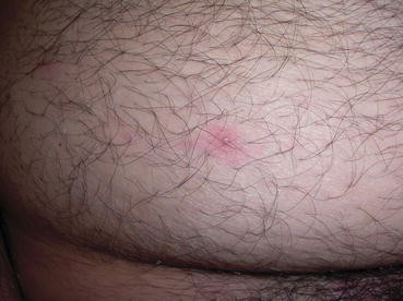
Folliculitis
-
Pseudofolliculitis (pili incarnati) results from the penetration into the skin of shaved hairs within follicles. This condition [38] affects people with curly hair. Papules and pustules may lead to discomfort and small scars or postinflammatory hyperpigmentation.
-
Furuncle (boil) is the consequence of an acute and sometimes necrotic PVL-positive staphylococcal infection of the entire hair follicle and presents as an inflammatory papule then a painful nodule with a central follicular pustule, evolving after a few days leading to spontaneous necrosis and suppuration with discharge of a necrotic core then a permanent scar. Lesions may be single or multiple, involving the face, buttocks, arms, thighs, and anogenital area. There is seldom fever and systemic complications are quite rare. However, with facial furuncles, cavernous sinus thrombosis may constitute a dangerous complication [39].
-
Chronic furunculosis is the recurrence of furuncles over a number of months and is, favored by obesity, diabetes mellitus, immunodepression, iron deficiency, alcoholism, and malnutrition. However, staphylococcal nasal carriage and exposure to an infected family member are the main risk factors, and antiseptic decontamination is most important in order to avoid recurrences [40–42].
-
Carbuncles are the aggregation of furuncles and form broad, swollen, painful, and often fluctuant nodules and deep masses with spontaneous multiple drainage tracts. Patients are often febrile may be unwell [43] (Fig. 1.4).
Fig. 1.4 
Carbuncle of the face


1.3.3 Treatment
1.3.3.1 Superficial Folliculitis
Any external causal agent should be removed or controlled if appropriate. Cleaning with water and soap and/or antiseptics, with a broad antimicrobial activity [44], is effective in most cases. Topical antibiotics such as mupirocin or fusidic acid are usually effective twice a day for 5–7 days with a low rate of resistance but are not always prescribed. Systemic antistaphylococcal antibiotics are recommended in more severe cases defined as extensive, persistent, and/or recurrent lesions, after bacteriological sampling [27, 29, 45].
1.3.3.2 Pseudofolliculitis
In the acute phase, shaving should be stopped for several weeks until improvement, and local treatments such as topic erythromycin or fusidic acid, antiseptics or tretinoin may be useful [46]. Shaving must subsequently be performed with individual adjustment, for example, electric razors preferentially over manual razors for beard folliculitis. Hair should be left 1 mm long. Hair removal with chemical depilatories may be effective, and laser, with or without eflornithine hydrochloride, could provide long-term remission, especially for beard folliculitis in patients with dark skin [46–49].
1.3.3.3 Gram-Negative Folliculitis
Lesions usually cure either spontaneously or with antiseptics such as acetic acid, chlorhexidine, povidone, and potassium permanganate within a few days. Silver sulfadiazine cream may also be useful, but other topical anti-gram-negative antibiotics such as polymyxin are disappointing.
Prevention is based on adequate maintenance of bathing facilities, chlorine level, and disinfecting procedures of public equipments [34].
The best treatment of tetracycline-induced gram-negative folliculitis occurring during the course of acne is isotretinoin [50].
1.3.3.4 Furuncles and Carbuncles
Systemic antistaphylococcal antibiotics such as penicillin M (oxacillin, cloxacillin, flucloxacillin) should be prescribed for 5–7 days [51], associated with topical antibiotics applied on the lesion and the surrounding skin. Occlusive clothes must be avoided. Hygiene measures should be reinforced (hand washing, nail brushing, repeated showers with soap and/or antiseptics).
In case of recurrences, an underlying condition must be sought (staphylococcal nasal and extranasal carriage in the patient and his household members, diabetes, immunodepression, etc.) and treated (see below). Nasal carriage is found in 60 % of patients with recurrent furunculosis [40].
Carbuncles require the same antibiotics approach as furuncles associated with surgical excision [52].
1.3.3.5 Staphylococcal Carriage
Due to the risk of spreading in the close environment of the patient, staphylococcal carriage must be sought and treated in the patient and his family but is not definitive, with progressive recolonization within 6 months [53, 54]. Prevention of the bacterial spread in the family or close contact persons (sportsmen [55]), especially in case of CA-MRSA, is based on hygiene, no sharing of personal clothes, and regular hand washing. Carriage decontamination procedures, based on antiseptics as chlorhexidine baths or showers associated with prolonged and intermittent topical antibiotics as mupirocin or fusidic acid [41, 56–58], remain unproven and have been mainly studied for MRSA in health-care facilities [42, 59–61].
1.4 Abscesses
1.4.1 Definition and Clinical Presentation
An abscess is a collection of pus. It is not clear in the literature which size of collection can define an abscess, but some consider 2 cm diameter as the lower limit for defining an abscess [62]. The abscess forms a nodule or painful and inflammatory erythematous plaque. After a few days of development, palpation reveals a soft consistency indicating a purulent collection (Fig. 1.5).

Abscess of the neck
Fever is rare, and lymphangitis and satellite nodes may be experienced. The majority of primary or spontaneous abscesses are caused by S. aureus producing Panton-Valentine leukocidin [63]. Secondary abscesses (accidental direct inoculation, addiction, septic injections, etc.) are most often due to S. aureus but not exclusively. The bacteriological analysis allows the identification of the bacteria. There is worldwide an emergence of S. aureus resistant in the community (CA-MRSA) responsible for suppurative infections including abscesses [64, 65].
1.4.2 Treatment
The first treatment is surgery, incision, and drainage [64, 65]. The benefit of antibiotic therapy is low. The Infectious Diseases Society of America (IDSA) recommends systemic antibiotics in the following cases: “critical” location (face for example, etc.), immunosuppression, large volume of the abscess (>5 cm), failure of drainage, extreme age, and the presence of systemic symptoms. In all other cases, antibiotic therapy is not indicated.
1.5 Suppurative Paronychia
1.5.1 Definition
Acute suppurative paronychia is an acute superficial infection or abscesses of the perionychium. Nail biting, finger sucking, aggressive manicuring, and trauma of the nail are the most frequent causes for a portal of entry of the bacteria [66]. S. aureus is the most frequent causative agent, followed by Streptococcus, Pseudomonas, anaerobes, and other microorganisms such as C. albicans and herpes. Acrodermatitis continua of Hallopeau is a differential diagnosis [67]. Acute paronychia must be differentiated from chronic paronychia, which is usually nonsuppurative, due to multiple physical external causes and/or Candida albicans.
1.5.2 Clinical Presentation
The patient complains of pain and tenderness of the perionychium that appears swollen, purulent, or even fluctuant. The nail coloration and consistency may be altered.
1.5.3 Treatment
If there is no abscess, treatment is based on warm water soaks three times a day and oral antistaphylococcal antibiotics such as amoxicillin-clavulanic acid or clindamycin, also effective on anaerobes, if persistence and after bacteriological culture. In case of abscess, surgical drainage becomes necessary in the absence of spontaneous evacuation of the pus [66].
1.6 Erythrasma
1.6.1 Definition
Erythrasma is a superficial infection of the skin usually localized in the large folds, especially axillae, due to coryneform bacteria (Corynebacterium minutissimum), belonging to the normal skin flora [68]. Warm and humid climate, age, and diabetes mellitus [69] are predisposing factors.
1.6.2 Clinical Presentation
Lesions are large, red or brown sharply marginated and in extensive patches (Fig. 1.6). They may be either asymptomatic or pruritic and complicated by lichenification. Coral-red fluorescence with Wood’s light, due to coproporphyrin III, strongly suggests the diagnosis, although it does not necessarily indicate active infection. Systemic septic complications are extremely rare [70]. Main differential diagnoses include pityriasis versicolor, fungal intertriginous infections (dermatophytosis, candidiasis), eczema, and psoriasis. Thus, mycological and bacteriological scraping and/or skin biopsy may be required if there is any doubt, but usually clinical and Wood’s light features are typical enough to make the diagnosis.

Erythrasma of the axillary fold
1.6.3 Treatment
There is no clear agreement on the best treatment. Both topical (fusidic acid) and systemic antibiotics (erythromycin 14 days, possibly best avoided because of cardiac side effects such as QT interval prolongation [31], clarithromycin single dose) are effective without significant difference [68, 71–73]. Azole antifungal creams are also effective [74]. A photodynamic treatment using red light without any exogenous agent, based on the fact that corynebacteria produce endogenous porphyrins, has been demonstrated in some cases [75].
1.7 Erysipelas
1.7.1 Definition
Erysipelas is an acute superficial dermal infection that usually affects the leg and is commonly caused by streptococci [76]. In contrast with the life-threatening condition necrotizing fasciitis, erysipelas does not involve deep fascia and muscles. However, there is confusion in the literature between erysipelas and cellulitis which is, sensu stricto, an inflammation limited to the subcutaneous tissue but often refers to more severe cases than classical erysipelas [77]. Incidence has increased in the past decades and is now estimated to be 2–2.5/1,000 persons/year [77, 78]. Whereas necrotizing fasciitis often has a multibacterial origin, almost all erysipelas are due to Streptococcus A (the most frequent), C, and G. However, Streptococcus B may be involved in perineal cellulitis [79] or in newborns [80]. A small number of bacteria are present in the affected tissue, as shown by needle aspiration, but blood cultures remain negative in most cases [81, 82]. Legs are the main site involved, followed by the face and the upper limb, especially in women treated for breast cancer [83, 84]. In erysipelas of the leg, a portal of entry, i.e., disruption of the cutaneous barrier (leg ulcer, wound, fissurated toe-web intertrigo, chronic dermatomycoses of the foot [85], pressure ulcer), but also lymphedema, are risk factors [86]. In contrast, no association was observed with diabetes, alcohol, or smoking [86, 87]. Furthermore, a previous history of cellulitis is a main risk factor for a subsequent recurrence, which affects up to 29 % of patients. However, recurrent erysipelas shares the same risk factors as single episodes [78, 88–90]. The thigh and the gluteal region are less frequently involved and are favored by metabolic syndrome and previous surgical interventions [91].
1.7.2 Clinical Presentation
The onset of the disease is in general sudden with fever, chills, malaise, and altered general conditions. Locally, erysipelas is characterized by a large erythematous, swollen, well-demarcated, and usually raised lesion [92] (Figs. 1.7 and 1.8). Locoregional adenopathy is frequent. Superficial blistering secondary to edema, sometimes with superficial hemorrhage, may be observed [93], especially in patients on anticoagulants or in old people with skin atrophy. Local and/or general signs are suggestive of necrotizing fasciitis and must not be ignored because of the risk of a rapid worsening: hemodynamic instability; intensive pain or, in contrast, local anesthesia; muscular pain or functional impairment; deep dermal blisters with no bleeding after incision; livedo or extensive ecchymosis; necrosis; crepitance; or gas effusion [92] (Fig. 1.9). However, in routine practice, intermediate presentations between classical erysipelas and necrotizing fasciitis are frequent, requiring a very close clinical monitoring and sometimes limited surgery [77, 94], (Fig. 1.10). Risk factors for abscesses formation are alcohol abuse and delayed antibiotics initiation [95].

Erysipelas of the leg

Erysipelas of the breast

Necrotizing fasciitis

Erysipelas of intermediate severity
1.7.3 Biological and Imaging Investigations
-
No laboratory test is warranted in uncomplicated cases. However, in case of hospitalization and/or in patients with underlying diseases or local signs evocative of intermediate severity cellulitis, the following laboratory tests should be considered [92]: blood count (white blood cells), creatinine, C-reactive protein, and creatine phosphokinase (CK) if there is any doubt about muscle involvement. Laboratory Risk Indicator for Necrotizing Fasciitis (LRINEC) score is based on total white cell count, hemoglobin, sodium, glucose, serum creatinine, and C-reactive protein and showed very good positive and negative predictive values for the diagnosis of necrotizing fasciitis [96]. Streptococcal serological studies have no value in practical routine management of classical erysipelas, especially as antibody response may be limited by an early use of antibiotics.
-
No imaging is necessary in typical cases of classical erysipelas. Similarly, in necrotizing fasciitis, no imaging is warranted before surgery in typical cases. However, in atypical or intermediate cases, imaging may be useful – plain films showing gas, ultrasounds in case of underlying abscesses, and CT scan or MRI if there is any doubt about myositis or fasciitis – but the clinician should always keep in mind that imaging should not lead to any delay in surgical decision and that sometimes only surgical exploration can confirm or not the presence of necrotic tissues [94, 97].
-
Bacteriological cultures from superficial swab sampling and fluid-filled bullae, including subcutaneous needle aspiration, are usually negative in common erysipelas but could be performed in immunocompromised patients or in case of failure on empiric treatment in order to investigate another bacteria than Streptococcus [81, 92], as previously discussed. Bacteriological sampling of associated wounds or ulcers, representing the portal of entry of Streptococcus, leads is of uncertain pathogenic significance, showing frequently gram-negative bacteria or Staphylococcus sp. Culture of a skin biopsy is not superior to superficial swabs [92].
1.7.4 Treatment
Hospitalization is necessary if there are local or general signs of severity, as described above [98]. Some underlying conditions such as diabetes mellitus, old age, bad social conditions (homeless people) or immunosuppression, and no possibility of medical reevaluation after 48 h are other criteria that help to determine the need for hospitalization. The best treatment is difficult to define in the absence of large comparative trials [99]. In hospitalized patients, intramuscular or intravenous antibiotics are preferred in severe cases or after failure of ambulatory treatment, whereas oral route remains possible in less severe cases, but the better efficacy of the parenteral route has not been proven [99]. The first-intent treatment in classical erysipelas is antistreptococcal antibiotics. Oral pristinamycin showed its non-inferiority compared to standard intravenous then oral penicillin [76]. However, because of better gastrointestinal tolerance, amoxicillin is usually preferred. In hospitalized patients, even if intravenous penicillin G was previously recommended as first-intent treatment, the currently recommended choice is intravenous amoxicillin 50 mg/kg/day, followed by oral administration after improvement of local signs and disappearance of fever [98]. In case of abscess formation, penicillin M or amoxicillin-clavulanic acid is indicated. In cases of allergy to penicillin, pristinamycin (if available), clindamycin, first generation cephalosporin, or macrolides excluding erythromycin are good choices [76, 98, 100].
The classical recommended duration of the treatment is 10–20 days [98]. Short treatments with levofloxacin (5 days) and tedizolid phosphate (6 days) have been described [101, 102] but are not commonly used in routine practice.
Atypical erysipelas should be treated first with intravenous then oral antistreptococcal and staphylococcal antibiotics such as amoxicillin-clavulanic acid or penicillin M or clindamycin in case of allergy. A surgical treatment may become necessary, especially in case of secondary abscess formation.
There is no indication for anticoagulant therapy for erysipelas. Prophylaxis of deep venous thrombosis should be considered depending on the patient’s other risk factors [103]. Venous compression is recommended during the acute phase and the following weeks to reduce the risk lymphedematous sequelae, which is a main risk factor of recurrence. The portal of entry must be treated as appropriate.
1.7.5 Prevention of Recurrent Erysipelas
Patients who experience two or more erysipelas should receive secondary prevention of recurrences. However, although prevention by penicillin has shown some efficacy [90, 104], strong data concerning the best therapeutic scheme are lacking, and recurrences may occur despite the prophylaxis [105, 106]. Intramuscular benzathine-penicillin G 2.4 MU at 14–21-day intervals is a good choice, but oral penicillin V (250 mg twice a day) [104] or amoxicillin (500–1,000 mg a day) is also possible. In cases of allergy, macrolides have been suggested [107], but erythromycin must be avoided (see above). The optimal duration of preventive treatment is unknown. Recently, a duration of 6 months has shown effectiveness against subsequent relapses compared to placebo [104]. However, some patients need prolonged prophylaxis.
Treatment and secondary prevention of portals of entry such as chronic toe-web maceration, fungal intertrigo, and other exacerbating factors, such as lymphedema or venous insufficiency, are of key importance [85, 98, 108, 109].
References
Del Giudice P, Chosidow O. Superficial pyodermas: advances, recommendations and needs. Dermatol Basel Switz. 2005;210:367–9.
Lorette G, Beaulieu P, Bismuth R, et al. Community-acquired cutaneous infections: causal role of some bacteria and sensitivity to antibiotics. Ann Dermatol Vénéréol. 2003;130:723–8.
Gong JQ, Lin L, Lin T, et al. Skin colonization by Staphylococcus aureus in patients with eczema and atopic dermatitis and relevant combined topical therapy: a double-blind multicentre randomized controlled trial. Br J Dermatol. 2006;155:680–7.
Balci DD, Duran N, Ozer B, Gunesacar R, Onlen Y, Yenin JZ. High prevalence of Staphylococcus aureus cultivation and superantigen production in patients with psoriasis. Eur J Dermatol EJD. 2009;19:238–42.
Talpur R, Bassett R, Duvic M. Prevalence and treatment of Staphylococcus aureus colonization in patients with mycosis fungoides and Sézary syndrome. Br J Dermatol. 2008;159:105–12.
Kluytmans J, van Belkum A, Verbrugh H. Nasal carriage of Staphylococcus aureus: epidemiology, underlying mechanisms, and associated risks. Clin Microbiol Rev. 1997;10:505–20.
Durupt F, Mayor L, Bes M, et al. Prevalence of Staphylococcus aureus toxins and nasal carriage in furuncles and impetigo. Br J Dermatol. 2007;157:1161–7.
Boyle-Vavra S, Daum RS. Community-acquired methicillin-resistant Staphylococcus aureus: the role of Panton-Valentine leukocidin. Lab Investig J Tech Methods Pathol. 2007;87:3–9.
Lina G, Piémont Y, Godail-Gamot F, et al. Involvement of Panton-Valentine leukocidin-producing Staphylococcus aureus in primary skin infections and pneumonia. Clin Infect Dis Off Publ Infect Dis Soc Am. 1999;29:1128–32.
Del Giudice P, Bes M, Hubiche T, et al. Panton-Valentine leukocidin-positive Staphylococcus aureus strains are associated with follicular skin infections. Dermatol Basel Switz. 2011;222:167–70.
Amagai M, Matsuyoshi N, Wang ZH, Andl C, Stanley JR. Toxin in bullous impetigo and staphylococcal scalded-skin syndrome targets desmoglein 1. Nat Med. 2000;6:1275–7.
Hanakawa Y, Stanley JR. Mechanisms of blister formation by staphylococcal toxins. J Biochem (Tokyo). 2004;136:747–50.
King MD, Humphrey BJ, Wang YF, Kourbatova EV, Ray SM, Blumberg HM. Emergence of community-acquired methicillin-resistant Staphylococcus aureus USA 300 clone as the predominant cause of skin and soft-tissue infections. Ann Intern Med. 2006;144:309–17.
Iyer S, Jones DH. Community-acquired methicillin-resistant Staphylococcus aureus skin infection: a retrospective analysis of clinical presentation and treatment of a local outbreak. J Am Acad Dermatol. 2004;50:854–8.
Del Giudice P, Bes M, Hubiche T, et al. Clinical manifestations and outcome of skin infections caused by the community-acquired methicillin-resistant Staphylococcus aureus clone ST80-IV. J Eur Acad Dermatol Venereol JEADV. 2011;25:164–9.
Del Giudice P, Blanc V, Durupt F, et al. Emergence of two populations of methicillin-resistant Staphylococcus aureus with distinct epidemiological, clinical and biological features, isolated from patients with community-acquired skin infections. Br J Dermatol. 2006;154:118–24.
Oehmcke S, Shannon O, Mörgelin M, Herwald H. Streptococcal M proteins and their role as virulence determinants. Clin Chim Acta Int J Clin Chem. 2010;411:1172–80.
Hung C-H, Tsao N, Zeng Y-F, et al. Synergistic effects of streptolysin S and streptococcal pyrogenic exotoxin B on the mouse model of group A streptococcal infection. Med Microbiol Immunol (Berl). 2012;201:357–69.
Seppälä H, Nissinen A, Järvinen H, et al. Resistance to erythromycin in group A streptococci. N Engl J Med. 1992;326:292–7.
Bingen E, Bidet P, Mihaila-Amrouche L, et al. Emergence of macrolide-resistant Streptococcus pyogenes strains in French children. Antimicrob Agents Chemother. 2004;48:3559–62.
D’ Humières C, Cohen R, Levy C, et al. Decline in macrolide-resistant Streptococcus pyogenes isolates from French children. Int J Med Microbiol IJMM. 2012;302:300–3.
Cocuzza CE, Mattina R, Mazzariol A, et al. High incidence of erythromycin-resistant Streptococcus pyogenes in Monza (North Italy) in untreated children with symptoms of acute pharyngo-tonsillitis: an epidemiological and molecular study. Microb Drug Resist Larchmt N. 1997;3:371–8.
Michos AG, Bakoula CG, Braoudaki M, et al. Macrolide resistance in Streptococcus pyogenes: prevalence, resistance determinants, and emm types. Diagn Microbiol Infect Dis. 2009;64:295–9.
Montseny JJ, Meyrier A, Kleinknecht D, Callard P. The current spectrum of infectious glomerulonephritis. Experience with 76 patients and review of the literature. Medicine (Baltimore). 1995;74:63–73.
Hay RJ, Steer AC, Engelman D, Walton S. Scabies in the developing world – its prevalence, complications, and management. Clin Microbiol Infect Off Publ Eur Soc Clin Microbiol Infect Dis. 2012;18:313–23.
Cole C, Gazewood J. Diagnosis and treatment of impetigo. Am Fam Physician. 2007;75:859–64.
Agence Française de Sécurité Sanitaire des Produits de Santé. [Topical antibiotic prescription in primary and secondary bacterial cutaneous infections]. Ann Dermatol Vénéréol. 2004;131:1018–21.
Villiger JW, Robertson WD, Kanji K, et al. A comparison of the new topical antibiotic mupirocin (‘Bactroban’) with oral antibiotics in the treatment of skin infections in general practice. Curr Med Res Opin. 1986;10:339–45.
Machet L, Wolkenstein P, Vaillant L. Topical antibiotics used in dermatology: efficiency, indications and adverse events. Ann Dermatol Vénéréol. 2000;127:425–31.
Chosidow O, Bernard P, Berbis P, et al. Cloxacillin versus pristinamycin for superficial pyodermas: a randomized, open-label, non-inferiority study. Dermatol Basel Switz. 2005;210:370–4.
Tschida SJ, Guay DR, Straka RJ, Hoey LL, Johanning R, Vance-Bryan K. QTc-interval prolongation associated with slow intravenous erythromycin lactobionate infusions in critically ill patients: a prospective evaluation and review of the literature. Pharmacotherapy. 1996;16:663–74.
Bidet P, Plainvert C, Doit C, et al. Streptococcus pyogenes or group A streptococcal infections in child: French national reference center data. Arch Pédiatrie Organe Off Sociéte Française Pédiatrie. 2010;17:201–8.
Empinotti JC, Uyeda H, Ruaro RT, Galhardo AP, Bonatto DC. Pyodermitis. An Bras Dermatol. 2012;87:277–84.
Ratnam S, Hogan K, March SB, Butler RW. Whirlpool-associated folliculitis caused by Pseudomonas aeruginosa: report of an outbreak and review. J Clin Microbiol. 1986;23:655–9.
Centers for Disease Control and Prevention (CDC). Pseudomonas dermatitis/folliculitis associated with pools and hot tubs – Colorado and Maine, 1999–2000. MMWR Morb Mortal Wkly Rep. 2000;49:1087–91.
Mazza J, Borkin M, Buchholz R, Deleo V. Pseudomonas folliculitis contracted from rubber gloves: a public health concern. J Am Acad Dermatol. 2013;69:e93–4.
Poli F, Prost C, Revuz J. Gram-negative bacteria folliculitis. Ann Dermatol Vénéréol. 1988;115:797–800.
Perry PK, Cook-Bolden FE, Rahman Z, Jones E, Taylor SC. Defining pseudofolliculitis barbae in 2001: a review of the literature and current trends. J Am Acad Dermatol. 2002;46(2 Suppl):S113–9.
Rohana AR, Rosli MK, Nik Rizal NY, Shatriah I, Wan Hazabbah WH. Bilateral ophthalmic vein thrombosis secondary to nasal furunculosis. Orbit Amst Neth. 2008;27:215–7.
Demos M, McLeod MP, Nouri K. Recurrent furunculosis: a review of the literature. Br J Dermatol. 2012;167:725–32.
Van Rijen M, Bonten M, Wenzel R, Kluytmans J. Mupirocin ointment for preventing Staphylococcus aureus infections in nasal carriers. Cochrane Database Syst Rev. 2008;CD006216.
Davido B, Dinh A, Salomon J, et al. Recurrent furunculosis: efficacy of the CMC regimen – skin disinfection (chlorhexidine), local nasal antibiotic (mupirocin), and systemic antibiotic (clindamycin). Scand J Infect Dis. 2013;45:837–41.
Stulberg DL, Penrod MA, Blatny RA. Common bacterial skin infections. Am Fam Physician. 2002;66:119–24.
McDonnell G, Russell AD. Antiseptics and disinfectants: activity, action, and resistance. Clin Microbiol Rev. 1999;12:147–79.
Bernard P, Jarlier V, Santerre-Henriksen A. Antibiotic susceptibility of Staphylococcus aureus strains responsible for community-acquired skin infections. Ann Dermatol Vénéréol. 2008;135:13–9.
Mahé A. Treatment of pseudofolliculitis barbae: recommendations. Ann Dermatol Vénéréol. 1999;126:543–4.
Battle Jr EF, Hobbs LM. Laser-assisted hair removal for darker skin types. Dermatol Ther. 2004;17:177–83.
Schulze R, Meehan KJ, Lopez A, et al. Low-fluence 1,064-nm laser hair reduction for pseudofolliculitis barbae in skin types IV, V, and VI. Dermatol Surg Off Publ Am Soc Dermatol Surg Al. 2009;35:98–107.
Xia Y, Cho S, Howard RS, Maggio KL. Topical eflornithine hydrochloride improves the effectiveness of standard laser hair removal for treating pseudofolliculitis barbae: a randomized, double-blinded, placebo-controlled trial. J Am Acad Dermatol. 2012;67:694–9.
Böni R, Nehrhoff B. Treatment of gram-negative folliculitis in patients with acne. Am J Clin Dermatol. 2003;4:273–6.
Ladhani S, Garbash M. Staphylococcal skin infections in children: rational drug therapy recommendations. Paediatr Drugs. 2005;7:77–102.
Iyer SP, Kadam P, Gore MA, Subramaniyan P. Excision of carbuncle with primary split thickness skin grafting as a new treatment modality. Int Wound J. 2012;10:697–702.
Carré N, Sillam F, Dabas J-P, et al. Staphylococcus aureus carrying Panton-Valentine leukocidin genes nasal colonization and skin infection: screening in case of outbreak in a school environment. Médecine Mal Infect. 2008;38:483–8.
Carré N, Herbreteau N, Askeur N, et al. Outbreak of skin infections due to Staphylococcus aureus carrying Panton-Valentine leukocidin genes in pupils and their relatives. Médecine Mal Infect. 2011;41:364–71.
Cohen PR. Cutaneous community-acquired methicillin-resistant Staphylococcus aureus infection in participants of athletic activities. South Med J. 2005;98:596–602.
Watanakunakorn C, Axelson C, Bota B, Stahl C. Mupirocin ointment with and without chlorhexidine baths in the eradication of Staphylococcus aureus nasal carriage in nursing home residents. Am J Infect Control. 1995;23:306–9.
Doebbeling BN, Reagan DR, Pfaller MA, Houston AK, Hollis RJ, Wenzel RP. Long-term efficacy of intranasal mupirocin ointment. A prospective cohort study of Staphylococcus aureus carriage. Arch Intern Med. 1994;154:1505–8.
Smith CH, Goldman RD. Staphylococcus aureus decolonization for recurrent skin and soft tissue infections in children. Can Fam Phys Méd Fam Can. 2012;58:1350–2.
Loveday HP, Pellowe CM, Jones SRLJ, Pratt RJ. A systematic review of the evidence for interventions for the prevention and control of methicillin-resistant Staphylococcus aureus (1996-2004): report to the Joint MRSA Working Party (Subgroup A). J Hosp Infect. 2006;63 Suppl 1:S45–70.
Coia JE, Duckworth GJ, Edwards DI, et al. Guidelines for the control and prevention of methicillin-resistant Staphylococcus aureus (MRSA) in healthcare facilities. J Hosp Infect. 2006;63 Suppl 1:S1–44.
Dow G, Field D, Mancuso M, Allard J. Decolonization of methicillin-resistant Staphylococcus aureus during routine hospital care: efficacy and long-term follow-up. Can J Infect Dis Med Microbiol J Can Mal Infect Microbiol Médicale AMMI Can. 2010;21:38–44.
Del Giudice P, Blanc V, de Rougemont A, et al. Primary skin abscesses are mainly caused by Panton-Valentine leukocidin-positive Staphylococcus aureus strains. Dermatol Basel Switz. 2009;219:299–302.
DeLeo FR, Otto M, Kreiswirth BN, Chambers HF. Community-associated methicillin-resistant Staphylococcus aureus. Lancet. 2010;375:1557–68.
Duong M, Markwell S, Peter J, Barenkamp S. Randomized, controlled trial of antibiotics in the management of community-acquired skin abscesses in the pediatric patient. Ann Emerg Med. 2010;55:401–7.
Schmitz GR, Bruner D, Pitotti R, et al. Randomized controlled trial of trimethoprim-sulfamethoxazole for uncomplicated skin abscesses in patients at risk for community-associated methicillin-resistant Staphylococcus aureus infection. Ann Emerg Med. 2010;56:283–7.
Rockwell PG. Acute and chronic paronychia. Am Fam Physician. 2001;63:1113–6.
Mooser G, Pillekamp H, Peter RU. [Suppurative acrodermatitis continua of Hallopeau. A differential diagnosis of paronychia]. Dtsch Med Wochenschr 1946. 1998;123:386–90.
Holdiness MR. Management of cutaneous erythrasma. Drugs. 2002;62:1131–41.
Montes LF, Dobson H, Dodge BG, Knowles WR. Erythrasma and diabetes mellitus. Arch Dermatol. 1969;99:674–80.
Dalal A, Likhi R. Corynebacterium minutissimum bacteremia and meningitis: a case report and review of literature. J Infect. 2008;56:77–9.
Hamann K, Thorn P. Systemic or local treatment of erythrasma? A comparison between erythromycin tablets and Fucidin cream in general practice. Scand J Prim Health Care. 1991;9:35–9.
Avci O, Tanyildizi T, Kusku E. A comparison between the effectiveness of erythromycin, single-dose clarithromycin and topical fusidic acid in the treatment of erythrasma. J Dermatol Treat. 2013;24:70–4.
Chodkiewicz HM, Cohen PR. Erythrasma: successful treatment after single-dose clarithromycin. Int J Dermatol. 2013;52:516–8.
Grigoriu D, Grigoriu A. Double-blind comparison of the efficacy, toleration and safety of tioconazole base 1% and econazole nitrate 1% creams in the treatment of patients with fungal infections of the skin or erythrasma. Dermatologica. 1983;166 Suppl 1:8–13.
Darras-Vercambre S, Carpentier O, Vincent P, Bonnevalle A, Thomas P. Photodynamic action of red light for treatment of erythrasma: preliminary results. Photodermatol Photoimmunol Photomed. 2006;22:153–6.
Bernard P, Chosidow O, Vaillant L, French Erysipelas Study Group. Oral pristinamycin versus standard penicillin regimen to treat erysipelas in adults: randomised, non-inferiority, open trial. BMJ. 2002;325:864.
Gabillot-Carré M, Roujeau J-C. Acute bacterial skin infections and cellulitis. Curr Opin Infect Dis. 2007;20:118–23.
Bartholomeeusen S, Vandenbroucke J, Truyers C, Buntinx F. Epidemiology and comorbidity of erysipelas in primary care. Dermatol Basel Switz. 2007;215:118–22.
James WD. Cutaneous group B streptococcal infection. Arch Dermatol. 1984;120:85–6.
Brady MT. Cellulitis of the penis and scrotum due to group B streptococcus. J Urol. 1987;137:736–7.
Lebre C, Girard-Pipau F, Roujeau JC, Revuz J, Saiag P, Chosidow O. Value of fine-needle aspiration in infectious cellulitis. Arch Dermatol. 1996;132:842–3.
Gunderson CG, Martinello RA. A systematic review of bacteremias in cellulitis and erysipelas. J Infect. 2012;64:148–55.
Bonnetblanc J-M, Bédane C. Erysipelas: recognition and management. Am J Clin Dermatol. 2003;4:157–63.
Masmoudi A, Maaloul I, Turki H, et al. Erysipelas after breast cancer treatment (26 cases). Dermatol Online J. 2005;11:12.
Roujeau J-C, Sigurgeirsson B, Korting H-C, Kerl H, Paul C. Chronic dermatomycoses of the foot as risk factors for acute bacterial cellulitis of the leg: a case-control study. Dermatol Basel Switz. 2004;209:301–7.
Mokni M, Dupuy A, Denguezli M, et al. Risk factors for erysipelas of the leg in Tunisia: a multicenter case-control study. Dermatol Basel Switz. 2006;212:108–12.
Dupuy A, Benchikhi H, Roujeau JC, et al. Risk factors for erysipelas of the leg (cellulitis): case-control study. BMJ. 1999;318:1591–4.
Björnsdóttir S, Gottfredsson M, Thórisdóttir AS, et al. Risk factors for acute cellulitis of the lower limb: a prospective case-control study. Clin Infect Dis Off Publ Infect Dis Soc Am. 2005;41:1416–22.
Jorup-Rönström C, Britton S. Recurrent erysipelas: predisposing factors and costs of prophylaxis. Infection. 1987;15:105–6.
Leclerc S, Teixeira A, Mahé E, Descamps V, Crickx B, Chosidow O. Recurrent erysipelas: 47 cases. Dermatol Basel Switz. 2007;214:52–7.
Glatz M, Degen D, French LE, Aberer W, Müllegger RR. Erysipelas of the thigh and the gluteal region: retrospective multicenter analysis of a very rare entity in 39 patients. Dermatol Basel Switz. 2012;225:277–83.
Gunderson CG. Cellulitis: definition, etiology, and clinical features. Am J Med. 2011;124:1113–22.
Crickx B, Chevron F, Sigal-Nahum M, et al. Erysipelas: epidemiological, clinical and therapeutic data (111 cases). Ann Dermatol Vénéréol. 1991;118:11–6.
Chosidow O. Subacute forms of necrotizing fasciitis and necrotizing cellulitis: diagnosis criteria and surgical decision-making. Ann Dermatol Vénéréol. 2001;128:390–3.
Picard D, Klein A, Grigioni S, Joly P. Risk factors for abscess formation in patients with superficial cellulitis (erysipelas) of the leg. Br J Dermatol. 2013;168:859–63.
Wong C-H, Khin L-W, Heng K-S, Tan K-C, Low C-O. The LRINEC (Laboratory Risk Indicator for Necrotizing Fasciitis) score: a tool for distinguishing necrotizing fasciitis from other soft tissue infections. Crit Care Med. 2004;32:1535–41.
Rahmouni A, Chosidow O, Mathieu D, et al. MR imaging in acute infectious cellulitis. Radiology. 1994;192:493–6.
Derancourt C. Management of erysipelas and necrotizing fasciitis [Management of erysipelas and necrotizing fasciitis]. Ann Dermatol Venereol. 2001;128:458–62.
Kilburn SA, Featherstone P, Higgins B, Brindle R. Interventions for cellulitis and erysipelas. Cochrane Database Syst Rev. 2010;CD004299.
Bernard P, Plantin P, Roger H, et al. Roxithromycin versus penicillin in the treatment of erysipelas in adults: a comparative study. Br J Dermatol. 1992;127:155–9.
Hepburn MJ, Dooley DP, Skidmore PJ, Ellis MW, Starnes WF, Hasewinkle WC. Comparison of short-course (5 days) and standard (10 days) treatment for uncomplicated cellulitis. Arch Intern Med. 2004;164:1669–74.
Prokocimer P, De Anda C, Fang E, Mehra P, Das A. Tedizolid phosphate vs linezolid for treatment of acute bacterial skin and skin structure infections: the ESTABLISH-1 randomized trial. JAMA J Am Med Assoc. 2013;309:559–69.
Perrot JL, Perrot S, Laporte SS. Is anticoagulant therapy useful when treating erysipelas? Ann Dermatol Vénéréol. 2001;128:352–7.
UK Dermatology Clinical Trials Network’s PATCH Trial Team, Thomas K, Crook A, et al. Prophylactic antibiotics for the prevention of cellulitis (erysipelas) of the leg: results of the UK Dermatology Clinical Trials Network’s PATCH II trial. Br J Dermatol. 2012;166:169–78.
Koster JB, Kullberg BJ, van der Meer JWM. Recurrent erysipelas despite antibiotic prophylaxis: an analysis from case studies. Neth J Med. 2007;65:89–94.
Vignes S, Dupuy A. Recurrence of lymphoedema-associated cellulitis (erysipelas) under prophylactic antibiotherapy: a retrospective cohort study. J Eur Acad Dermatol Venereol JEADV. 2006;20:818–22.
Kremer M, Zuckerman R, Avraham Z, Raz R. Long-term antimicrobial therapy in the prevention of recurrent soft-tissue infections. J Infect. 1991;22:37–40.
Cox NH. Oedema as a risk factor for multiple episodes of cellulitis/erysipelas of the lower leg: a series with community follow-up. Br J Dermatol. 2006;155:947–50.
Chosidow O, Le Cleach L. Prophylactic antibiotics for the prevention of cellulitis (erysipelas) of the leg. A commentary. Br J Dermatol. 2012;166:6.
Author information
Authors and Affiliations
Corresponding author
Editor information
Editors and Affiliations
Rights and permissions
Copyright information
© 2016 Springer International Publishing Switzerland
About this chapter
Cite this chapter
Ingen-Housz-Oro, S., Del Giudice, P., Chosidow, O. (2016). Common Skin Bacterial Infections. In: Ólafsson, J., Hay, R. (eds) Antibiotic and Antifungal Therapies in Dermatology. Springer, Cham. https://doi.org/10.1007/978-3-319-39424-4_1
Download citation
DOI: https://doi.org/10.1007/978-3-319-39424-4_1
Published:
Publisher Name: Springer, Cham
Print ISBN: 978-3-319-39422-0
Online ISBN: 978-3-319-39424-4
eBook Packages: MedicineMedicine (R0)