
Abstract
Care means responsibility and zeal, so caring for something or someone has a process that should be interactive with development actions, attitudes, and behaviors that are based on scientific knowledge, experience, and professional intuition. To treat chronic wounds one should know its etiology and include a multidisciplinary team in decision making. Healing of a wound depends on many factors and a slow healing process may lead people to isolate themselves socially. The choice of therapy should be systematized in its many components, including the identification of a dressing that promotes healing and physical comfort and leads to improved quality of life of affected persons.
Access provided by CONRICYT-eBooks. Download chapter PDF
Keywords
FormalPara Key Points Summary-
Wound and wound care – definition and context
-
Evaluation of wound and its relevance for diagnosis and treatment
-
Possible findings following evaluation of a wound
-
The edge of the wound
-
Etiology: arterial, venous, mixed, neuropathic wounds
-
Evaluation of wounds: acute, chronic, complex; depth; infection; wound healing; phases of healing; the wound bed
-
Treatment of wounds/types of coverage: dressings, ointments, and oils
-
For the successful treatment of wounds, a complete assessment by a multidisciplinary team is necessary, leading to a comprehensive care plan for treatment and long-term monitoring of patients
Concepts
The word care means “to pay attention,” “take care of,” “be responsible for,” and denotes a dynamic action, thought, reflection. Thus, care has a connotation of responsibility and zeal, and the interactive care process includes actions, attitudes, and behaviors that are based on scientific knowledge, experience, and professional intuition, using critical thinking as its main tool. These behaviors are performed for and together with the individual needing care in order to promote, maintain, and/or restore their dignity and wholeness.
Concern in caring for wounds is ancient. Many civilizations evaluated and treated with existing resources and achieved results satisfactory for that time. The Egyptian civilization lent greater prominence to scientific and technical creations that even empirically treated wounds. It was they who concluded that infected areas closure with debridement healed rapidly and also classified the types of skin lesions and detailed treatment of each. Hippocrates in 300 BC suggested treatment with local heat, ointments, and removal of necrotic material. However, wound care has evolved, and in the late nineteenth century arose the concept that wounds should be kept dry and treated with antimicrobial substances to prevent contamination and infection. In the 1950s the first studies of wound healing in a moist environment appeared [1,2,3].
For the successful treatment of wounds the patient should be assessed by a multidisciplinary team who should establish a comprehensive plan for the treatment and long-term follow-up of the affected person [4, 5]. The concept of the wound is shown in Box 72.1.
Box 72.1 Wound
Wound is a disruption of skin integrity caused by physical, chemical, or biological agent, may reach the epidermis, dermis, subcutaneous tissue, fascia, muscle tissue, bones, organ cavities, and any other body structure [6, 7].
Clinical Presentation
Structured evaluation with clinical history, physical examination, collection of material for biopsy, culture, and log data is important to make the differential diagnosis and define systemic and local treatment, ensuring the proper selection of techniques and products to be used.
The evaluation of the perilesional area and the border is as important as evaluation of the wound bed. The perilesional area is the area that surrounds or encircles the wound. Its size depends on the etiology and the degree of compromise, as well as on the location of the wound. This area of the skin is exposed to exudative action resulting from both the wound itself and the application of products [8] (Fig. 72.1).

Perilesional area with scaly skin (arrow)
Particular findings are possible following evaluation of a wound:
-
Ocher dermatitis: Occurs due to extravasation of red blood cells in the dermis along the deposit of hemosiderin in macrophages, and presents as signs resulting from the confluence of purpuric spots, punctate, with hyperpigmented regions (gray-brown appearance) (Fig. 72.2).
Fig. 72.2 
Venous wound with ocher dermatitis (arrow) in perilesional area
-
Eczema: Skin injury resulting from inflammation that begins with erythema and edema. Fluids may accumulate in small vesicles. The secretion of a serous fluid may favor the formation of crusts [9].
-
Lipodermatosclerosis: Characterized as the concomitant presence of hard edema and slight pitting (sclerosis), diffuse pigmentation (ocher dermatitis), superficial scar areas, absence of hair, and hypohidrosis.
-
Maceration: Softening of the surrounding skin due to excess drainage or contact with fluids on intact skin. It can occur due to improper treatment or increased exudate due to changes in wound tissue.
-
Erythema: Redness of the skin due to increased blood flow in superficial capillaries; the color may range from pink to bright red that disappears by digital pressure. It can be due to irritation by exudate or substances resulting from covers and adhesives.
-
Excoriation: Superficial injury caused by trauma or skin erosion.
-
Scaling: Detachment of skin in scales resulting from local drying.
-
Itching: Can be due to drying of the skin in contact with chemicals [10].

The edge of the wound is its external limit. It may present as attached, detached, undermined, hyperkeratotic, or macerated.
-
Edge bonded and flat: indicates good evolution of the wound.
-
Detached: indicates the need to pack for healing.
-
Undermined: worn, bumpy, lumpy, with friable tissue that may rupture spontaneously or by any friction.
-
Maceration: softening of the surrounding skin due to excessive exudate or constant contact with fluids.
-
Hyperkeratotic: when there is excessive production of keratin leading to thickening of the outermost layer of skin (stratum corneum and epidermis) forming hard whitish tissue, which is due to mechanical irritation or pressure, usually produced to protect the site [10].
Etiology
The most frequent chronic wounds are those with vascular or neuropathic origin. Vascular wounds can be of arterial or venous background.
Arterial wounds are the result of a chronic ischemic process. Usually the wound bed is not exudative; the surrounding skin is atrophic and has no hair. There are severe pain, numbness, and muscle stiffness due to lack of irrigation of the sensory nerves, as well low temperature in the extremities due to poor blood circulation in peripheral tissues. It represents 5% of vascular wounds [11].
Venous wound may occur from failure in the venous pump, valve incompetence or thrombophlebitis history, and chronic edema of the affected limb. The wound bed is red with presence of excessive exudate. It is usually superficial, multiple in number, and extensive in area. The surrounding skin shows changes such as atrophy, hyperpigmentation, and ocher-hardened cellulite. It accounts for 80–90% of extremity ulcers (Fig. 72.2).
Mixed wounds have venous and arterial components in the genesis of the process and for its treatment it is important to define the predominant factor [11].
Neuropathic wound is caused by peripheral neuropathy resulting from diseases such as leprosy, diabetes mellitus, or alcoholism. Many factors are associated with the development of neuropathic ulcers: altered skin sensation, continued unaware pressure, loss of autonomic fibers that are responsible for maintaining the sebaceous and sweat glands, allowing the inelastic skin and dryness, which may easily cause cracks and injuries [4, 5].
Etiologically, wounds can also be classified as:
-
Accidental or traumatic: when it occurs unpredictably, being caused by sharp or, blunt objects, piercing, lacerating, inoculation of poisons, abrasions, crushing, bites, burns, and others.
-
Intentional or surgical: when caused by a proposed therapeutic purpose.
-
Pathologic: these are secondary injuries to a particular underlying disease (diabetes mellitus, hypertension, leprosy, gangrenous pyoderma, venous disease, arterial, oncologic, and others).
-
Iatrogenic: inappropriate result from medical procedures or treatments.
-
External factors: lesions that appears as a result of continuous pressure exerted by body weight, friction, shear, and moisture, such as pressure ulcers.
It is important to define the etiology of the wound because the treatment is an integrated and continuous process involving a multidisciplinary team [12]. In this connection, the reader is referred to the management chart that follows. Another important issue is to highlight the importance of the affected individual as a singular being who should not be addressed out of his/her familiar and social context. As important as the etiology are the factors that slow the healing process, such as age, obesity, smoking, impaired nutrition, stress, anemia, hypertension, diabetes, and use of medications that interfere with coagulation, with platelet function, or in the immune responses [13].
Management Chart for Chronic Wounds

Diagnosis: Evaluation of Wounds
Taking into consideration the time span, wounds can be considered as acute and chronic.
Acute wounds originate from surgery or trauma, and repair occurs in a timely manner, without complications.
Chronic wounds are those that fail to progress through the orderly healing process, exceeding 3 months.
Complex wound is a new concept which aims to identify those chronic or acute wounds that are difficult to resolve using conventional and simple dressings. Complex wounds have high morbidity and mortality and have been identified as a serious public health problem in many centers, and must be approached by a multidisciplinary team [1, 14].
There are other aspects of the wound to be covered during the evaluation: depth, etiology, presence of infection, wound type, and phases of the healing process.
Depth
-
Superficial: affects only the epidermis and dermis.
-
Deep superficial: destroys the epidermis, the dermis, and the subcutaneous tissue (Fig. 72.3).
Fig. 72.3 
Wound with superficial depth with silver impregnation points, indiscriminate use of silver coverage
-
Total deep: reaches the level of muscle tissue and adjacent structures.

Presence of Infection
-
Clean or aseptic: free of pathogenic microorganisms; produced without glitches under aseptic conditions through an incision in sterile tissue or of easy decontamination, without evidence of signs of inflammation.
-
Colonized or contaminated clean: the presence of microorganisms on its surface that proliferate due to a favorable environment, but do not trigger infection, which may or may not contribute to delay in the healing process. No obvious clinical manifestations.
-
Contaminated: those recent accidental wounds that remain open for a period of time longer than 6 h and are invaded by considerable bacterial microbiota, although not virulent.
-
Infected or septic: features over 100,000 colonies per gram of tissue or presenting an obvious infectious process, such as devitalized tissue, purulent exudate, and characteristic odor. They are potentially colonized by microorganisms such as parasites, bacteria, viruses, or fungi, due to reduced immune defenses.
An infection is the invasion and proliferation to deeper tissues layers of microorganisms from the wound bed, producing a reaction of the host as response of polymorphonuclear leukocytes causing local clinical symptoms (pain, heat, redness, odor, and purulent or seropurulent) and/or systemic (fever, loss of appetite, malaise).
The line that separates colonization and infection is not always clear, and in recent years the concept of bacterial load, defined as the concentration of microorganisms per gram of tissue of the wound, has been granted great importance.
The biofilm is a challenge in the treatment of chronic wounds. The biofilm is a community of microorganisms surrounded by a mucopolysaccharidic extracellular matrix creating a cohesive film in the wound bed that serves as a common defense strategy of these settler microorganisms.
Control of the biofilm is a fundamental part of the treatment of chronic wounds. Debridement, maintenance, and use of topical antimicrobials are more effective than systemic antibiotics. These should be reserved for the treatment of systemic infection (osteomyelitis, cellulitis, bacteremia, among others), because of the continuous increase of antimicrobial resistance to antibiotics [10, 11, 15].
Regarding the Type of Wound Healing
-
First intention: occurs when the edges are rough, with minimal loss of tissue, absence of infection, and mild edema. Granulation tissue formation is not visible. Example: a surgical wound that is sutured.
-
Second intention: occurs when there is excessive loss of tissue with the presence or absence of infection. The primary approximation of the edges is not possible. The wound is left open and closes by means of contraction and epithelialization.
-
Third intention: occurs when there is an approximation of the wound edges (skin and subcutaneous) with suture after infection control and formation of granulation tissue, for better functional and aesthetic results.
Phases of the Healing Process [4]
-
Hemostatic and inflammatory phase: vasoconstriction occurs shortly after the trauma; bleeding stops due to the presence of platelets; there are fibrin clots that activate the coagulation cascade, resulting in the release of substances forming the provisional extracellular matrix. This matrix is the support for the migration of inflammatory cells, followed by activation of protection mechanisms and tissue preparation for the development of healing. The clinical manifestations are pain, heat, swelling, redness, and loss of function. These signals can be minimal, transient, or lasting.
-
Intermediate or proliferative phase: includes granulation tissue (neoangiogenesis, proliferation and migration of fibroblasts, collagen synthesis) and epithelialization (maturation of the extracellular matrix consists of basic elements of the basal membrane, such as structural and specialized proteins).
-
Maturation or remodeling phase: starts with the formation of granulation tissue and the reorganization of collagen fibers. This phase can extend for months after the re-epithelialization.
Although these phases are clearly divided for pedagogic needs, on the histologic level they overlap, showing different wound bed phases simultaneously. The correct identification of the stages of healing leads to the accurate choice of coverage.
Tissues Found in the Bed of Wounds
There are two types of tissue that need to be considered in the evaluation of a wound bed.
Viable, which is composed of tissue formed in the healing process aiming at the epithelial reconstruction of the damaged area. These are:
-
Granulation tissue: a granulose tissue, bright red, shiny, moist, richly vascularized (Fig. 72.4).
Fig. 72.4 
Wound with granulation tissue and exposed tendon
-
Epithelialization fabric: new flooring, pink and fragile (Fig. 72.5).
Fig. 72.5 
Neuropathic wound with nonviable tissue, granulation tissue, and epithelialization


Nonviable, which is the devitalized tissue composed of different organic materials. It can occur in different ways:
-
Coagulation necrosis or dry necrosis (eschar): characterized by compressed layer of crusts of hard consistency; usually it can be dried and soft depending on the degree of hydration thereof (Fig. 72.6).
Fig. 72.6 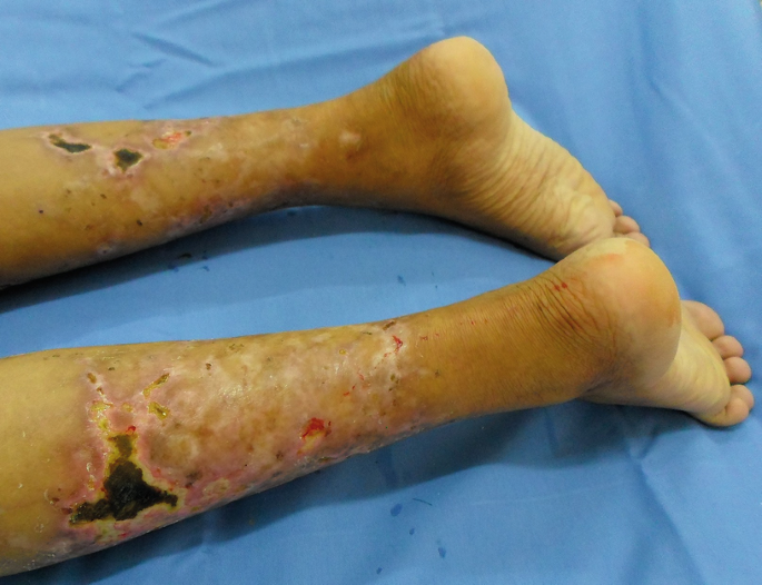
Leprous wound (reaction) with necrosis
-
Necrosis liquefaction or wet or crumbling necrosis: yellowish tissue, whitish, greyish, more slender, soft consistency, and can be tightly or loosely adhered to the bed and borders, presented as strings, crusting, or mucinous being formed by bacteria, fibrin, elastin, collagen, intact leukocytes, cellular debris, and large amounts of exudate DNA.

There are several instruments used to categorize these types of tissue. We will quote the RYB system,MEASURE, PUSH tool, TIME.
-
RYB (Red/Yellow/Black), proposed in 1988 by Cuzzel, which reveals the condition of the tissues: viable and nonviable. Red means that the wound bed is red with a predominance of granulation tissue and new epithelium; the aim of the treatment is to favor the humid environment, protecting the tissue and preventing infection. Yellow: the bed is yellow in color due to the presence of fibrous exudate, devitalized soft tissue, which may be colonized. The goal is to identify the presence or absence of infection; if present institute systemic therapy and promote debridement and cleansing. Black: presence of necrotic tissue with thick eschar formation and requiring removal of this tissue quickly and effectively through debridement [16].
-
The acronym MEASURE, where M stands for Measure (measure the length, width, depth, and surface area); E for Exudate (quantity and quality of exudate); A for Appearance (appearance of the wound bed type and amount of tissue); S for Suffering (type and intensity of pain); U for Undermining (presence or absence of detachment); R for Re-evaluation (periodic review of all parameters); E for Edge (conditions of the edges and the adjacent skin) [17].
-
The PUSH tool scale (Pressure Ulcer Scale for Healing) is a tool developed in 1996 by NPUAP (National Pressure Ulcer Advisory Panel) that considers the following parameters for the assessment of pressure ulcer: area, amount of exudate, and appearance of the wound bed [18].
-
TIME, the T assesses the viability/nonviability of the tissue in the wound bed; I relates to infection, colonization; M to the moisture imbalance; and E the edge of the wound [10].
Therapeutic Approach for Wounds
The care process is quite comprehensive, particularly when it comes to persons with chronic wounds, as they, most of the time, may feel fragile, embarrassed, and ashamed of his/her appearance, all of which goes beyond the physical aspect, affecting the psychological and social demeanor. Therefore, the type of reception the health team grants to these individuals as an attitude has a direct effect on treatment. In fact, a person properly hosted, to whom one pays attention to what they have to say, feels part of the care process. This sort of kindly approach should be used by all professionals working in the institution.
For the successful treatment of wounds, it is necessary for the person to be fully assessed by the multidisciplinary team. The nurse must establish a comprehensive care plan for the treatment and long-term monitoring of these patients [4].
After careful evaluation of the wound it must be cleansed using fluids to remove loosely adherent debris and necrotic tissue on the surface [19,20,21].
A fluid commonly used for wound cleansing is physiologic solution at 0.9%. Polyhexamethylene biguanide (PHMB) is one of the most advanced technologies for cleaning wounds. PHMB is an antimicrobial belonging to the group of chlorhexidines (biguanide), active against a large number of microorganisms, among them methicillin-resistant Staphylococcus aureus (MRSA), vancomycin-resistant Enterococcus (VRE), and the Acinetobacter baumannii that are responsible for multidrug-resistant infections. It is importantly to note that it is biocompatible, i.e., it has no toxicity on living tissues [22].
Other local antiseptics are not recommended. Usually they are chemical products with cytotoxic action that inhibits cell proliferation. Among them one can cite iodine-povidone, acetic acid, chlorhexidine, hydrogen peroxide, and hypochlorite solutions.
Debridement
The debridement consists of removing nonviable tissue, such as necrotic tissue, devitalized tissue, and colonized bodies and foreign matter, optimizing the healing process and preventing infection.
Types of Debridment (Fig. 72.7)
Enzymatic or chemical debridement is based on the use of proteolytic enzymes (papain, collagenase, fibrinolysin/DNase) which are capable of dissolving devitalized tissues. The choice of enzyme is dependent on the type of existing tissue in the wound.

Types of debridment
The application of the selected enzyme should be restricted to the devitalized tissue, avoiding contact with the perilesional areas. For protection of these areas hydrocolloid powder, essential fatty acid oil, or creams that promote a protective barrier can be used. The wound should be covered with a bandage to retain the moisture needed for enzyme action.
Autolytic debridement is obtained by using hydrogels and hydrocolloids that act naturally and selectively. Through the maintenance of moisture, phagocytic cells and proteolytic enzymes are activated, promoting the degradation of nonviable tissue.
Surgical debridement is the method indicated for wounds with large amounts of devitalized tissues. Depending on the severity and extent of the wound, this procedure should be performed by surgeons in the operating room under anesthesia. In superficial wounds, which usually do not require anesthesia, it can be performed on an outpatient basis by medical professionals and trained nurses. Although aggressive, results are faster than with other methods.
Mechanical debridement is also indicated for wounds with large amounts of devitalized tissue. This technique consists in the removal of tissues by application of mechanical strength. This procedure, however, can damage the granulation tissue or already existing epithelialization, and cause pain. Mechanical debridement methods include negative pressure therapy, wet–dry dressings, hydrotherapy, and irrigation.
Negative pressure therapy involves intermittent and continuous application of subatmospheric pressure at the wound surface. The mechanism of action in promoting the healing is by reducing the superficial edema; improvement of local blood circulation; reduction of excess exudate; stimulating the proliferation of fibroblasts, endothelial cells, and vascular smooth muscle cells; reduction of bacterial load; and favoring wound contraction.
Wet–dry dressings: moistened gauze with saline solution is applied to the wound bed and left until dry. The gauze when removed takes out necrotic tissue. It is a painful method that requires analgesia.
Hydrotherapy uses pulsed and pressurized irrigation. The water is delivered in a continuous or intermittent base under low, intermediate, and high pressure flow.
Irrigation is the steady flow of a solution across a wound bed with the use of a 40 × 12 needle in a 20 mL syringe or a 25 × 8 needle in a 20 mL syringe. The syringe must be positioned in a 45–90° angles and at a distance of 2.5–5.0 cm, which respectively ensure the pressure of 9.5 psi and 13.5 psi, capable of promoting the removal of cell debris, exudates with pathogens, and residues of topically applied creams and ointments which remain in the wound bed. The choice of pressure should vary according to the amount of debris to be removed. In the case of wounds with the largest amount of debris, one should use the 20 mL syringe and a 25 × 8 needle. It is also recommended to use a heated solution to prevent temperature reduction in the wound bed, since a constant temperature of 37 °C stimulates mitosis during granulation and re-epithelialization [23, 24].
Maggot or biological debridement therapy, or maggot therapy, requires the application in the wound bed of larvae reared in the laboratory. The larvae feed on necrotic/devitalized tissue and make selective debridement [10].
Based on the experience of the authors, topical therapy should be decided upon by evaluating the type of tissues, according to the algorithm that follows (Figs. 72.8 and 72.9):

Algorithm for viable tissue

Algorithm for non-viable tissue
Dressings: A Rapid Guide
EFA (Essential Fatty Acids)
-
Composition: polyunsaturated vegetable oils, linoleic acid, caprylic acid, capric acid, vitamin A and E, soybean lecithin, and lanolin.
-
Function: transporting materials across cell membranes, which ensures the life of the cell through balanced flow of nutrients and waste products in biological activity.
-
Indication: protection, hydration, skin restoration, and receiving area graft.
-
Contraindication: injury to tissue necrosis without debridement, sensitivity to the product.
-
Change frequency: every 24 h
AGin (Fatty Acids, Unsaturated)
-
Composition: oleic acid, vitamin A palmitate, DL-α-tocopherol, Vitamin E.
-
Function: antioxidant activity; protects the cell’s DNA particularly in those who are in training; inhibits the free radicals produced by lymphocytes that hinder the process of tissue repair in chronic injuries.
-
Indication: maintain or restore normal skin characteristics; prevention of skin breaks; moisturizing dry skin.
-
Contraindication: sensitivity to product.
-
Change frequency: every 24 h
Hydrogel
-
Composition: deionized water, glycerin, sodium carboxymethylcellulose, allantoin (natural hydrocolloid).
-
Function: interacts with the exudate creating moist environment that favors the autolysis, pain relief by moistening the exposed nerve endings in the wound.
-
Indication: wounds with granulation tissue; with necrotic tissue, venous ulcers, arterial and pressure; second-degree burns of small extent; wounds with partial or total loss of tissue; post-traumatic areas.
-
Contraindication: patients with sensitivity to product.
-
Change frequency: 24 h to 3 days depending on the exudate
Hydrogel with Calcium Alginate and Sodium
-
Composition: calcium alginate and carboxymethylcellulose sodium in an aqueous, transparent, viscous excipient.
-
Function: keep moist medium, helps in autolysis.
-
Indication: soften and rehydrate necrotic and devitalized areas of pressure ulcers, stasis, burning of first and second grade, cut, abrasions, and lacerations.
-
Contraindication: sensitivity to the product, damage to infection and/or secretion.
-
Change frequency: 24 h for necrotic or very exudative lesions up to 3 days to clean lesions with granulation tissue
Silver Sulfadiazine with Cerium Nitrate
-
Composition: 0.4% cerium nitrate and silver sulfadiazine 1%.
-
Function: sulfadiazine (bactericidal and bacteriostatic acting on the bacterial cytoplasmic membrane) effective against Gram-positive and -negative viruses, dermatophyte fungi; cerium nitrate enhances the bactericidal effect.
-
Indication: devitalized tissue, burn, colonization prevention.
-
Contraindication: sensitivity to components.
-
Change frequency: daily in infected lesions and up to 3 days in fresh lesions.
Collagenase
-
Composition: collagenase, clostridiopeptidase A, and proteolytic enzymes.
-
Function: acts to selectively degrade the native collagen (necrolysis).
-
Indication: devitalized tissue: necrosis, fibrosis.
-
Contraindication: wound healing by first intention, hypersensitivity to enzyme.
-
Change frequency: every 12 or 24 h.
Fibrinolysin
-
Composition: bovine emollients, fibrinolysins, deoxyribonucleases, and 1% chloramphenicol.
-
Function: lytic action of fibrinolysin and deoxyribonuclease dissolve the exudate and necrotic waste.
-
Indication: devitalized tissue (necrosis, fibrosis).
-
Contraindication: hypersensitivity to bovine substances.
-
Change frequency: every 12 or 24 h.
Cadexomer Iodine
-
Composition: Cadexomer, polyethylene glycol, poloxamer, and iodine.
-
Function: Cadexomer beads are biodegradable, remove excess exudate and fibrin in the wound base, and reduce bacterial contamination on the surface.
-
Indication: topical treatment of chronic exuding wounds, infected wounds.
-
Contraindication: dry necrotic tissue or with known sensitivity to iodine or any of its components; pregnant women, breastfeeding women, children, people with diabetes insipidus, thyroid disorder.
-
Change frequency: fluid saturation; 72 h.
Zinc Oxide
-
Function: antiseptic, drying, and anti-inflammatory action, decreased odor and exudate, 25% concentration increases the mitotic index.
-
Indication: devitalized tissue, abundant exudation, fetid odor.
-
Contraindication: Hypersensitivity to the product and painful.
-
Change frequency: every 24–72 h.
Polyhexamethylene Biguanide
-
Composition: amorphous hydrogel polyhexamethylene biguanide 0.1%, pectin, cellulose.
-
Function: antimicrobial action (Gram-positive yeasts and fungi), autolytic, wound deodorization, promotes moist environment, facilitates mechanical debridement, stimulates epithelialization.
-
Indication: chronic wounds or acute, with devitalized tissue, exudative, infected.
-
Contraindication: none.
-
Change frequency: daily or up to 3–7 days
Biopolymer
-
Composition: glyceryl monostearate, cetostearyl alcohol, stearic acid, N-coconut acyl derivatives of L-glutamic acid, sodium acrylate copolymer, hydrogenated polyisobutene, phospholipids, polyglyceryl-10-esters, sunflower oil, hydrogenated olive oil, olive ester, phenoxyethanol, hydrogenated vegetable oil, behenyl alcohol, lecithin, soy sterols, shea butter, fatty acid triglycerides, of Camelina sativa oil, dimethicone, hydroxyethylcellulose, disodium EDTA, serum and water Hevea brasiliensis.
-
Function: Gel-cream for skin restoration, has angiogenic activity and accelerating the healing process.
-
Indication: skin ulcers of various pathologies: arterial, diabetic, mixed, venous, neuropathic, leprous, by pressure, in acute and chronic stages, little exudate.
-
Change frequency: daily or up to 3 days
Amorphous Collagen Gel
-
Composition: collagen hydrolysate in a hydrogel matrix.
-
Function: protective barrier against external contaminants preventing fungal and bacterial proliferation, accelerates healing, and promotes rapid regeneration of the skin after therapeutic and aesthetic dermatologic procedures.
-
Indication: beaten skin for dermatologic procedures (laser, peeling, or minor dermatologic surgery) or external factors.
-
Contraindication: sensitivity of formula.
-
Change frequency: up to 3 days.
Barrier Cream
-
Function: Protects, moisturizes, and restores the skin’s pH, and against maceration.
-
Indication: perilesional skin for protection against excess moisture that can cause maceration and delayed wound-healing process.
-
Contraindication: not shown.
-
Change frequency: daily.
Hydrocolloid Powder
-
Composition: synthetic resin powder, microgranules, consisting of nonadherent hydrocolloids.
-
Function: Protects skin with exudate, absorbs moisture caused by chafing.
-
Indication: to be used in the peristomal skin. It is also indicated in bruised skin.
-
Contraindication: none.
-
Change frequency: daily.
Nonadherent Mash Dressing
-
Function: maintains moist environment, which prevents dehydration of the granulation tissue.
-
Indication: First- and second-degree burns, grafts, venous ulcers, pressure, eczema, surgical incisions.
-
Contraindication: sensitivity to components.
-
Change frequency: several days, depending on amount of exudate.
Sodium Alginate and Calcium
-
Function: calcium ions and sodium in the blood and exudates interact with the same ions found in the healing, inducing hemostasis, assists in autolytic debridement, promotes absorption of exudate and maintains the humidity (gel formation).
-
Indication: pressure ulcers, venous, arterial, diabetic, donor area, and other skin lesions with no hemorrhagic bleeding.
-
Contraindication: dried injuries and sensitivity to product.
-
Change frequency: daily changes in infected lesions and lesions cleaned according to saturation.
Coverage Collagen and Alginate
-
Function: hemostatic properties, stimulates granulation tissue formation, epithelialization, makes healing occur faster.
-
Indication: treatment of wounds with low to high levels of exudate (pressure ulcers, venous, arterial, diabetic, donor area, and other skin lesions with no hemorrhagic bleeding).
-
Contraindication: dried injuries and sensitivity to product, and carcinomas.
-
Change frequency: daily change on exudative lesions and lesions cleaned according to saturation.
Hydrofiber
-
Function: retains the fluid forming a gel around the fibers, keeps the wound bed moist and warm.
-
Indication: chronic lesions (pressure ulcers, lower limb), acute injuries (lacerations, incisions, donor area, first- and second-degree burns, and control of small bleeding), highly exudative lesions requiring autolytic debridement.
-
Contraindication: sensitivity to the product.
-
Change frequency: up to 7 days.
Hydrofiber with Silver
-
Function: retain the fluid forming a gel around the fibers, keeps the bed of the wound moist, warm, free of bacteria and odors.
-
Indication: small abrasions, cuts, lacerations, superficial burns, infections.
-
Contraindication: sensitivity to the product.
-
Change frequency: up to 7 days
Activated Carbon and Silver
-
Function: carbon attracts bacteria and silver combats microorganisms and reduces bacterial colonization and infection control and odor.
-
Indication: chronic wounds, traumatic and surgical lesions, with or without infection, odor and fibrin.
-
Contraindication: necrosis, bone exposure.
-
Change frequency: 7 days.
Natural Biological Film
-
Function: retains moist environment, does not allow entry of microorganisms.
-
Indication: burns, wounds, skin graft donor sites.
-
Contraindication: infected wounds.
-
Change frequency: single use.
Hydrocolloids
-
Function: promoting moisture forming a gel that provides for autolysis debridement. It stimulates neogenesis, pH maintenance.
-
Indication: without infection lesions without exudate, necrosis, prevention of decubitus ulcer.
-
Contraindication: lesions with infection and/or exudate.
-
Change frequency: 7 days in clean lesions with granulation tissue.
Hydropolymers
-
Function: maintain moisture, absorb and retain excess exudate, adhered to the wound bed, preventing maceration, stimulates autolytic debridement.
-
Indication: no infected lesions, slightly exudative, in granulation tissue and for preventing decubitus ulcer.
-
Contraindication: lesions with infection and/or secretion.
-
Change frequency: up to 7 days and, in case of prevention, up to 10 days.
Hydropolymer with Silver
-
Function: antibacterial, releasing silver ions continuously; exudate is absorbed protecting perilesional area. It has selective permeability (water controls the output and input bacteria), minimizing the risk of infection.
-
Indication: wounds with moderate to high exudate with delayed healing, with risk or clinical signs of infection, such as leg ulcers, pressure ulcers, second-degree burns, diabetic ulcers.
-
Contraindication: individuals with allergic reactions to any component of the product.
-
Change frequency: up to 7 days.
Polyurethane Foam
-
Function: exchange of ions occurs between the ingredients and silver alginate matrix and wound exudate, leading to the presence of Ag+ and Ca++ ions in the wound bed.
-
Indication: moist and thermal healing of wounds, infected or colonized; pressure ulcers, venous ulcers, and burns.
-
Contraindication: hypersensitivity known to alginate or silver, the presence of metals, ulcers due to infectious processes (tuberculosis, syphilis, deep mycoses or third degree burns).
-
Change frequency: up to 7 days.
Papain
-
Function: chemical debridement, bacteriostatic, bactericidal, and anti-inflammatory, provides alignment of collagen fibers.
-
Indication: chemical debridement and facilitator of the healing process, supporting systemic antibiotic treatment of infected wounds. Use concentration of 2% in wound granulation tissue; 4–6% when there is purulent exudate; and 10% when there is presence of necrotic tissue.
-
Contraindication: sensitivity to the substance or other component of the formulation.
-
Change frequency: every 12 h.
Growth Factor Gel
-
Function: acts on the cell membrane (activates the tyrosine kinase that comes into contact with DNA, stimulating cell division and proliferation).
-
Note: poorly healing ulcers with just partial damage, but with adequate blood supply.
-
Change frequency: daily, always at the same time.
Unna Boot
-
Function: reduces edema through the venous pump movement, facilitating venous return, aiding healing.
-
Indication: venous ulcers of the lower limbs.
-
Contraindication: arterial and mixed ulcers
-
Change frequency: every 7 days since there is no discharge or dirt.
Elastic Bandage
-
Function: reduces edema by means of pump action, making easier venous return and promoting healing.
-
Indication: treatment of venous leg ulcers and associated conditions where compression therapy indicated.
-
Contraindication: arterial ulcers and mixed ulcers, leg with circumference of 18 cm below the ankle.
Multilayer Compression Therapy
-
Function: promotes a recommended therapeutic pressure of 40 mmHg, facilitating venous return and reducing edema.
-
Indication: venous ulcer.
-
Contraindication: arterial and mixed ulcer.
-
Change frequency: up to 7 days.
References
Jorge AS, Dantas SRPE. Abordagem multiprofissional no tratamento de feridas. São Paulo: Atheneu; 2003.
Blanes L. Tratamento de feridas. In: Baptista-Silva JCC, editor. Cirurgia vascular: guia ilustrado. São Paulo; 2004. http://files.artedecuidar.webnode.com.br/200000015-0ad7c0b337/Tratamento%20de%20Feridas.pdf. Accessed 4 July 2016.
Santos AAR. O ensino da temática feridas no curso de graduação em enfermagem da Universidade Federal da Paraíba. [dissertação]. João Pessoa: UFP; 2012.
Ministério da Saúde (BR). Manual de condutas para tratamento de úlceras em hanseníase e diabetes. 2a ed. revisada e ampliada. Brasília: Ministério da Saúde; 2008. 92 p.
Puri V, Venkateshwaran N, Khare N. Trophic ulcers - practical management guidelines. Indian J Plast Surg. 2012;45(2):340–51. doi:10.4103/0970-0358.101317.
Oda RM, Galan NGA, Opromolla DVA. Úlceras de perna na hanseníase. In: Opromolla DAV, Baccarelli R, editors. Prevenção de Incapacidades e reabilitação em hanseníase. Bauru: Instituto Lauro de Souza Lima; 2003. p. 130–3.
Barros ALBL. Anamnese e exame físico: avaliação diagnóstica de enfermagem no adulto. 3rd ed. Artmed: Porto Alegre; 2016.
Fornells MG, González RFG. Cuidados de la piel perilesional. Espana; 2006. http://www.fundacionsergiojuan.org/pdf_gneaupp/libro_piel_perilesional.pdf. Accessed 4 July 2016.
Dealey C. Cuidando de feridas: um guia prático para as enfermeiras. 3a ed. Atheneu: São Paulo; 2008.
Malagutti W, Kakihara CT. Curativos, estomia e dermatologia: uma abordagem multiprofissional. 2a ed. São Paulo: Martinari; 2011.
Agredda JJS, Bou JET. Atenção integral nos cuidados das Feridas Crônicas. Petrópolis: EPUB; 2012.
Vries JCH, Groot R, van Brakel WH. Social participation of diabetes and ex leprosy -patients in the Netherlands and patient preference for combined self-care groups. Front Med. 2014;1(21). 10.3389/fmed.2014.00021.
Robb C. Module 1784: chronic wound management. Chem Drug. 2016;26:12.
Ferreira MC, Tuma P Jr, Carvalho VF, Kamamoto F. Complex wounds. Clinics. 2006;61(6):571–8.
Leaper D, Assadian O, Edmiston CE. Approach to chronic wound infections. Br J Dermatol. 2015;173(2):351–8. doi:10.1111/bjd.13677.
Cuzzel JZ. The new RYB color code. Am J Nurs. 1988;88(10):1342–6.
Keast DH, et al. MEASURE. A proposed assessment framework for developing practice recpmmedatios for wound assessment. Wound Rep Reg. 2007;51(12):S1–S17.
Santos VLCG, Azevedo MAJ, Silva TS, Carvalho VMJ, Carvalho VF. Adaptação transcultural do Pressure Ulcer Scale for Healing (PUSH), para a língua portuguesa. Rev Lat Am Enfermagem. 2005;13(3):305–13.
Joanna Briggs Institute. Solutions, techniques and pressure in wound cleansing. Nurs Stand. 2008;22(27):35–9.
Bee TS, et al. Wound bed preparation: cleansing techniques and solutions: a systematic review. Singap Nurs J. 2009;36:17–22.
Fernandez R, Griffiths R. Water for wound cleansing. Cochrane Database Syst Rev. 2012;(2):CD003861. doi:10.1002/14651858.CD003861.pub3.
Santos EJF, Silva MANCGMM. Tratamento de feridas colonizadas/infetadas com utilização de polihexanida. Rev Enf Ref [online]. 2011;serIII(4):135–42. http://www.scielo.mec.pt/scielo.php?script=sci_arttext&pid=S0874-02832011000200014&lng=pt&nrm=i&tlng=pt. Accessed 4 July 2016.
Martins PAE. Avaliação de três técnicas de limpeza do sítio cirúrgico infectado utilizando soro fisiológico para remoção de microrganismos [dissertação]. São Paulo: Escola de Enfermagem da Universidade de São Paulo; 2000.
Gall TT, Monnet E. Evaluation of fluid pressures of common wound-flushing techniques. Am J Vet Res. 2010;71(11):1384–6. doi:10.2460/ajvr.71.11.1384.
Author information
Authors and Affiliations
Corresponding author
Editor information
Editors and Affiliations
Glossary
- Iatrogenic
-
Inappropriate result from medical procedures or treatments.
- Biofilm
-
Community of microorganisms surrounded by a mucopolysaccharidic extracellular matrix, creating a cohesive film in the wound bed that serves as a common defense strategy of these settler microorganisms.
- Lipodermatosclerosis
-
Concomitant presence of hard edema and slight pitting (sclerosis).
- Maggot therapy
-
Application in the wound bed of larvae of the species Lucilia sericata or Phaenicia sericata in order to selectively remove the necrotic/devitalized tissue.
- Ocher dermatitis
-
Extravasation of red blood cells in the dermis along the deposit of hemosiderin in macrophages.
Enzymes that promote protein breakdown.
Rights and permissions
Copyright information
© 2018 Springer International Publishing Switzerland
About this chapter
Cite this chapter
Guimarães, H.C.Q.C.P., Bassoli, S.R.B., Bernardo, R.M.P., da Cunha Lopes Virmond, M. (2018). Care of Wounds: Dressings. In: Bonamigo, R., Dornelles, S. (eds) Dermatology in Public Health Environments. Springer, Cham. https://doi.org/10.1007/978-3-319-33919-1_72
Download citation
DOI: https://doi.org/10.1007/978-3-319-33919-1_72
Published:
Publisher Name: Springer, Cham
Print ISBN: 978-3-319-33917-7
Online ISBN: 978-3-319-33919-1
eBook Packages: MedicineMedicine (R0)