
Abstract
Various common injuries in the wrist and hand are investigated in this chapter. Significant attention is paid not only to proper diagnostic examination and imaging, but also to optimal nonoperative and operative treatment options to manage these conditions.
Access provided by CONRICYT-eBooks. Download chapter PDF
Keywords
- Hand
- Wrist
- Finger
- Phalanx
- Scaphoid
- Distal radius
- Fracture
- Flexor tendon
- Extensor tendon
- Infection
- Laceration
- Amputation
- Carpal fractures
- Hand infections
- Perilunate dislocation
- Scaphoid fracture
- Scapho-lunate instability
- Tendon lacerations
Treatment of finger injuries depends on integrity of bony and soft tissue structures.
Overview
Metacarpal Fractures
-
Categories include metacarpal head, neck, shaft, and base fractures
-
Metacarpal neck fractures will typically present apex dorsal deformity secondary to pull of intrinsics
-
Fracture of the fifth metacarpal neck is referred to as a “boxer’s fracture”
-
-
Fractures of the base of the metacarpal are often associated with dislocations (Bennett fracture dislocation for base of first digit, along with “Reverse Bennett” for base of fifth digit)
-
Obtain oblique hand X-rays to better identify these injuries
-
Phalanx Fractures
-
Deformity is dependent on fracture location
-
Proximal phalanx fracture typically exhibit apex volar deformity because of intrinsic pull
-
Middle phalanx fractures proximal to FDS insertion display apex dorsal deformity, while those fracture distal to the FDS insertion display apex volar deformity
-
Malrotation and extensor lag are typical findings in displaced fractures of the digit
History
-
Mechanism and timing of injury
-
Presence of open or contaminated (e.g., bite to finger) injury
Physical Exam
-
Must assess for presence of open fracture
-
Detailed neurovascular exam
-
Visual inspection to assess alignment
-
Malrotation must be assessed—can assess when asking patient to flex hand and compare to contralateral side, or assess orientation of the fingernails in comparison to other digits on the ipsilateral or contralateral hand
-
Assess active and passive range of motion where indicated
Diagnosis
Imaging/Hand X-Rays
-
Obtain full series of X-rays including hand and involved digit (AP, lateral, and oblique)
-
Ensure that true lateral of the isolated digit is taken
-
MC base fractures and CMC dislocations, often require oblique views to identify fracture
Treatment Plan (According to Particular Injury)
First CMC Dislocation
-
Reduce, place in thumb spica splint
First MC Fracture/Dislocation
-
Reduce, immobilize in thumb spica splint
First Digit UCL Sprain (“Game Keeper”/“Skiier’s Thumb”)
-
Injury to the ulnar collateral ligament at the first digit MCP joint (Fig. 1)
Fig. 1 
Thumb UCL injury (MRI)
-
Diagnosed with pain at origin, along with ligamentous laxity upon stress testing
-
Increased laxity to ulnar stress at MCP in flexed position when compared to contralateral
-
-
Treat acutely with thumb spica splint/cast

2nd–5th MC Fractures (Figs. 2–4)

Fracture dislocation of base of fifth MC (“reverse Bennett’s fracture ”)

Metacarpal fracture (fifth MC head/neck and fourth MC base)

Pin stabilization of multiple metacarpal fractures
-
Following reduction (hematoma block), can immobilize in intrinsic plus short arm splint
-
Can also consider metacarpal mitt splint
-
10, 20, 30, 40° angulation acceptable with 2nd, 3rd, 4th and 5th MC shaft fractures, with up to an additional 10° acceptable at each digit for neck fractures. About 7° extensor lag expected for every 2 mm MC shortening, but some shortening is often well tolerated.
Consider surgery for poly-trauma patient, multiple fractures in the same hand or rotated, or severely angulated fractures
MCP Dislocation (Simple If No Interposed Tissue or Associated Fracture)
-
Dorsal/volar dislocation: reduction and immobilization with buddy tape and intrinsic plus splint
-
For complex dislocations or with interposed tissue (volar plate for irreducible dorsal MCP dislocation), will need to go to operating room on urgent basis
Phalanx Fractures
-
Reduction (can lever with pen in between adjoining digits) followed by buddy taping
-
Should buddy tape to adjacent fingers to help maintain reduction
-
AlumaFoam splinting generally sufficient
-
Acceptable non-operative treatment sufficient for proximal and middle phalanx fractures if able to maintain stable reduction in extra-articular fractures with less than 10° angulation without rotational overlap and <50 % overlap with the adjacent finger. About 10–12° extensor lag expected for every 1 mm phalangeal shortening. Distal phalanx fractures rarely require surgical intervention, although displaced transverse fractures can be considered for closed reduction and percutaneous pinning.
PIP Dislocations (Simple)
-
Dorsal dislocations most common (Fig. 5)—disruption of volar plate. Axial traction for reduction, buddy taping, and early ROM after 3–4 days immobilization. Missed injury leads to volar plate attenuation, which can lead to swan neck deformity.
Fig. 5 
Simple PIP dorsal dislocation
-
Volar dislocation—rupture of the central slip. Following reduction, will need to be immobilized in extension for 4–6 weeks for central slip repair. Missed/chronic central slip injury leads to boutonniere deformity

PIP Fracture Dislocations
-
If >40 % joint surface involved, usually requires surgical fixation; provisionally splint in ER
-
Technique dependent on amount of bone preserved (Figs. 6, 7, and 8)
Fig. 6 
Dorsal PIP fracture dislocation
Fig. 7 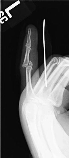
Persistent dislocation despite extension splinting
Fig. 8 
Volar PIP fracture dislocation


-
In general, nonoperative interventions listed above used in emergency room setting.
-
Surgical management reserved for irreducible fractures/fracture dislocations, open injuries, those with neurovascular compromise (not always), and digit injuries with malrotation.
Author information
Authors and Affiliations
Corresponding author
Editor information
Editors and Affiliations
Rights and permissions
Copyright information
© 2017 Springer International Publishing Switzerland
About this chapter
Cite this chapter
Makhni, M.C., Makhni, E.C., Swart, E.F., Day, C.S. (2017). Finger Fractures and Dislocations. In: Makhni, M., Makhni, E., Swart, E., Day, C. (eds) Orthopedic Emergencies. Springer, Cham. https://doi.org/10.1007/978-3-319-31524-9_49
Download citation
DOI: https://doi.org/10.1007/978-3-319-31524-9_49
Published:
Publisher Name: Springer, Cham
Print ISBN: 978-3-319-31522-5
Online ISBN: 978-3-319-31524-9
eBook Packages: MedicineMedicine (R0)