
Abstract
Radiology is a rapidly evolving specialty of medicine with new technologies and innovations emerging at a constant pace. This tutorial provides a concise overview of future developments in imaging with a focus on artificial intelligence, radiation dose reduction and advances in imaging techniques.
Access provided by Autonomous University of Puebla. Download chapter PDF
Keywords
Aims and Guidance for Tutors
Radiology is a rapidly evolving specialty of medicine with new technologies and innovations emerging at a constant pace. This tutorial provides a concise overview of future developments in imaging with a focus on artificial intelligence, radiation dose reduction and advances in imaging techniques.
Introduction
-
Radiology is a specialty that has progressed at an unprecedented speed over the past two decades due to a variety of technological advances such as a transition from film-based to fully digitised reporting environments, increase in computational power and refinements in scanner technology and image processing techniques.
-
In tandem with advances in diagnostic imaging, minimally-invasive Interventional Radiology (IR) procedures have also showed similar progress over this time and includes examples such as endovascular stroke thrombectomy, tumour embolisation and endovascular treatment of vascular diseases, made possible by new techniques and tools available to the Radiologist.
-
Computational power has advanced to such a level that has allowed machines to run very complex algorithms which can assist the Radiologist in performing certain tasks such as semi-automated image interpretation, workflow optimisation and clinical decision making.
Artificial Intelligence/Machine Learning
-
Artificial intelligence in its broadest sense is defined as the ability of computers to evaluate external data in the environment, learn from the data and respond to achieve a particular aim.
-
Machine learning (ML) is a form of artificial intelligence and refers to a set of complex data modelling and analysis techniques with the potential to learn from experience. These algorithms can be supervised or unsupervised by the human operator.
-
With ML, the process is semi-automated—the computer is provided the data, creates complex analytical models by utilising a learning framework to refine and optimise the accuracy of prediction.
-
A wide array of ML algorithms exist, for which the specific details are beyond the scope of this chapter. However, some examples that may be encountered in the medical literature include different types of artificial neural networks and support vector machine. Readers are directed to recent review articles of ML in medicine for further detail.
Relevance to radiology:
-
ML has attracted a lot of attention in Radiology due to the increasing volume and complexity of imaging work.
-
As a speciality that deals with high level interpretation of large volumes of data in the form of digital images, reports and electronic clinical information, there is great potential for ML to support the growth of Radiology into the future.
-
Pattern recognition and automated analyses of medical images by computers has emerged, partly due to the increase in number of imaging modalities and to the increased resolution and number of images generated from modern scanners, in an effort to reduce the workload within Radiology.
-
Examples of ML applications include the automated detection, segmentation and analysis of lesions within an organ system (e.g. lung nodules, breast or liver lesions), either automatically generating a summary report or presenting these results to the reporting Radiologist for validation and cross checking.
-
When used appropriately, ML has the potential to minimise error and improve reporting efficiency within Radiology departments—an important consideration given the exponential increase in demands on medical imaging, both in terms of volume and complexity.
-
A potential future will see Radiologists engaging more in clinical work, supervising and validating ML assisted image reads and increasing their participation in ML assisted multidisciplinary meetings where management decisions are made. The latter decisions will be based on multidimensional big data analysis resulting in a tailored management plan for each individual patient.
Examples of current applications in radiology:
-
Imaging analytics and Computer Aided Detection of lesions (lung nodules, breast and liver lesions, automated analysis of brain perfusion imaging studies).
-
Automated measurement of tumours in Oncologic imaging studies to accurately assess response to therapy and to improve reporting efficiency and reduce errors in the reproducibility of measurements.
-
Worklist optimisation—ML algorithm continuously monitors new studies and prioritises patients with urgent clinical conditions (e.g. intracranial haemorrhage or stroke) to be presented next on the Radiologist’s reporting workflow.
-
Keyword detection in electronic medical records and radiology information systems—ML algorithm detects keywords relevant to the current imaging examination (e.g. Abdominal ultrasound in a patient with jaundice) and presents relevant clinical information (such as a history of cholestatic liver biochemistry results and cholecystectomy for gallstones) from the medical record to the Radiologist.
-
Providing intelligent decision support for Interventional Radiology procedures incorporating data from imaging studies, patient demographics, risk factors and co-morbidities to present a selection of management strategies (e.g. management of intracranial aneurysms or peripheral vascular disease; Fig. 1).
Fig. 1 
Behind the scene of an example of a supervised learning ML algorithm to assist decision making for management of peripheral arterial disease. This algorithm consists of a number of ‘nodes’ and is programmed to study a database of patients with disease, risk factors and clinical outcomes. The aim in this example was to uncover complex relationships between patient and treatment variables using an artificial neural network to predict clinical outcomes and present it to the user in the form of a decision tree (not shown)

Radiation Dose Reduction
-
Many imaging tests involve exposure of a patient to ionising radiation with substantial differences in effective patient dose depending on the modality (e.g. conventional radiographs versus computed tomography [CT]) and sensitivity of the body part imaged to radiation (e.g. breast tissue versus bone).
-
Due to the rapid expansion and utilisation of CT imaging, there has been increasing concern regarding radiation exposure, particularly in younger patients or those requiring repeated imaging studies over their lifetime (e.g. young patients with inflammatory bowel disease).
-
Radiation dose reduction strategies are now incorporated in most modern CT scanners employing a combination of scanner based and image reconstruction techniques to reduce the dose to the patient.
-
Scanner based strategies include the use of efficient imaging detectors and tailored x-ray tube output based on patient body habitus (e.g. lower x-ray beam kilovoltage and current setting based on body mass index or body part).
-
Image reconstruction techniques make use of modern computing power allowing very complex algorithms to be used to produce diagnostic image quality for much lower radiation exposure parameters compared to older scanners. These techniques are commonly referred to as iterative reconstruction which represent a major advancement compared to older filtered back projection algorithms utilised since the early days of CT.
-
Modern scanners are now able to acquire scans at very low dose—for example, CT coronary angiography can now be performed with doses of around 1 mSv on a modern scanner (based on prospective ECG acquisition, high scanner pitch, low tube exposure and iterative reconstruction) compared to 10–15 mSv on older scanners. Ultra-low dose chest CT can also be performed (Fig. 2) with some scanners and centres achieving doses comparable to a combined PA and lateral chest radiograph (0.2 mSv).
Fig. 2 
a Ultra-low dose chest CT showing bronchiectasis in the left lower lobe (white arrow) at a dose of 0.2 mSv. b Previous study on an older CT scanner shows the same bronchiectatic changes (white arrow) at a dose of 2.5 mSv. There is increased image noise in the ultra-low dose scan, but image detail is sufficient for diagnosis

Advances in Imaging Techniques
-
Innovative developments in CT technology has allowed imaging to expand beyond the traditional confines of anatomic imaging. It is now possible to gain further information on tissue characteristics or tissue blood flow (perfusion) through dual-energy CT scanning.
-
Dual-energy CT exploits the principle of differing tissue interactions with x-ray beams of different energy levels (e.g. 80 and 140 kVp). These differences in tissue interaction allow tissues of different compositions to be characterised—for example, the composition of renal calculi detected on CT or the detection of uric acid deposition in periarticular soft tissues in patients with gout (Fig. 3).
Fig. 3 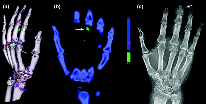
a 3D volumetric reconstruction from a dual-energy CT study showing areas of uric acid deposition (white arrows) in a patient with gout. b Coronal dual-energy CT reconstruction showing uric acid deposition (arrow). c Hand radiograph of the same patient showing dactylitis of the middle finger (arrow) but no visible tophi
-
High field strength MRI scanners (7–9 Tesla) and ultra-high field strength scanners (ranging from 11–21 Tesla) are under development and some are starting to enter clinical use. Some of the advantages of higher field strength scanners include the ability to scan patients quicker or obtain images with superior spatial resolution and detail compared to current mainstream scanners which typically operate at a field strength of 1.5 or 3 Tesla.
-
In addition to improvements in MRI scanner hardware, similar advances have been made in scanner software and sequences leading to a range of advanced imaging techniques. Recent examples include functional MRI which can measure brain activity based on the detection in blood flow alteration and oxygen consumption in different parts of the brain when activated.

Suggested Reading
-
Handelman GS, Kok HK, Chandra RV, Razavi AH, Huang S, Brooks M, Lee MJ, Asadi H. Peering Into the black box of artificial intelligence: evaluation metrics of machine learning methods. AJR Am J Roentgenol. 2019;212 (1):38–43.
-
Handelman GS, Kok HK, Chandra RV, Razavi AH, Lee MJ, Asadi H. eDoctor: machine learning and the future of medicine. J Intern Med. 2018;284 (6):603–619.
Author information
Authors and Affiliations
Corresponding author
Editor information
Editors and Affiliations
Rights and permissions
Copyright information
© 2020 Springer Nature Switzerland AG
About this chapter

Cite this chapter
Kok, H.K., Asadi, H. (2020). Tutorial 15: Emerging Imaging Technologies. In: Redmond, C., Lee, M. (eds) Tutorials in Diagnostic Radiology for Medical Students. Springer, Cham. https://doi.org/10.1007/978-3-030-31893-2_15
Download citation
DOI: https://doi.org/10.1007/978-3-030-31893-2_15
Published:
Publisher Name: Springer, Cham
Print ISBN: 978-3-030-31892-5
Online ISBN: 978-3-030-31893-2
eBook Packages: MedicineMedicine (R0)