
Abstract
Recombinant hypoallergenic derivative is the next generation of tolerogen replacing the natural allergen extract to increase safety and efficacy. Japanese cedar pollinosis is the predominant seasonal allergy disease in Japan. A rice seed-based oral vaccine containing the recombinant hypoallergens derived from these allergens was developed. Efficacy of this rice-based allergy vaccine was evaluated by oral administration in animal models.
Access provided by CONRICYT – Journals CONACYT. Download protocol PDF
Key words
1 Introduction
Allergen-specific immunotherapy (AIT) is the only curative treatment for allergic diseases. The conventional AIT treatment is achieved by subcutaneous or sublingual administration using natural allergen extract over the period of more than 3 years. This treatment is sometimes accompanied by side effects of anaphylaxis shock. To resolve these poor outcomes, safer, effective, and more convenient treatment is desired. Japanese cedar pollinosis is the predominant seasonal allergy disease in Japan. Major allergens are identified to be Cry j 1 and Cry j 2. A rice-seed-based oral vaccine containing the recombinant hypoallergens derived from these major allergens was developed as an alternative to the conventional therapies. The tertiary structures of allergens required for these specific IgE binding were destructed by fragmentation and molecular shuffling in the form of tail-to-head orientation. Low allergenicity for these modified allergens was evaluated by reduced binding activity to the specific IgE and reduced beta-glucosaminidase release from basophils. The destructed Cry j 1 and Cry j 2 were expressed and deposited in ER-derived protein bodies (PB-Is) in endosperm of transgenic rice seed. Efficacy of rice-based allergy vaccines and ability to induce immune tolerance to the allergens were confirmed by oral administration of transgenic rice seeds to mouse models. The success of the allergy vaccine is attributed to high dose of accumulation and deposition in protein bodies which exhibit high resistance to harsh conditions and digestive enzymes in gastrointestinal tract, providing an efficient oral allergen delivery system.
2 Materials
2.1 Reagents for Production of Transgenic Rice Lines
-
1.
Cfr9I restriction enzyme.
-
2.
Entry clone plasmids designed for Multisite Gateway LR reaction (Fig. 1).
Fig. 1 
Schematic drawing of entry clones and expression binary vector. The target gene DNAs are sub-cloned into the Cfr9I site of the entry clone plasmids (pKS4-1/pKS221/pKS2-3). For the expression of multiple target gene DNAs, these expression cassettes are integrated into a binary vector (pCSPmALS43GW) by the MultiSite Gateway LR clonase reaction. att Gateway attachment region, 16k-P 16 kDa prolamin promoter, mGluB modified glutelin B (GluB)-coding region, 16k-T 16 kDa prolamin terminator, GluB4-P glutelin B4 (GluB4) promoter, mGluA modified glutelin A (GluA)-coding region, GluB4-T glutelin B4 (GluB4) terminator, 10k-P 10 kDa prolamin promoter, mGluC modified glutelin C (GluC)-coding region, 10k-T 10 kDa prolamin terminator, CS-P callus-specific promoter, mALS mutated acetolactate synthase-coding region, LB left border, RB right border
-
3.
A binary vector designed for Multisite Gateway LR reaction (Fig. 1).
-
4.
Gateway LR clonase II plus enzyme mix (Fig. 1).

2.2 Reagents and Antibodies for Allergenicity Tests
-
1.
Nitrocellulose membrane.
-
2.
Blocking solution (Block Ace, DS Pharma Biomedical, Japan).
-
3.
TBS-T solution (25 mM Tris, 138 mM NaCl, 2.7 mM KCl, 0.1 % Tween-20, pH 7.4).
-
4.
Anti-mouse IgE secondary antibodies conjugated to biotin (Southern Biotech, USA).
-
5.
Streptavidin-horseradish peroxidase (HRP) conjugate.
-
6.
Enhanced chemiluminescence (ECL)-HRP substrate solution.
-
7.
Deionized water.
-
8.
Dulbecco’s modified Eagle’s medium (DMEM).
-
9.
Fetal bovine serum (FBS) (HyClone, USA).
-
10.
Penicillin-streptomycin-glutamine mixed solution.
-
11.
Tyrode’s solution (Sigma-Aldrich, USA).
-
12.
Bovine serum albumin (BSA).
-
13.
HEPES.
-
14.
p-Nitrophenyl N-acetyl-beta-d-glucosaminide (Sigma-Aldrich, USA).
-
15.
Sodium citrate.
-
16.
Glycine.
2.3 Instruments, Reagents, Antigens, and Antibodies for Efficacy Tests
-
1.
Multi-beads shocker (Yasui Kikai, Osaka, Japan).
-
2.
Deionized water.
-
3.
Phosphate-buffered saline (PBS) (137 mM NaCl, 2.68 mM KCl, 8.1 mM Na2HPO4·12H2O, 1.47 mM KH2PO4, pH 7.4).
-
4.
Total protein extract of Japanese cedar pollen (LSL, Japan).
-
5.
Aluminium hydroxide (Alum).
-
6.
Recombinant mouse IL-4.
-
7.
CD4 (L3T4) MicroBeads (Miltenyi Biotec, Germany).
-
8.
autoMACS Pro Separator (Miltenyi Biotec, Germany).
-
9.
Complete medium (DMEM/F-12 supplemented with 10 % FBS, 1× PBS, 50 μM 2-mercaptoethanol, 10 mM HEPES, and 1× penicillin-streptomycin-glutamine mixed solution).
-
10.
Celltiter 96 aqueous one solution cell proliferation assay (Promega, USA).
-
11.
Anti-mouse IgE capture antibody (Southern Biotech, USA).
-
12.
Anti-mouse IgG capture antibody (Southern Biotech, USA).
-
13.
Carbonate-bicarbonate buffer (Sigma-Aldrich, USA).
-
14.
PBS-T solution (1× PBS containing 0.1 % Tween-20, pH 7.4).
-
15.
Blocking solution (Block Ace, DS Pharma Biomedical, Japan).
-
16.
Streptavidin-HRP conjugate.
-
17.
TMB solution (BD Biosciences, USA).
-
18.
HCl.
-
19.
Histamine ELISA test kit (Neogen, USA).
-
20.
Cytospin3 (Thermo Shandon, USA).
-
21.
Diff-Quik stain kit (Dade Behring, Germany).
3 Methods
3.1 Production of Transgenic Rice Seed-Based Allergy Vaccines
-
1.
Design and PCR-amplify the target gene DNAs (see Note 1 ).
-
2.
Sub-clone the target gene DNAs into the Cfr9I site of the entry clone plasmids (Fig. 1) (see Note 2 ).
-
3.
For the expression of multiple target gene DNAs, integrate the expression cassettes (endosperm-specific promoters/signal peptide/target gene DNAs/terminators) into a binary vector such as pCSPmALS43GW by the MultiSite Gateway LR clonase reaction (Fig. 1) (see Note 3 ).
-
4.
Electrotransform Agrobacterium cells with the expression binary vector (see Note 4 ).
-
5.
Perform Agrobacterium-mediated transformation of rice calli to integrate the expression binary vector into the rice genome (see Note 4 ).
-
6.
Select and regenerate calli to obtain transgenic rice plants (see Note 4 ).
3.2 Evaluation of the Allergenicity of Antigens
3.2.1 Dot-Blot IgE Binding Assay
-
1.
Prepare the target antigen (see Note 5 ).
-
2.
Spot 1–2 μl of the antigen sample onto the nitrocellulose membrane.
-
3.
Block the membrane in the blocking solution at room temperature (RT) for 1 h.
-
4.
Incubate the membrane in the sera from mice immunized with Japanese cedar pollen allergen (as described in Subheading 3.3) at RT for 1–2 h or at 4 °C for 16–24 h.
-
5.
Wash the membrane in TBS-T solution 3–5 times.
-
6.
Incubate the membrane in anti-mouse IgE secondary antibodies conjugated to biotin at RT for 1–2 h or at 4 °C for 16–24 h.
-
7.
Wash the membrane as described in step 5.
-
8.
Incubate the membrane in the streptavidin-HRP conjugate solution at RT for 30–60 min.
-
9.
Wash the membrane as described in step 5.
-
10.
Incubate the membrane in the ECL-HRP substrate solution at RT for 5–10 min to let the signal develop.
-
11.
Rinse the membrane 2–3 times with deionized water and detect the HRP-derived signal. An experimental example is shown in Fig. 2.
Fig. 2 
An experimental example of dot-blot IgE binding assay [1]. Purified native Cry j 1 and Cry j 2 (Asahi Food & Healthcare Co., Japan), recombinant Cry j 1-F1 [1], and shuffled Cry j 2 [1] were dotted onto a nitrocellulose membrane at 200 ng, 50 ng, 10 ng, 5 ng, 1 ng, and 0 ng, and incubated with sera from BALB/c mice immunized with Japanese cedar pollen. IgE bound to Cry j 1/2, Cry j 1-F1, and shuffled Cry j 2 were detected with anti-mouse IgE-biotin and streptavidin-HRP, and then visualized by autoradiography. The result shows that IgE reactivity of Cry j 1-F1 and shuffled Cry j 2 is markedly reduced when compared to that of native Cry j 1/2

3.2.2 RBL-2H3 Basophil Degranulation Test
-
1.
Prepare the target antigen (see Note 5 ).
-
2.
Maintain RBL-2H3 cells in DMEM containing 10 % FBS and 1× penicillin-streptomycin-glutamine mixed solution at 37 °C/5 % CO2.
-
3.
Seed the RBL-2H3 cells at a 1 × 10⁵/well into a 96-well cell culture plate.
-
4.
Sensitize the RBL-2H3 cells with 1/100 volume of sera from mice immunized with Japanese cedar pollen allergen (as described in Subheading 3.3) for 16–24 h at 37 °C/5 % CO2.
-
5.
Wash the RBL-2H3 cells three times with 100 μl Tyrode’s solution containing 0.1 % BSA and 25 mM HEPES (pH 7.4).
-
6.
Add the target antigen in 200 μl Tyrode’s solution containing 0.1 % BSA and 25 mM HEPES (pH 7.4).
-
7.
Incubate the RBL-2H3 cells for 1 h at 37 °C/5 % CO2.
-
8.
Aliquot 50 μl supernatant to a new 96-well plate.
-
9.
Add 50 μl of 3 μM p-nitrophenyl N-acetyl-beta-d-glucosaminide (a substrate for beta-glucosaminidase) in 40 mM sodium citrate (pH 4.5) to the supernatant.
-
10.
Incubate the solution for 1–3 h at 37 °C.
-
11.
Add 10 μl of 2 M glycine (pH 10) to terminate the enzyme reaction.
-
12.
Read the optical density at 405 nm.
-
13.
Calculate the percentage of beta-hexosaminidase released from the RBL-2H3 cells (see Note 6 ).
3.3 Oral Feeding of Rice and Sensitization of Mice
-
1.
Prepare transgenic rice seed accumulating antigens and/or non-transgenic control rice seed.
-
2.
Remove the hull of rice seed.
-
3.
Ground unpolished brown rice seed to a fine powder using a mill or multi-beads shocker.
-
4.
Suspend the rice powder in an appropriate volume of deionized water or PBS.
-
5.
Mix well the rice powder solution just before the administration to mice.
-
6.
Administer the rice powder solution to mice using a gastric tube.
-
7.
For ad libitum feeding of rice seed, feed the rice powder using a feeding container (see Note 7 ).
-
8.
Following the rice feeding period, sensitize the mice by intraperitoneal injection of 0.1 mg total protein extract of Japanese cedar pollen mixed with 5 mg alum and 0.1 μg recombinant mouse IL-4 (see Note 8 ).
-
9.
At 7 days after the first sensitization, boost the mice with the intraperitoneal injection of 0.1 mg total protein extract of Japanese cedar pollen mixed with 5 mg alum.
-
10.
At 14 days after the second sensitization, examine the efficacy of rice seed-based allergy vaccines as described in Subheading 3.4.
3.4 Evaluation of the Efficacy of Rice Seed-Based Allergy Vaccines
3.4.1 Splenic CD4+ T Cell Responses and Cytokines
-
1.
At 14 days after the second sensitization as in Subheading 3.3, step 10, sacrifice the mouse, remove the spleen, and isolate splenic cells.
-
2.
To isolate CD4+ T cells, stain the splenic cells by CD4 (L3T4) MicroBeads according to the manufacturer’s instructions.
-
3.
Magnetically isolate splenic CD4+ T cells using the autoMACS Pro Separator according to the manufacturer’s instructions.
-
4.
Seed the cells at 1 × 10⁵/well into a 96-well cell culture plate in 100 μl complete medium.
-
5.
Add 100 μl antigen -presenting cells prepared from spleens of naive BALB/c mice at 3 × 10⁵/well in the complete medium.
-
6.
Add filter-sterilized total protein extract of Japanese cedar pollen dissolved in the complete medium at 10 μg/ml.
-
7.
Incubate the plate for 4–6 days at 37 °C/5 % CO2.
-
8.
Aliquot 100 μl cell culture supernatant to a new tube for cytokine assay (see Note 9 ).
-
9.
Using the celltiter 96 aqueous one solution cell proliferation assay solution, determine the number of viable cells in the proliferation assay according to the manufacturer’s instructions.
3.4.2 Allergen-Specific IgE, IgG Antibodies and Histamine
-
1.
Coat the wells of a 96-well microplate with 50–100 μl capture antibody solution at a concentration of 2 μg/ml in 50 mM carbonate-bicarbonate buffer.
-
2.
Incubate the plate at 4 °C for 16–24 h.
-
3.
Wash the plate 3–5 times with 200 μl PBS-T solution.
-
4.
Block the coated wells by adding 200 μl blocking solution.
-
5.
Incubate the plate at RT for 1–2 h.
-
6.
Add 50–100 μl of appropriately diluted serum sample to the plate.
-
7.
Incubate the plate at RT for 1–2 h.
-
8.
Wash the plate as in step 3.
-
9.
Add 50 μl biotinylated total protein extract of Japanese cedar pollen (see Note 10 ).
-
10.
Incubate the plate at RT for 1–2 h.
-
11.
Wash the plate as in step 3.
-
12.
Add 50 μl of 1/5000 diluted streptavidin-HRP conjugate.
-
13.
Incubate the plate at RT for 0.5–1 h.
-
14.
Wash the plate as in step 3.
-
15.
Add 50 μl TMB solution.
-
16.
Incubate the plate at RT for 10–30 min to let the signal develop.
-
17.
Add 50 μl of 1 N HCl solution to terminate the HRP-TMB reaction.
-
18.
Read the optical density at 450 nm.
-
19.
Measure the levels of serum histamine by using histamine ELISA test kit according to the manufacturer’s instructions.
3.4.3 Inflammatory Granulocytes in the Nasal Lavage Fluid
-
1.
Fix the mouse in a supine position and perform a tracheotomy.
-
2.
Inject 150 μl PBS from the larynx to the nasal cavity.
-
3.
Collect the nasal passage recovered from the anterior nares.
-
4.
Cytospin the nasal passage onto a glass slide using Cytospin3.
-
5.
Air-dry the glass slide briefly.
-
6.
Stain the glass slide with Diff-Quik stain kit.
-
7.
Count eosinophils and neutrophils with a microscope. An experimental example is shown in Fig. 3.
Fig. 3 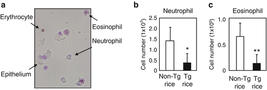
Inflammatory granulocytes in the nasal lavage fluid of mice [1]. (a) A representative staining of nasal lavage fluid. The number of neutrophils (b) and eosinophils (c) in the nasal lavage was significantly lower in the group of mice fed allergy vaccine rice (Tg rice) when compared to that in the control group of mice fed non-transgenic rice (Non-Tg rice). **p < 0.01 and *p < 0.05 for the group of mice fed Tg rice in comparison with the group of mice fed Non-Tg rice

3.4.4 Sneezing
-
1.
Sensitize mice twice with the total protein extract of Japanese cedar pollen as described in Subheading 3.3, steps 8 and 9.
-
2.
Dissolve the total protein extract of Japanese cedar pollen at 1 μg/ml in PBS.
-
3.
At 14 days after the second sensitization, administer 20 μl pollen protein prepared as in step 2 via the intranasal route (10 μl to each nostril).
-
4.
Repeat the intranasal challenge as in step 3 once a day for 1 week.
-
5.
After the last intranasal challenge, count the number of sneezing observed in 2–5 min.
4 Notes
-
1.
For optimized translation in rice seed, the target gene DNAs can be designed using codons frequently used in the genes coding for rice seed storage proteins [1].
-
2.
In the entry clone plasmids, the Cfl9I site locates at the variable region of the C-terminal of glutelin acidic subunits; therefore the target gene DNAs are expressed as a fusion protein with glutelin acidic subunits.
-
3.
The binary plasmid CSP mALS 43 GW contains the mutated rice acetolactate synthase (mALS) marker gene, which is expressed under the control of the rice callus-specific promoter (CSP). Compared to the hygromycin-based selection, CSP:mALS-based callus-specific selection offers higher accumulation levels of the target gene products in transgenic rice seed [2].
-
4.
The details for the management of Agrobacterium cells and rice calli for rice transformation are described previously [3].
-
5.
The target antigen should be prepared as a soluble form in non-denaturing buffer conditions, since the allergen-IgE binding is thought to be conformation dependent [4].
-
6.
The total amount of beta-hexosaminidase released from the RBL-2H3 cells is obtained by lysing the RBL-2H3 cells in the presence of 0.2 % Triton-X100, and is used for the calculation of the percentage of beta-hexosaminidase release in each test.
-
7.
Approximately 2.0–2.5 g rice powder is daily consumed by each BALB/c mouse at age of 6–10 weeks when the mouse can access to normal chow and drinking water ad libitum.
-
8.
Recombinant mouse IgE can enhance the induction of Japanese cedar allergen-specific IgE responses [5].
-
9.
Cytokines, including IL-4, IL-5, IL-6, IL-10, IL-17, IFN-gamma, and TGF-beta, can be measured using commercially available ELISA kits according to the manufacturer’s instructions.
-
10.
Total protein extract of Japanese cedar pollen can be biotinylated by using a biotin protein labeling kit according to the manufacturer’s instructions.
References
Wakasa Y, Takagi H, Hirose S et al (2013) Oral immunotherapy with transgenic rice seed containing destructed Japanese cedar pollen allergens, Cry J 1 and Cry J 2, against Japanese cedar pollinosis. Plant Biotechnol J 11:66–76
Wakasa Y, Ozawa K, Takaiwa F (2009) Higher-level accumulation of foreign gene products in transgenic rice seeds by the callus-specific selection system. J Biosci Bioeng 107:78–83
Wakasa Y, Takaiwa F (2012) Use of a callus-specific selection system to develop transgenic rice seed accumulating a high level of recombinant protein. Methods Mol Biol 847:467–479
Midoro-Horiuti T, Schein CH, Mathura V et al (2006) Structural basis for epitope sharing between group 1 allergens of cedar pollen. Mol Immunol 43:509–518
Takagi H, Hiroi T, Yang L et al (2005) A rice-based edible vaccine expressing multiple T cell epitopes induces oral tolerance for inhibition of Th2-mediated IgE responses. Proc Natl Acad Sci U S A 102:17525–17530
Author information
Authors and Affiliations
Corresponding author
Editor information
Editors and Affiliations
Rights and permissions
Copyright information
© 2016 Springer Science+Business Media New York
About this protocol
Cite this protocol
Takagi, H., Takaiwa, F. (2016). Production of Rice Seed-Based Allergy Vaccines. In: Thomas, S. (eds) Vaccine Design. Methods in Molecular Biology, vol 1403. Humana Press, New York, NY. https://doi.org/10.1007/978-1-4939-3387-7_40
Download citation
DOI: https://doi.org/10.1007/978-1-4939-3387-7_40
Published:
Publisher Name: Humana Press, New York, NY
Print ISBN: 978-1-4939-3385-3
Online ISBN: 978-1-4939-3387-7
eBook Packages: Springer Protocols