
Abstract
Chemoresistance is a major challenge for cancer therapy and drives tumor relapse. The emergence, within the treated tumor mass, of specific cancer cell subpopulations endowed with high tolerance to the microenvironment stress induced by therapy is being growingly recognized as a mechanism of tumor progression. To obtain detailed information with regard to the pathways underlying survival, expansion, and microenvironmental cross talk of such chemoresistant cell subpopulations may be instrumental for cancer chemoprevention. Additionally, the obtained cell subpopulations may be used for direct screening of cancer chemopreventive compounds, in appropriate experimental settings. Here we report detailed experimental procedures that we and others have setup in order to obtain cell cultures enriched for chemoresistant cells from both malignant pleural mesothelioma specimens and primary cell cultures. We provide indications for the purification and characterization of those chemoresistant cell populations and to generally validate the obtained enriched cell populations for their chemoresistance.
Access provided by CONRICYT – Journals CONACYT. Download protocol PDF
Key words
1 Introduction
Chemotherapy treatment represents a very important aspect of cancer management. However, it appears more and more evident that development of resistance to therapy greatly influences tumor relapse and negatively shapes patient prognosis. One mechanism of tumor resistance which is gaining growing attention is the emergence of specific chemoresistant cell subpopulations within the tumor mass [1]. This is of translational relevance, since combined therapies including stemlike cell-targeting agents exhibit improved efficacy [2, 3]. A hierarchical organization and micro environmental control may underlie the function of such cell subpopulations. We have shown that pemetrexed and cisplatin treatment of malignant pleural mesothelioma (MPM) cell lines and primary cultures trigger the emergence of cell subpopulations endowed with chemoresistance properties (MPM-CICs-chemotherapy-induced cells). The latter represent a small fraction of unsorted, untreated MPM cell populations and their number is increased by pemetrexed (and cisplatin) treatment [4] (Fig. 1). MPM-CICs exhibit high levels of aldehyde dehydrogenase (ALDH) activity and can be tracked in unfractionated tumor cell populations by means of this (Fig. 1). Purified ALDHbright MPM cells are chemoresistant as compared to ALDHlow cells (Fig. 2): additionally, their number inversely correlates with survival of xenografted host mice [4]. The present chapter aims at describing the protocols we and others have recently setup to identify, enrich and characterize chemoresistant cell subpopulations (ALDHbright cells) from both cell lines and primary cultures . We (and others) have found that the criteria listed below identify cell subpopulations with chemoresistance properties in vitro and in vivo, enrichment for early-differentiation stemlike markers, and ability to reconstitute tumor heterogeneity in vitro and in vivo. The protocols reported here have been applied to successfully isolating mesothelioma [4, 5] and lung cancer (unpublished) cell subpopulations from both cell lines and primary samples. Additionally, we report procedures for the stable selection and enrichment for chemoresistant cell subpopulations.

Pemetrexed treatment induces chemoresistant mesothelioma cell subpopulations. Upper. Representative micrographs of vehicle- or pemetrexed-treated mesothelioma cells. Lower. Quantitation of ALDHbright cells by FACS from the same cells as in upper panel

Features of purified ALDHbright mesothelioma cells. (a) Scheme used for FACS -based sorting of ALDHbright and ALDHlow cells. (b) The ALDHbright cells are chemoresistant. Clonogenic assay . Representative micrographs of CFA assays. Adapted with permission from Oncogene. 2012;31(26):3148–3163
2 Materials
This is a general protocol for the isolation, selection, and maintenance of primary MPM and lung cultures of chemoresistant cells. The resulting cell subpopulations can be used as a tool for the identification of tumor-initiating cells and early progenitor-targeting drugs [6]. The protocol is suitable for both solid specimens and pleural effusion which will be discusses in separate sections below.
2.1 Disaggregation and Cell Culture Reagents
-
1.
PBS: Phosphate-buffered saline. Dissolve the following in 800 ml distilled H2O: 8 g of NaCl, 0.2 g of KCl 1.44 g of Na2HPO4, 0.24 g of KH2PO4. Adjust pH to 7.4 with HCl. Adjust volume to 1000 ml with additional distilled H2O. Sterilize by autoclaving.
-
2.
Collagenase type IV (300 U/ml). Weigh 100 mg of Collagenase Type IV powder and transfer to 150 ml DMEM-F12 medium. When the collagenase is completely dissolved filter-sterilize the solution (0.22 μm) and tighten the cap (the solution is stable for 14 days after preparation at 4 °C). Prepare the working dilution in DMEM-F12 + 50 mM HEPES cell culture medium according to the specific activity indicated from the manufacturer.
-
3.
Hyaluronidase (100 U/ml). Use Type IV-S from bovine testes (cell culture or embryo-tested). Prepare a stock solution at 10 mg/ml in DMEM-F12 + 50 mM HEPES cell culture medium. Filter-sterilize (0.22 μm), aliquot, and store at −20 °C. The stock solution is stable for 3 months. Prepare the working dilution in DMEM-F12 + 50 mM HEPES cell culture medium according to the specific activity indicated from the manufacturer.
-
4.
Red blood lysis buffer. Dissolve the following in 100 ml distilled H2O:NH4Cl 8.02 g; NaHCO3 (sodium bicarbonate) 0.84 g; EDTA (disodium) 0.37 g. Store at 4 °C for 6 months. Prepare the working solution by diluting 10n times the stock solution in distilled water. Keep cold until use.
-
5.
FBS: Non-heat-inactivated fetal bovine serum.
-
6.
Digestion medium: DMEM-F12 (1:1) + GLUTAMAX supplemented with 1 % BSA-FAF and 5 μg/ml human insulin.
-
7.
Growth medium: DMEM F12 (1:1) + GLUTAMAX supplemented with 5 % non-heat-inactivated FBS, insulin (5 μg/ml).
-
8.
Selection medium: DMEM F12 (1:1) + GLUTAMAX supplemented with 5 % non-heat-inactivated FBS.
-
9.
Freezing medium: 90 % non-heat-inactivated FBS-10 % DMSO.
-
10.
Human recombinant insulin. Dissolve insulin in cell culture grade water at 1–10 mg/ml. Adjust the pH to 2.0–3.0 with diluted HCl. Filter using a low protein-binding filter with a pore size of 0.2 μm. Store at −20 °C for 2 months.
-
11.
BSA-FAF: Bovine serum albumin-fatty acid free.
-
12.
Ciprofloxacin.
-
13.
ACCUTASE Cell detachment solution.
-
14.
Trypan Blue Cell Staining Reagent. Weight 0.2 g Trypan Blue in 99.8 ml distilled water. Filter at 0.22 μm. Dilute the stock solution five times in PBS1× before cell staining.
-
15.
ALDEFLUOR kit (Stem Cell Technologies).
-
16.
SYTOX Dead cell staining reagent.
-
17.
Pemetrexed. Dissolve pemetrexed disodium initially in DMSO at a concentration of 4 mg/ml and further dilute with cell culture medium to the desired concentration.
-
18.
Cisplatin. Dissolve in DMSO at 50 mg/ml and further dilute with cell culture medium to the desired concentration.
2.2 Equipment
-
1.
Scalpels and microdissecting forceps.
-
2.
5 ml Pasteur pipette.
-
3.
15 ml centrifuge tubes.
-
4.
50 ml centrifuge tubes.
-
5.
Centrifuge capable of running at ≥300 × g.
-
6.
Nylon mesh (70 μm).
-
7.
Cell culture setup.
-
8.
CORNING #3261 for 100 mm Ultralow attachment dishes or alternatively, sterile Petri dishes not treated for cell culture.
-
9.
Polycarbonate FACS tubes.
-
10.
A suitable cytofluorimeter for FACS analysis.
-
11.
A suitable FACS -based sorter.
3 Methods
3.1 Isolating MPM Cells from Clinical Specimens
3.1.1 Procedure for Isolating MPM Cells from Surgical Specimens ( See Notes 1 and 2 )
-
1.
Wash the tumor specimen three times with PBS1× supplemented with ciprofloxacin 4 mg/ml. Submerge three times for 5 min the sample in three different 50 ml tubes filled with 25 ml of antibiotic solution. To disaggregate the solid tumor follow three sequential steps (in a tissue-culture sterile hood) (see Note 3 ).
-
2.
Manually cut the solid tumor into ≤1.5 mm pieces with scalpels in a sterile 60 mm Petri dish with 1 ml PBS1×.
-
3.
Enzymatic disaggregation: Resuspend tumor pieces in a T-25 cell culture flask with 5 ml of digestion medium. After resuspension, add collagenase (final concentration 50 U/ml) and hyaluronidase (final concentration 20 U/ml) to the tumor suspension and leave cells in the incubator for 2 h at 37 °C, 5 % CO2. Every 15 min resuspend the semi-digested tumor with a 5 ml sterile Pasteur pipette by gently pipetting up and down to disperse tumor pieces.
-
4.
Filter the digested material through a sterile nylon mesh (70 μm) in a 50 ml tube. Wash the filter with PBS1× and collect the flow-through.
-
5.
Transfer the filtered material to a 15 ml centrifuge tube.
-
6.
Spin at 300 × g for 10 min at room temperature (RT).
-
7.
Resuspend the pellet in growth medium supplemented with ciprofloxacin (4 μg/ml) (see Note 4 ). Assess the number of live cell with Trypan Blue exclusion method.
-
8.
Seed cells in low-adhesion cell culture dishes at a cell density ≥1–1.5 × 10⁶ cells/ml (see Note 5 ). Grow cells for 10 days by adding 25 % fresh medium every 3 days. After 10 days, a relatively homogeneous population of mesothelioma cells (virtually devoid of adhering macrophages, lymphocytes, fibroblasts) [5] can be observed in culture (see Note 6 ).
3.1.2 Procedure for Isolating MPM Cells from Pleural Effusions
-
1.
Collect the pleural effusion in 15 ml FALCON tubes diluted 1:1 with PBS1× supplemented with ciprofloxacin 4 μg/ml.
-
2.
Harvest cells by centrifugation at 300 × g for 10 min at room temperature (RT). Keep the cell-free medium (supernatant–pleural effusion) (see Note 7 ). Filter (0.22 μM) the supernatant-pleural effusion for subsequent use.
-
3.
Resuspend cells in red blood lysis buffer (10 bed pellet volumes). Incubate for 5 min at room temperature (RT).
-
4.
Centrifuge (300 × g for 10 min) and discard supernatant.
-
5.
Resuspend the pellet in growth medium supplemented with ciprofloxacin (4 μg/ml) and add 30 % (vol/vol) of the previously collected cell-free conditioned medium (from step 2). Count total live cell number with Trypan Blue.
-
6.
Seed cells in low-adhesion cell culture dishes at a cell density ≥1–1.5 × 10⁶ live cells/ml. Size of the dish must be chosen according to the available number of cells in order to achieve the desired concentration. Grow cells for 10 days by adding 25 % fresh medium every 3 days. After 10 days, a relatively homogeneous population of mesothelioma cells (virtually devoid of adhering macrophages, lymphocytes, fibroblasts but still comprising both adherent and floating elements) can be observed in culture (Fig. 3).
Fig. 3 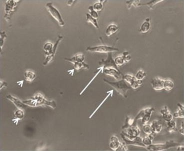
Representative micrograph of a MPM cell culture (from a malignant pleural effusion) at 2 weeks after seeding. Arrows: adherent, fibroblast-like cells. Arrowheads: loosely adherent, rounded cells. Reproduced with permission from http://www.bio-protocol.org/e285
-
7.
The MPM primary cultures can be propagated for a limited length of time (8–12 weeks) as follows.

3.2 Propagation of MPM Cultures
-
1.
Collect cell culture medium in a centrifuge tube (see Note 8 ). Wash the adherent cells with 3–5 ml of PBS1× and add it to the collected cell culture medium. This contains loosely adherent or floating cells. PBS1× should be ≤30 % final volume in the collection tube.
-
2.
Wash cells again with PBS1×, discard PBS1× and add Accutase (1.5 ml/dish).
-
3.
Incubate cells in the incubator for 5 min at 37 °C, 5 % CO2. Harvest the detached cells with the collected cell culture medium/PBS1× washing from step 1. Collect cells by centrifugation at 300 × g for 10 min at room temperature (RT). Do not discard the supernatant.
-
4.
Resuspend the pellet in growth medium. Count total live cell number with Trypan Blue exclusion method.
-
5.
Seed cells in low-adhesion cell culture dishes at a density ≤0.5 × 10⁶ live cells/ml. To achieve the required cell concentrations dilute the harvested cells with the previously collected supernatant. The dilution medium must represent ≥of the 30 % of the final cell culture volume in the dish.
3.3 Selection of Chemoresistant Cell Subpopulations
3.3.1 Determining the CC50 (See Note 9 )
-
1.
Seed a small aliquot of the obtained cell populations (pooled: adherent and floating cells—“probe cells”) in 96 wells at 1500-cell well in selection medium. Treat the cells 24 h later with at least 9–12 points of doses (we usually test cisplatin + pemetrexed, a current line of treatment for MPM) in duplicate wells (see Note 10 ).
-
2.
At 24, 48, and 72 h from the treatment evaluate viability by either Trypan Blue counting of detached cells or by SYTOX staining of the detached cells by FACS (see Notes 11 and 12 ).
-
3.
Calculate CC50 (see Note 13 ).
3.3.2 Start Selection for Chemoresistant Cell Subpopulation
-
1.
After having empirically determined the CC50, add the appropriate volume of selecting agents (for MPM: cisplatin + pemetrexed) to the larger cell culture without further addition of fresh cell culture medium. Incubate for 7 dd without renewing the culture medium.
-
2.
After 7 dd, a sharp increase of chemoresistant cell subpopulations is expected [4]. Such cell subpopulations may be analyzed by FACS and enriched by FACS sorting for their aldehyde dehydrogenase (ALDH) activity components as follows.
3.4 FACS-Based Identification of ALDHbright Cells
-
1.
Collect whole-cell populations (adherent + loosely attached cells) by centrifugation. Detach the adherent fraction by Accutase treatment as previously described.
-
2.
Centrifuge the cell suspension at 300 × g for 10 min to pellet the cells.
-
3.
Discard the supernatant.
-
4.
Resuspend the cell pellet with 10 ml of PBS1× to remove residual Accutase.
-
5.
Centrifuge the cell suspension as before. Aspirate and discard the rinse solution.
-
6.
Resuspend the cells at 1 × 10⁶ cells/ml (range 0.5–1 × 10⁶ cells/ml) at room temperature (15–25 °C) with ice cold ALDEFLUOR® assay buffer.
-
7.
For each sample, have ready a test (ALDH substrate, BAA: BODIPY®-aminoacetate, 1:200) and a “control” (15 UM) ALDH inhibitor:DEAB, diethylaminobenzaldehye + BAA).
-
8.
Incubate the “test” and “control” samples between for 45 min (range 30–60 min) in a 37 °C 5% CO2 incubator, light protected. Allow free diffusion of the oxygen/CO2 (keep the tubes uncapped!).
-
9.
Following incubation, centrifuge the “test” and “control” tubes at 4 °C for 5 min at 300 × g. Carefully aspirate the supernatant without disturbing the cell pellet. Resuspend the cell pellet in 0.5 ml of ice cold ALDEFLUOR® assay buffer and place samples immediately on ice. To exclude dead cells from data acquisition, add SYTOX® Red Dead Cell Stain (Life Technologies) which allows nonproblematic detection of both ALDHbright and dead/dying cells with compromised cell membrane permeability.
-
10.
For details on flow cytometer setup and data acquisition, refer to the product information sheet provided with the ALDEFLUOR® Assay Kit or go to: http://www.stemcell.com/technical/01700-PIS.pdf. (see Note 14 ). Briefly, the cells endowed with high ALDH activity will be gated as the brightest cells on the FITC axis (test tube) whose fluorescence disappears after inhibition of the ALDH enzyme with DEAB (“control” tube). Cells acquiring fluorescence independently of DEAB treatment do not possess ALDH activity but rather fluoresce because of passive diffusion of the ALDH substrate (BAA). FACS-based purification of ALDHbright cells. Perform FACS-sorting according to the manufacturer’s instructions of your FACS sorting instrument.
3.5 Freezing or Propagation of Purified ALDHbright Cells
The obtained cells can be used immediately after sorting for gene expression/microRNA profiling , frozen or seeded for propagation and further selection.
-
1.
Freezing of FACS-sorted cells.
Freshly sorted cells can be collected by centrifugation and directly resuspended in freezing medium (see Note 15 ).
-
2.
Propagation of the FACS-sorted ALDHbright cells (see Notes 16 – 20 ).
For the propagation of the sorted cells, please refer to point Subheading 3.2.
4 Notes
-
1.
Please note that all the procedures described below involving the use of patient samples must be pre-approved by the Ethical Committee.
-
2.
Critical step: The time interval from tumor resection and processing must be kept to a minimum, ideally ≤2 h.
-
3.
Critical step: To prevent undesired contamination, work under sterile condition at all steps and whenever possible. Do not allow alcohol, staining reagents or disinfectants to come in contact with the specimen.
-
4.
Ciprofloxacin prevents contamination of the material from non-sterile handling of the tumor specimens during the harvesting of the sample.
-
5.
The obtained tumor digests consist initially of a heterogeneous population, comprising but not limited to mesothelioma cells, macrophages, immune infíltrate, stromal cells, adipocytes, and remnant red blood cells. However, within 72–96 h from seeding most of the cells in culture consist of mesothelioma cells, since the mentioned accompanying cell subpopulations will not propagate in the experimental conditions used here, as revealed by morphological observations and clonogenic assays (55) (and unpublished observations). The obtained populations have been shown to originate MPM-like tumors when injected into NOD/SCID mice with very high resemblance to the originating tumor [4].
-
6.
A typical yield of 1 × 10⁶ cells can be obtained from a 100 mg solid specimen. A typical yield of 10 × 106 cells can be obtained from 30 to 50 ml freshly collected pleural effusion (after removal of red blood cells, RBC).
-
7.
The reason to keep the conditioned medium during propagation of the MPM cultures is its enrichment for growth factors and cytokines produced by the MPM cells which favors survival and propagation of the cells especially at early steps of establishing the culture [7].
-
8.
For the propagation of the primary cell cultures, always collect both floating (or loosely adherent) and adherent cell subpopulations. It is very important to keep conditioned medium during harvesting of the cells and to add it back to cell culture during the establishment of primary cultures. All the volumes listed below refer to 100 mm dishes. Please vary volumes of solutions according the size of the cell culture dishes used.
-
9.
This protocol requires, first, to determine the sensitivity (CC50) of the isolated cell cultures (a probe aliquot of the obtained culture) to the chemotherapy agents. The CC50 is defined as the concentration of drug (alone or in combination) capable of eliciting a 50 % reduction in the number of viable cells. After that, a larger fraction of the culture will be selected, at the identified CC50 dosages, for 7 dd to elicit the emergence of chemoresistant cell subpopulations
-
10.
Please include appropriate vehicles and do not exceed 0.5 % final concentration if DMSO or other organic solvents are used as a vehicle.
-
11.
Please note that this assay will not distinguish, at this stage, between cytostatic or cytotoxic effects of the treatments.
-
12.
Other viability assays can be used to evaluate the effect of the treatments. However, we have empirically found the mentioned to be of broader utility as being not influenced by metabolic status of the cells or changes in plasma membrane composition induced by some chemotherapy agents.
-
13.
CC50 can be calculated manually or by using commercially available software, such as KALEIDAGRAPH, SIGMAPLOT, or GRAPHPAD. In case of combined treatment, the drug interactions must be taken in consideration to unravel “additive” or synergistic interaction between the treatments.
-
14.
The aldehyde dehydrogenases (ALDH) are enzymes ubiquitously expressed in developing tissues and adult liver where they act as either as a detoxifying enzymes and as a metabolic modulators. Cancer cells endowed with high ALDH activity (generally named ALDHbright because of the intracellular accumulation of a fluorescent ALDH substrate, as opposed to ALDHlow, which do not exhibit substrate accumulation) have been isolated from a variety of solid tumors, including breast, lung, ovary, prostate, osteosarcoma, and glioblastoma and shown to contribute protumorigenic features [7–9].
-
15.
Alternatively, a serum-free, methylcellulose containing-freezing medium can be used. No differences in the biological features of the sorted cells (clonogenicity and chemoresistance) were observed when comparing the two methods. However, the lack of DMSO in the freezing medium significantly affects the viability of the frozen cells (from four representative MPM primary cultures).
-
16.
This protocol may be generally applicable to other solid tumors (breast, prostate, glioblastoma) proven that important differences residing in the different biological context of the tissues examined will be taken into consideration (i.e., hormone sensitivity). In order to fulfill the definition of “chemoresistant,” the isolated cell subpopulations must exhibit resistance (time dependent and over a broad range of concentrations) to the currently used chemotherapeutics, specific for the tumor/tissue studied. Resistance to therapeutics should be defined as relative to untransformed nonimmortalized (if available) cell cultures derived from apparently unaffected tissue or at least from immortalized, untransformed cell cultures.
-
17.
The isolated cell subpopulations should be at least partially propagable under the selective conditions mentioned. The selected cell subpopulations should exhibit ability to reconstitute the cell culture heterogeneity in vitro or the tissue heterogeneity in vivo. This indicates potential for tumor relapse.
-
18.
Inter-patient variability. Please keep in mind that working with patient samples is intrinsically complex. First, the rate of successful isolation and propagation can vary (we obtain 70 % of successful isolation/maintenance of primary cultures). Second. A significant inter-patient variability (due to different underlying health status, to different stages of the disease, to the presence of potentially undiagnosed lesions impinging on altered tumor microenvironment) is normally observed when patient-derived primary cell cultures for a specific biological function. The latter problem can be overcome by using larger number of specimens in order to obtain statistical support to the observed phenomena.
-
19.
The FACS-based selection for high aldehyde dehydrogenase-expressing cells (ALDHbright) identifies a heterogeneous population of chemoresistant cells. The ALDHbright cell population is intrinsically heterogeneous, in terms of cell cycle status, expression of membrane markers and, possibly, pathway activation. However, the described procedure undoubtedly enriches for chemoresistant cell subpopulations, as described by us and others in MPM, lung, breast, GBM, and prostate.
-
20.
Be aware that, as shown by us and others [4, 8, 9], the isolated ALDHbright cells do spontaneously generate ALDHlow cells in culture in a unidirectional way. This is partially independent of the drug selection process. For MPM, ALDHbright cell number drops significantly within the first 2 weeks of culture, after FACS sorting.
References
Abdullah LN, Chow EK (2013) Mechanisms of chemoresistance in cancer stem cells. Clin Transl Med 2(1):3
Cojoc M, Mabert K, Muders MH et al (2015) A role for cancer stem cells in therapy resistance: Cellular and molecular mechanisms. Semin Cancer Biol 31:16–27
Vidal SJ, Rodriguez-Bravo V, Galsky M et al (2014) Targeting cancer stem cells to suppress acquired chemotherapy resistance. Oncogene 33(36):4451–4463
Canino C, Mori F, Cambria A et al (2012) SASP mediates chemoresistance and tumor-initiating-activity of mesothelioma cells. Oncogene 31(26):3148–3163
Cioce M, Canino C, Goparaju C et al (2014) Autocrine CSF-1R signaling drives mesothelioma chemoresistance via AKT activation. Cell Death Dis 5:e1167
Cioce M, Gherardi S, Viglietto G et al (2010) Mammosphere-forming cells from breast cancer cell lines as a tool for the identification of CSC-like- and early progenitor-targeting drugs. Cell Cycle 9(14):2878–2887
Lanfrancone L et al (1992) Human peritoneal mesothelial cells produce many cytokines (granulocyte colony-stimulating factor [CSF], granulocyte-monocyte-CSF, macrophage-CSF, interleukin-1 [IL-1], and IL-6) and are activated and stimulated to grow by IL-1. Blood 80(11):2835–2842
Cortes-Dericks L, Froment L, Boesch R et al (2014) Cisplatin-resistant cells in malignant pleural mesothelioma cell lines show ALDH(high)CD44(+) phenotype and sphere-forming capacity. BMC Cancer 14:304
Luo Y, Dallaglio K, Chen Y et al (2012) ALDH1A isozymes are markers of human melanoma stem cells and potential therapeutic targets. Stem Cells 30(10):2100–2113
Acknowledgement
M.C. was supported by the NYU Cancer Institute Cancer Center Support Grant’s Developmental Project Program (P30CA016087).
Author information
Authors and Affiliations
Corresponding author
Editor information
Editors and Affiliations
Rights and permissions
Copyright information
© 2016 Springer Science+Business Media New York
About this protocol
Cite this protocol
Canino, C., Cioce, M. (2016). Isolation of Chemoresistant Cell Subpopulations. In: Strano, S. (eds) Cancer Chemoprevention. Methods in Molecular Biology, vol 1379. Humana Press, New York, NY. https://doi.org/10.1007/978-1-4939-3191-0_13
Download citation
DOI: https://doi.org/10.1007/978-1-4939-3191-0_13
Publisher Name: Humana Press, New York, NY
Print ISBN: 978-1-4939-3190-3
Online ISBN: 978-1-4939-3191-0
eBook Packages: Springer Protocols